Advertisement

Main menu

  • Home
  • Content
    • Current Issue
    • Accepted Manuscripts
    • Article Preview
    • Past Issue Archive
    • AJNR Case Collection
    • Case of the Week Archive
    • Classic Case Archive
    • Case of the Month Archive
  • Special Collections
    • Spinal CSF Leak Articles (Jan 2020-June 2024)
    • 2024 AJNR Journal Awards
    • Most Impactful AJNR Articles
  • Multimedia
    • AJNR Podcast
    • AJNR Scantastics
    • Video Articles
  • For Authors
    • Submit a Manuscript
    • Author Policies
    • Fast publishing of Accepted Manuscripts
    • Graphical Abstract Preparation
    • Manuscript Submission Guidelines
    • Imaging Protocol Submission
    • Submit a Case for the Case Collection
  • About Us
    • About AJNR
    • Editorial Board
  • More
    • Become a Reviewer/Academy of Reviewers
    • Subscribers
    • Permissions
    • Alerts
    • Feedback
    • Advertisers
    • ASNR Home
  • Other Publications
    • ajnr

User menu

  • Alerts
  • Log in

Search

  • Advanced search
American Journal of Neuroradiology

American Journal of Neuroradiology

ASHNR American Society of Functional Neuroradiology ASHNR American Society of Pediatric Neuroradiology ASSR
  • Alerts
  • Log in

Advanced Search

  • Home
  • Content
    • Current Issue
    • Accepted Manuscripts
    • Article Preview
    • Past Issue Archive
    • AJNR Case Collection
    • Case of the Week Archive
    • Classic Case Archive
    • Case of the Month Archive
  • Special Collections
    • Spinal CSF Leak Articles (Jan 2020-June 2024)
    • 2024 AJNR Journal Awards
    • Most Impactful AJNR Articles
  • Multimedia
    • AJNR Podcast
    • AJNR Scantastics
    • Video Articles
  • For Authors
    • Submit a Manuscript
    • Author Policies
    • Fast publishing of Accepted Manuscripts
    • Graphical Abstract Preparation
    • Manuscript Submission Guidelines
    • Imaging Protocol Submission
    • Submit a Case for the Case Collection
  • About Us
    • About AJNR
    • Editorial Board
  • More
    • Become a Reviewer/Academy of Reviewers
    • Subscribers
    • Permissions
    • Alerts
    • Feedback
    • Advertisers
    • ASNR Home
  • Follow AJNR on Twitter
  • Visit AJNR on Facebook
  • Follow AJNR on Instagram
  • Join AJNR on LinkedIn
  • RSS Feeds

Welcome to the new AJNR, Updated Hall of Fame, and more. Read the full announcements.


AJNR is seeking candidates for the position of Associate Section Editor, AJNR Case Collection. Read the full announcement.


Review ArticleSpine
Open Access

Spinal Vascular Shunts: A Patterned Approach

M.P. Kona, K. Buch, J. Singh and S. Rohatgi
American Journal of Neuroradiology December 2021, 42 (12) 2110-2118; DOI: https://doi.org/10.3174/ajnr.A7312
M.P. Kona
aFrom the Division of Neuroradiology (M.P.K.), Department of Radiology, Hunter Holmes McGuire VA Medical Center, Richmond, Virginia
  • Find this author on Google Scholar
  • Find this author on PubMed
  • Search for this author on this site
  • ORCID record for M.P. Kona
K. Buch
bDivision of Neuroradiology (K.B.), Department of Radiology, Massachusetts General Hospital, Harvard Medical School, Boston, Massachusetts
  • Find this author on Google Scholar
  • Find this author on PubMed
  • Search for this author on this site
  • ORCID record for K. Buch
J. Singh
cDivision of Neuroradiology (J.S., S.R.), Department of Radiology, University of Massachusetts Medical School, Worcester, Massachusetts
  • Find this author on Google Scholar
  • Find this author on PubMed
  • Search for this author on this site
  • ORCID record for J. Singh
S. Rohatgi
cDivision of Neuroradiology (J.S., S.R.), Department of Radiology, University of Massachusetts Medical School, Worcester, Massachusetts
  • Find this author on Google Scholar
  • Find this author on PubMed
  • Search for this author on this site
  • ORCID record for S. Rohatgi
  • Article
  • Figures & Data
  • Info & Metrics
  • Responses
  • References
  • PDF
Loading

Abstract

SUMMARY: Spinal vascular shunts, including fistulas and malformations, are rare and complex vascular lesions for which multiple classification schemes have been proposed. The most widely adopted scheme consists of 4 types: type I, dural AVFs; type II, intramedullary glomus AVMs; type III, juvenile/metameric AVMs; and type IV, intradural perimedullary AVFs. MR imaging and angiography techniques permit detailed assessment of spinal arteriovenous shunts, though DSA is the criterion standard for delineating vascular anatomy and treatment planning. Diagnosis is almost exclusively based on imaging, and features often mimic more common pathologies. The radiologist’s recognition of spinal vascular shunts may improve outcomes because patients may benefit from early intervention.

ABBREVIATIONS:

ASA
anterior spinal artery
CS
classification scheme
IPAVF
intradural perimedullary arteriovenous fistula
PSA
posterior spinal artery
SDAVF
spinal dural arteriovenous fistula
SGAVM
spinal “glomus” arteriovenous malformation
SJAVM
spinal juvenile (metameric) arteriovenous malformation
SVS
spinal vascular shunt

Spinal vascular shunts (SVSs) including arteriovenous fistulas (AVFs) and malformations (AVMs) are rare lesions accounting for 3%–4% of intradural spinal lesions.1⇓⇓-4 The SVS presents a challenge from both an imaging and management perspective. Since the first SVS classification scheme (CS) was proposed in 1971 by Di Chiro et al,5 physicians from various specialties have proposed CSs on the basis of anatomy and/or management considerations typically relevant to the authors’ fields of expertise.5⇓⇓⇓⇓-10 The current, most widely referenced CS is derived from the works of multiple authors.5⇓⇓-8 This article offers a review of the commonly referenced historic classification schemes, clinical symptomology, imaging findings, differential diagnoses, and management considerations.

Normal Spinal Vascular Anatomy

The cord is supplied by intramedullary sulcal branches of the anterior spinal artery (ASA) and radial perforators from the pial-based vasacorona (Fig 1).11 The ASA and paired posterior spinal arteries (PSAs) originate from the intradural vertebral arteries and sometimes the posterior inferior cerebellar arteries. The ASA, PSA, and vasocorona receive collateral flow from approximately 7 or 8 medullary arteries, which share a common origin with the radicular and dural arteries supplying the nerve roots and dural sleeves. These radiculomedullary arteries arise from ventral spinal branches of segmental arteries. The well-known radiculomedullaris magna artery of Adamkiewicz provides collateral flow to the thoracolumbar cord, typically arising on the left between T9 and T12.2

FIG 1.
  • Download figure
  • Open in new tab
  • Download powerpoint
FIG 1.

A, Normal arterial anatomy: 1, intramedullary branches; 2, sulcal artery; 3, anterior spinal artery; 4, radial perforators; 5, pial arterial plexus; 6, posterior spinal arteries; 7, segmental artery; 8, spinal artery; 9, dorsal branch of the dorsospinal artery; 10, ventral branch of dorsospinal artery; 11, ventral epidural plexus; 12, dorsal epidural plexus; 13, dural artery; 14, radicular artery; 15, ventral radicular artery; 16, dorsal radicular artery; 17, medullary artery. B, Normal venous anatomy: 1, Intramedullary veins; 2, sulcal vein; 3, radial veins; 4, pial venous plexus; 5, anterior spinal vein; 6, posterior spinal veins; 7, medullary vein; 8, ventral radicular vein; 9, dorsal radicular vein; 10, emissary vein; 11, intervertebral vein; 12, branches from ventral epidural venous plexus; 13, branches from dorsal epidural venous plexus.

Normal spinal venous anatomy grossly mirrors that of the arterial counterpart, with single anterior and dual posterior spinal veins coursing through a pial surface plexus. Medullary and radicular veins drain via transdural emissaries to intervertebral, longitudinal efferent, and other level-specific venous channels.4

Historical Classification Schemes

Various proposed SVS CSs have been historically based on anatomic, management, prognostic, or other considerations relevant to the authors’ fields of expertise (Table 1).5,9 Modified versions of earlier SVS CSs have also challenged the scope of included lesions and/or degrees of subclassification.6⇓-8,10 Although no proposal has been universally adopted, the recent emergence of a composite CS appears to seek a compromise.3,12⇓⇓⇓⇓⇓⇓-19 At the time of this publication, the most widely referenced CS reflects the contributions of multiple authors, beginning with types I–III of Di Chiro et al5 based on angiographic morphology (1971). The proposed type IV AVF addition of Heros et al6 (1986); the further subclassification by Gueguen et al,7 of type IV into 3 subtypes based on size and number of arterial feeders; and the proposed subdivision by Spetzler et al8 of type I AVF into subtypes A (single arterial feeder) and B (multiple feeders).

View this table:
  • View inline
  • View popup
Table 1:

Historical spinal vascular shunt classification schemes

View this table:
  • View inline
  • View popup
Table 2:

Imaging findings of spinal vascular shunts types I–IV

Type I: Spinal Dural AVF.

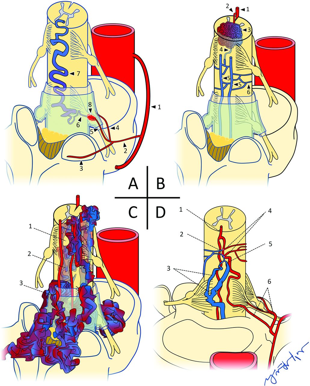
Spinal dural arteriovenous fistulas (SDAVFs) account for 70% of all SVSs (Fig 2A).3,12 Approximately 80% of SDAVFs are found along the dorsal lower thoracic cord surface between levels T6 and L2.6,8,13 Anatomically, an SDAVF is an anomalous intradural communication between a dural artery and radicular vein within the intervertebral foramen without an intervening capillary network. Drainage is typically retrograde via a medullary vein and the pial venous network.13,20,21 SDAVFs are slow-flow lesions, mostly acquired, possibly related to chronic fibrosis and/or venous thrombosis.1,18 Arterial flow is transferred to engorged venous collaterals, producing the angiographic “single coiled vessel” appearance.5

FIG 2.
  • Download figure
  • Open in new tab
  • Download powerpoint
FIG 2.

A, Type I spinal dural AVF: 1, Intercostal artery; 2, spinal artery; 3, dorsal/muscular branch; 4, radicular artery, ventral branch; 5, radicular artery, dorsal branch; 6, radicular vein; 7, engorged perimedullary vein; 8, dural AVF. B, Type II spinal glomus AVM: 1, anterior spinal artery; 2, feeding arterial branch; 3, intramedullary glomus/nidus; 4, draining branch to pial venous network; 5, posterior spinal veins; 6, pial venous network. C, Type III spinal juvenile/metameric AVM: 1, normal cord tissue within the nidal interstices; 2, intramedullary elements of AVM; 3, extramedullary elements of AVM. D, Type IV IPAVF: 1, anterior spinal artery; 2, fistula; 3, multiple dilated perimedullary veins; 4, multiple contributing arterial feeders; 5, medullary artery; 6, ventral and dorsal radicular arteries.

Type II: Spinal Glomus AVM.

Type II spinal “glomus” arteriovenous malformations (SGAVMs) account for 19%–45% of all SVSs. SGAVMs are congenital, high-flow lesions that may be thoracic (51%), cervical (29%), or lumbar/conus (20%) (Fig 2B).1,12,17,22 From a morphologic perspective, Di Chiro et al5 distinguished SGAVMs from SDAVFs by the presence of a “localized vascular plexus confined to a short cord segment.” This intramedullary and/or pial-based nidus of abnormal vascular channels may be supplied by ≥1 branch of the ASA, PSA, vertebral arteries, dural arteries, and/or vasocorona.23

Type III: Spinal Juvenile AVM.

Type III spinal juvenile (metameric) arteriovenous malformations (SJAVMs) are the least common3,12 type of SVS, with estimated frequencies between 5.4% and 9%. Most cases involve the thoracic spine, followed by the cervical spine (Fig 2C).16,24

These voluminous,5 high-flow lesions are sometimes characterized as possessing normal cord tissue within their interstices.2,25,26 Besides the cord, these lesions may involve the bony spine, paraspinal tissues, and skin, sharing a common metameric tissue origin.3,27

Type IV: Intradural Perimedullary AVF.

The intradural perimedullary AVF (IPAVF) was first described by Djindjian et al,28 in 1977, and later proposed as a type IV6 addition to the 1971 CS of Di Chiro et al5 (Fig 2D). IPAVF prevalence is approximately 8%–19% of SVSs.14 The IPAVFs are pial-based lesions supplied by the ASA and/or PSA, sometimes with a small intervening nidus. In 1987, Gueguen et al7 proposed IPAVF types I–III according to the size and number of arterial feeders, now frequently referenced as subtypes IVa, IVb, and IVc.

However, this morphologically based SVS CS is limited in scope because some SVSs remain outside their descriptive criteria. Recent proposals to address this limitation include a location-based CS consisting of 6 lesion types proposed by Zozulya et al9 and a type V extradural AVF proposed by Takai.10

Clinical Symptomology

Type I: Spinal Dural AVF.

The typical presentation is an older (range, 55–60 years of age) man (male/female ratio  = 5:1)13 with a history of nonspecific progressive myelopathic symptoms. The clinical picture is sometimes referred to as Foix-Alajouanine syndrome.4,29 Symptoms are related to cord edema and ischemia resulting from arterialized intravenous pressure competing with cord drainage, though venous thrombosis has also been described.30⇓-32

Type II: Spinal Glomus AVM.

Patients are typically in their second-to-fourth decades of life (mean age, 28 years) without sex predilection.11,23 Approximately 50%–75% of patients present with acute or subacute symptoms related to subarachnoid hemorrhage or, less commonly, venous thrombosis.23,27

Type III: Spinal Juvenile AVM.

Although the low frequency of SJAVMs limits available data, the typical presentation age is younger than 15 years, with a slight male predominance (male/female ratio  = 1.7:1).12,16,26 As reported in a 2014 meta-analysis of 51 cases of SJAVM,16 the most common presentation was a progressive neurologic deficit in 35%, followed closely by acute hemorrhage (31%) and an acute neurologic deficit without hemorrhage (22%). Twelve percent of cases were incidental discoveries. Cobb syndrome refers to the clinical manifestations of SJAVM involving all tissues of the involved metamere.33

Type IV: Intradural Perimedullary AVF.

Although the clinical presentation of IPAVF varies with subtype, approximately 93% of patients present with neurologic deficits, among which 29% are acute, typically resulting from subarachnoid hemorrhage.15,22 A 2013 meta-analysis15 consisting of 213 cases found subtype IVa to be more common in males (male/female ratio = 1.4:1), with a mean age of 46.9 years. Mean ages for subtypes IVb and IVc were 34.3 and 18.7 years, both without sex predilection.

Syndromic associations with IPAVFs have been reported, including Osler-Weber-Rendu, Proteus, Klippel-Trenaunay, and Down syndromes.3,6,15,22,34⇓⇓-37

Imaging Findings

Type I: Spinal Dural Arteriovenous Fistula.

T2-weighted MR imaging depicts serpiginous perimedullary flow voids, typically overlying the dorsal cord and conus, without an intramedullary nidus (Fig 3 and Table 2). Secondary congestive myelopathy is also best-depicted on routine T2-weighted TSE and STIR imaging as nonspecific, hyperintense signal within an expanded cord.38,39 Contrast-enhanced T1 fat-suppression MR imaging or CT may show cord enhancement related to chronic venous congestion and compromise of the blood−spinal cord barrier.40 The presence of a thin T2 hypointense rim may be more specific to SDAVFs, possibly due to the presence of deoxyhemoglobin within dilated peripheral capillaries of the distended cord.14,39⇓-41

FIG 3.
  • Download figure
  • Open in new tab
  • Download powerpoint
FIG 3.

A 79-year-old man with a type I spinal dural AVF. A, Sagittal T2 MR imaging of the thoracic spine shows extensive intramedullary edema as signal hyperintensity (white arrow) throughout the cord. The thin peripheral hypointense rim (yellow rectangle) may reflect deoxyhemoglobin within dilated peripheral capillaries. Serpiginous perimedullary flow voids (blue arrow) are most conspicuous along the dorsal aspect of the thoracic cord. B, Frontal view DSA injection of the left L3 segmental artery (white arrow) shows early filling of an ectatic spinal vein (black arrow).

First-pass MRA may depict the involved dural artery communicating with a prominent arterial network along the dural root sleeve at the level of the fistula, with a prominent early filling radicular vein. Multiphase time-resolved MRA depicts progressive enhancement through the same vascular structures and may increase the rate of successful SDAVF localization.42

Selective DSA injection of the involved segmental artery shows shunted contrast advancing through the fistulous connection with retrograde reflux into a dilated radicular vein and perimedullary venous network.38,39

Type II: Spinal Glomus AVM.

The eccentric (not centered) nidus of a SGAVM shows variable enhancement and may be partially or completely intramedullary (Fig 4 and Table 2).14,43 Surrounding T2-hyperintensity with regional cord expansion may represent edema, gliosis, and/or ischemia.11,26 Subarachnoid or intramedullary signal loss on gradient MR imaging may reflect hemorrhage.26 The presence of prominent radicular venous outflow depicted as intra- and perimedullary flow voids on T2-weighted MR imaging is variable.44

FIG 4.
  • Download figure
  • Open in new tab
  • Download powerpoint
FIG 4.

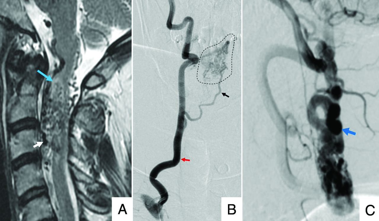
A 45-year-old man with a type II spinal glomus AVM. A, Sagittal T2 MR imaging of the cervical spine shows serpiginous intramedullary and perimedullary flow voids (white arrow), with adjacent cord hyperintensity (blue arrow). B, Frontal view DSA injection of the right vertebral artery (red arrow) shows opacification of a feeding arterial branch (black arrow) and nidal elements (dashed outline). C, Lateral view DSA shows early filling of ectatic perimedullary veins (blue arrow).

Selective DSA injections typically show ≥1 feeding branch originating from the ASA, PSA, or vertebral arteries directed toward the intramedullary nidus. Intranidal aneurysms may opacify in up to one-third of cases. Drainage is typically via veins of the coronal and epidural venous plexuses, which may or may not be engorged.44

Type III: Metameric Spinal AVM.

T2-weighted imaging depicts extensive ectatic intramedullary flow voids with variable involvement of the subarachnoid and epidural spaces, as well as surrounding spinal elements and extraspinal tissues in a metameric distribution (Fig 5 and Table 2).45 Parenchymal cord tissue may be visible within the interstices of the nidus.2,25,26 Cord compression from the markedly dilated extramedullary vascular architecture is also best-depicted on MR imaging.14

FIG 5.
  • Download figure
  • Open in new tab
  • Download powerpoint
FIG 5.

A 14-year-old girl with a type III spinal juvenile/metameric AVM. A, Sagittal T2 MR imaging of the lumbar spine shows numerous ectatic perimedullary and intramedullary flow voids (blue/white arrow). B, Frontal view DSA injection of the right L2 lumbar artery shows opacification of a prominent anterior spinal artery (red arrow), intramedullary and extramedullary nidal elements (black arrow), and early filing of an ectatic perimedullary vein (blue arrow).

Volume-rendered surface reconstructions from CTA source data permit further assessment of soft-tissue involvement, particularly with regard to the bony spinal elements, and are useful for surgical planning.45

Numerous ectactic high-flow intra- and extramedullary shunts are typically present with extremely rapid antegrade drainage via the often-dilated perimedullary and epidural venous pathways.45

Type IV: Intradural Perimedullary AVF.

T2-weighted MR imaging demonstrates prominent perimedullary flow voids typically along the ventral cord surface, in contrast to SDAVFs, which are typically dorsal (Fig 6 and Table 2). Depending on the degree of arterial and venous ectasia, cord compression and displacement may be present. Less consistently, T2 signal hyperintensity reflecting cord edema, ischemia, and/or gliosis may be present, as well as enhancement of the pia arachnoid.35,45

FIG 6.
  • Download figure
  • Open in new tab
  • Download powerpoint
FIG 6.

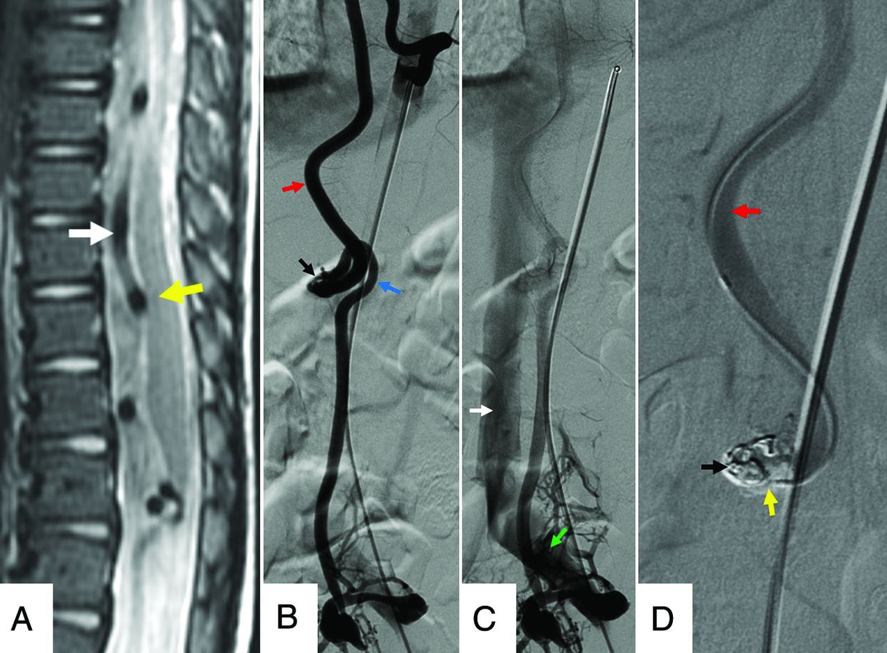
An 11-year-old boy with a type IV intradural-perimedullary spinal AVF. A, Sagittal T2 MR imaging of the thoracic spine shows enlarged, predominantly ventral, perimedullary flow voids (white arrow) indenting the ventral cord (yellow arrow). B and C, Sequential frontal view DSA images from injection of the left T9 segmental artery show contrast progression through the ASA (red arrow), nidus (black arrow), ectatic left radiculomedullary vein (blue arrow), left common iliac vein (green arrow), and inferior vena cava (white arrow). D, Frontal magnified DSA shows a catheter traversing the opacified ASA (red arrow) and embolization coil (black arrow) and Onyx (Covidien) (yellow arrow) material within the nidus. The draining vein no longer opacifies as a result of shunt obliteration.

The angioarchitectural findings of IPAVFs depicted on CTA or MRA depend on the subtype. Findings of subtype IVa lesions include enhancement of a normal-caliber ASA branch and a mildly dilated perimedullary vein along the ventral surfaces of the conus medullaris or filum terminale.45,46

Subtypes IVb and IVc IPAVFs both depict ≥1 arterial feeder and/or shunt, with ectatic perimedullary drainage. Subtype IVc generally has a greater number and size of involved vessels and shunts compared with subtype IVb.46 Selective DSA injection may depict this shunt/vessel multiplicity as an abrupt transition to a larger caliber vessel, with contrast dilution from unopacified converging feeders.47 The rate of contrast progression through the vascular elements increases with subtype order.7

Differential Diagnoses/Mimics

CSF Flow Artifacts.

Sequences using short-TE, thinner slices, and imaging planes perpendicular to the direction of CSF flow can create signal voids mimicking the vascular flow voids related to shunting (Fig 7A).39 Heavily T2-weighted sequences are most likely to distinguish vascular from CSF flow voids.14

FIG 7.
  • Download figure
  • Open in new tab
  • Download powerpoint
FIG 7.

A, A 41-year-old woman with normal CSF flow-related signal voids. Sagittal T2 MR imaging of the thoracic spine shows multiple prominent CSF flow voids (green arrows), mimicking the perimedullary vascular flow voids characteristic of spinal vascular shunts. B, A 71-year-old man with spinal stenosis. Sagittal T2 MR imaging of the lumbar spine shows multiple tortuous, redundant-appearing cauda equina nerve roots (yellow arrow) mimicking the perimedullary flow voids of a spinal vascular shunt above the levels of thecal sac stenoses (white and blue arrows).

Tortuous Redundant Roots.

Severe thecal sac stenosis can result in tortuous cauda equina roots appearing as serpentine areas of perimedullary low signal (Fig 7B).48 The close proximity of these low-signal areas to the level of stenosis can help distinguish this finding from vascular flow voids.

Spinal Vascular Masses.

Vascular masses of the cord may exhibit imaging features similar to the those of the nidus of a type II spinal AVM (Fig 8A).13,26,49 For example, hemangioblastomas may be accompanied by surrounding cord edema and a dilated draining vein. Distinction is made by lesion multifocality, avid enhancement, or syndromic associations such as Von Hippel-Lindau disease.26,50

FIG 8.
  • Download figure
  • Open in new tab
  • Download powerpoint
FIG 8.

A, A 61-year-old man with a cavernous malformation of the cervical cord. Sagittal T2 MR imaging of the cervical spine shows a small intramedullary mass with central hyperintense signal (white arrow) and a peripheral rim of low signal (black arrow), indicative of blood products in various stages of degradation. There is normal signal in the adjacent cord (yellow arrow). B, A 42-year-old woman with a history of Von Hippel-Lindau disease and cervical cord hemangioblastomas. Sagittal T2 MR imaging shows a small intramedullary mass (yellow arrow) with heterogeneous hyperintense internal signal and surrounding cord edema with cord expansion (blue arrow). C, Sagittal contrast-enhanced T1 with fat saturation depicts intense enhancement (white arrows) within multiple lesions.

Mass effect sometimes imparted by cavernous malformations may result in regional venous engorgement.40 However, a thin rim of susceptibility signal loss related to hemosiderin staining, as well as the characteristic “popcorn” signal pattern and the angiographically occult nature, may distinguish a cavernous malformation from a type II spinal AVM.13,51

Collateral Venous Flow.

Obstructive pathologies compromising normal venous drainage with resulting collateral venous engorgement may be difficult to distinguish from the vascular prominence of SVS types lacking an intervening nidus.52 Recognizing characteristic patterns of collateral venous engorgement and identifying a source of obstruction can help distinguish this process from an SVS.

Management

Type I: Dural AVF.

Operative management involves ligation of the fistulized vein through a laminectomy.21 Endovascular management typically involves cannulation of the involved segmental artery followed by embolization of the fistula and draining vein.

Type II: Glomus AVM.

Although the technique varies with lesion location and operator preferences, multilevel laminectomies typically afford sufficient lesion access23,53 for microsurgical resection of extramedullary SGAVM elements. Presurgical embolization may achieve partial lesion obliteration and facilitate intraoperative localization via casting of the embolic material.23,53

Stereotactic radiosurgery is an emerging treatment strategy, with 2 single-center studies demonstrating rates of lesion-size reduction between 50% and 100%, with complete obliteration rates up to 19%.17,54

Type III: Spinal Juvenile AVM.

SJAVMs are not frequently amenable to intervention, and benefits are often brief, with high rates of additional procedures required.1,26,34 When attempted, embolization is generally regarded as first-line therapy for SJAVMs due to high rates of intraoperative hemorrhage.

Type IV: Intradural Perimedullary AVF.

The treatment approach depends on the number and size of the involved arterial feeders and thus the IPAVF subtype. Therapy generally consists of endovascular embolization, surgery, or a combination of both.

CONCLUSIONS

The relatively infrequent and anatomically complex nature of spinal vascular shunts can present diagnostic and therapeutic challenges to radiologic and surgical teams. Assessment of these lesions can be further complicated by the lack of a universally accepted classification scheme. However, a working knowledge of the 4 most frequently described types of spinal vascular shunts permits a useful initial evaluation of these lesions. Imaging is critical, and radiologists play a pivotal role in suggesting a diagnosis and planning selective treatment options. Early diagnosis improves outcomes, because patients may benefit from early intervention.

Footnotes

  • Paper previously presented, in part, as an educational poster in a short digital format at: Annual Meeting of the European Congress of Radiology, July 15–19, 2020; Virtual.

Indicates open access to non-subscribers at www.ajnr.org

References

  1. 1.↵
    1. Bao Y,
    2. Ling F
    . Classification and therapeutic modalities of spinal vascular malformations in 80 patients. Neurosurgery 1997;40:75–81 doi:10.1097/00006123-199701000-00017 pmid:8971827
    CrossRefPubMed
  2. 2.↵
    1. Singh R,
    2. Lucke-Wold B,
    3. Gyure K, et al
    . A review of vascular abnormalities of the spine. Ann Vasc Med Res2016;3:1045 pmid:28191502
    PubMed
  3. 3.↵
    1. Endo T,
    2. Endo H,
    3. Sato K, et al
    . Surgical and endovascular treatment for spinal arteriovenous malformations. Neurol Med Chir (Tokyo) 2016;56:457–64 doi:10.2176/nmc.ra.2015-0327 pmid:26948701
    CrossRefPubMed
  4. 4.↵
    1. Flores BC,
    2. Klinger DR,
    3. White JA, et al
    . Spinal vascular malformations: treatment strategies and outcome. Neurosurg Rev 2017;40:15–28 doi:10.1007/s10143-016-0713-z pmid:27075861
    CrossRefPubMed
  5. 5.↵
    1. Di Chiro GJ,
    2. Doppman L,
    3. Ommaya AK
    1. Di Chiro GJ,
    2. Doppman L,
    3. Ommaya AK
    . Radiology of spinal cord arteriovenous malformations. In: Di Chiro GJ, Doppman L, Ommaya AK. Progress in Neurological Surgery. Karger Publishers; 1971;4:329–54 doi:10.1159/000391825
    CrossRef
  6. 6.↵
    1. Heros RC,
    2. Debrun GM,
    3. Ojemann RG, et al
    . Direct spinal arteriovenous fistula: a new type of spinal AVM. J Neurosurg 1986;64:134–39 doi:10.3171/jns.1986.64.1.0134 pmid:3941336
    CrossRefPubMed
  7. 7.↵
    1. Gueguen B,
    2. Merland JJ,
    3. Riche MC, et al
    . Vascular malformations of the spinal cord: intrathecal perimedullary arteriovenous fistulas fed by medullary arteries. Neurology 1987;37:969–79 doi:10.1212/wnl.37.6.969 pmid:3587648
    CrossRefPubMed
  8. 8.↵
    1. Spetzler RF,
    2. Detwiler PW,
    3. Riina HA, et al
    . Modified classification of spinal cord vascular lesions. J Neurosurg 2002;96:145–56 doi:10.3171/spi.2002.96.2.0145 pmid:12450276
    CrossRefPubMed
  9. 9.↵
    1. Zozulya YP,
    2. Slin’ko EI,
    3. Al-Qashqish II
    . Spinal arteriovenous malformations: new classification and surgical treatment. Neurosurg Focus 2006;20:E7 doi:10.3171/foc.2006.20.5.8 pmid:16711664
    CrossRefPubMed
  10. 10.↵
    1. Takai K
    . Spinal arteriovenous shunts: angioarchitecture and historical changes in classification. Neurol Med Chir (Tokyo) 2017;57:356–65 doi:10.2176/nmc.ra.2016-0316 pmid:28515372
    CrossRefPubMed
  11. 11.↵
    1. Krings T,
    2. Mull M,
    3. Gilsbach JM, et al
    . Spinal vascular malformations. Eur Radiol 2005;15:267–78 doi:10.1007/s00330-004-2510-2 pmid:15538580
    CrossRefPubMed
  12. 12.↵
    1. Ferch RD,
    2. Morgan MK,
    3. Sears WR
    . Spinal arteriovenous malformations: a review with case illustrations. J Clin Neurosci 2001;8:299–304 doi:10.1054/jocn.2000.0914 pmid:11437566
    CrossRefPubMed
  13. 13.↵
    1. Krings T,
    2. Geibprasert S
    . Spinal dural arteriovenous fistulas. AJNR Am J Neuroradiol 2009;30:639–48 doi:10.3174/ajnr.A1485 pmid:19213818
    Abstract/FREE Full Text
  14. 14.↵
    1. Higano S
    . Magnetic resonance imaging of spinal vascular lesions. Neurovascular Imaging. London: Springer; 2011:487–505
  15. 15.↵
    1. Gross BA,
    2. Du R
    . Spinal pial (type IV) arteriovenous fistulae: a systematic pooled analysis of demographics, hemorrhage risk, and treatment results. Neurosurgery 2013;73:141–51 doi:10.1227/01.neu.0000429848.91707.73 pmid:23615108
    CrossRefPubMed
  16. 16.↵
    1. Gross BA,
    2. Du R
    . Spinal juvenile (type III) extradural-intradural arteriovenous malformations. J Neurosurg Spine 2014;20:452–58 doi:10.3171/2014.1.SPINE13498 pmid:24527826
    CrossRefPubMed
  17. 17.↵
    1. Kalani MA,
    2. Choudhri O,
    3. Gibbs IC, et al
    . Stereotactic radiosurgery for intramedullary spinal arteriovenous malformations. J Clin Neurosci 2016;29:162–67 doi:10.1016/j.jocn.2015.12.005 pmid:26869363
    CrossRefPubMed
  18. 18.↵
    1. Singh B,
    2. Behari S,
    3. Jaiswal AK, et al
    . Spinal arteriovenous malformations: is surgery indicated? Asian J Neurosurg 2016;11:134 doi:10.4103/1793-5482.177663
    CrossRef
  19. 19.↵
    1. Eddleman CS,
    2. Jeong H,
    3. Cashen TA, et al
    . Advanced noninvasive imaging of spinal vascular malformations. Neurosurg Focus 2009;26:E9 doi:10.3171/FOC.2009.26.1.E9 pmid:19119895
    CrossRefPubMed
  20. 20.↵
    1. Sorenson T,
    2. Giordan E,
    3. Cannizzaro D, et al
    . Surgical ligation of spinal dural arteriovenous fistula. Acta Neurochir (Wien) 2018;160:191–94 doi:10.1007/s00701-017-3381-z pmid:29138973
    CrossRefPubMed
  21. 21.↵
    1. Goyal A,
    2. Cesare J,
    3. Lu VM, et al
    . Outcomes following surgical versus endovascular treatment of spinal dural arteriovenous fistula: a systematic review and meta-analysis. J Neurol Neurosurg Psychiatry 2019;90:1139–46 doi:10.1136/jnnp-2019-320648 pmid:31142659
    Abstract/FREE Full Text
  22. 22.↵
    1. Mourier KL,
    2. Gobin YP,
    3. George B, et al
    . Intradural perimedullary arteriovenous fistulae: results of surgical and endovascular treatment in a series of 35 cases. Neurosurgery 1993;32:885–91 doi:10.1227/00006123-199306000-00001 pmid:8327088
    CrossRefPubMed
  23. 23.↵
    1. Connolly E Jr.,
    2. Zubay GP,
    3. McCormick PC, et al
    . The posterior approach to a series of glomus (type II) intramedullary spinal cord arteriovenous malformations. Neurosurgery 1998;42:774–84 doi:10.1097/00006123-199804000-00057 pmid:9574642
    CrossRefPubMed
  24. 24.↵
    1. Samonenko YM,
    2. Shchehlov DV,
    3. Sviridyuk OE, et al
    . Endovascular and microsurgical treatment for spinal arteriovenous malformations: our experience. Endovasc Neuroradiol 2019;27:32–40 doi:10.26683/2304-9359-2019-1(27)-32-40
    CrossRef
  25. 25.↵
    1. Rosenblum B,
    2. Oldfield EH,
    3. Doppman JL, et al
    . Spinal arteriovenous malformations: a comparison of dural arteriovenous fistulas and intradural AVM’s in 81 patients. J Neurosurg 1987;67:795–802 doi:10.3171/jns.1987.67.6.0795 pmid:3681418
    CrossRefPubMed
  26. 26.↵
    1. Do-Dai DD,
    2. Brooks MK,
    3. Goldkamp A, et al
    . Magnetic resonance imaging of intramedullary spinal cord lesions: a pictorial review. Curr Probl Diagn Radiology 2010;39:160–85 doi:10.1067/j.cpradiol.2009.05.004 pmid:20510754
    CrossRefPubMed
  27. 27.↵
    1. Niimi Y,
    2. Berenstein A,
    3. Setton A, et al
    . Embolization of spinal dural arteriovenous fistulae: results and follow-up. Neurosurgery 1997;40:675–83 doi:10.1097/00006123-199704000-00004 pmid:9092840
    CrossRefPubMed
  28. 28.↵
    1. Djindjian M,
    2. Djindjian R,
    3. Rey A, et al
    . Intradural extramedullary spinal arterio-venous malformations fed by the anterior spinal artery. Surg Neurol 1977;8:85–93 pmid:888091
    PubMed
  29. 29.↵
    1. Wyburn-Mason R
    . The Vascular Abnormalities and Tumours of the Spinal Cord and Its Membranes. Henry Kimpton; 1943
  30. 30.↵
    1. Shinoyama M,
    2. Endo T,
    3. Takahash T, et al
    . Long-term outcome of cervical and thoracolumbar dural arteriovenous fistulas with emphasis on sensory disturbance and neuropathic pain. World Neurosurg 2010;73:401–08 doi:10.1016/j.wneu.2010.01.003 pmid:20849800
    CrossRefPubMed
  31. 31.↵
    1. Sasamori T,
    2. Hida K,
    3. Yano S, et al
    . Long-term outcomes after surgical and endovascular treatment of spinal dural arteriovenous fistulae. Eur Spine J 2016;25:748–54 doi:10.1007/s00586-015-3887-0 pmid:25801745
    CrossRefPubMed
  32. 32.↵
    1. Criscuolo GR,
    2. Oldfield EH,
    3. Doppman JL
    . Reversible acute and subacute myelopathy in patients with dural arteriovenous fistulas: Foix-Alajouanine syndrome reconsidered. J Neurosurg 1989;70:354–59 doi:10.3171/jns.1989.70.3.0354 pmid:2915243
    CrossRefPubMed
  33. 33.↵
    1. Cobb S
    . Haemangioma of the spinal cord: associated with skin naevi of the same metamere. Ann Surg 1915;62:641–49 doi:10.1097/00000658-191512000-00001 pmid:17863459
    CrossRefPubMed
  34. 34.↵
    1. Rubin MN,
    2. Rabinstein AR
    . Vascular diseases of the spinal cord. Neurol Clin 2013;31:153–81 doi:10.1016/j.ncl.2012.09.004 pmid:23186899
    CrossRefPubMed
  35. 35.↵
    1. Antonietti L,
    2. Sheth SA,
    3. Halbach VV, et al
    . Long-term outcome in the repair of spinal cord perimedullary arteriovenous fistulas. AJNR Am J Neuroradiol 2010;31:1824–30 doi:10.3174/ajnr.A2236 pmid:20813874
    Abstract/FREE Full Text
  36. 36.↵
    1. Cullen S,
    2. Alvarez H,
    3. Rodesch G, et al
    . Spinal arteriovenous shunts presenting before 2 years of age: analysis of 13 cases. Childs Nerv Syst 2006;22:1103–10 doi:10.1007/s00381-006-0075-x pmid:16586138
    CrossRefPubMed
  37. 37.↵
    1. Rohany M,
    2. Shaibani A,
    3. Arafat O, et al
    . Spinal arteriovenous malformations associated with Klippel-Trenaunay-Weber syndrome: a literature search and report of two cases. AJNR Am J Neuroradiol 2007;28:584–89 pmid:17353342
    PubMed
  38. 38.↵
    1. Kular S,
    2. Tse G,
    3. Budu A, et al
    . Transarterial CT angiography for surgical planning of spinal dural arteriovenous fistula. Br J Radiol 2020;93:20200020 doi:10.1259/bjr.20200020 pmid:32228302
    CrossRefPubMed
  39. 39.↵
    1. Jeng Y,
    2. Chen DY,
    3. Hsu HL, et al
    . Spinal dural arteriovenous fistula: imaging features and its mimics. Korean J Radiol 2015;16:1119–31 doi:10.3348/kjr.2015.16.5.1119 pmid:26357504
    CrossRefPubMed
  40. 40.↵
    1. Krings T
    . Vascular malformations of the spine and spinal cord. Clin Neuroradiol 2010;20:5–24 doi:10.1007/s00062-010-9036-6 pmid:20229203
    CrossRefPubMed
  41. 41.↵
    1. Hurst RW,
    2. Grossman RI
    . Peripheral spinal cord hypointensity on T2-weighted MR images: a reliable imaging sign of venous hypertensive myelopathy. AJNR Am J Neuroradiol 2000;21:781–86 pmid:10782797
    Abstract/FREE Full Text
  42. 42.↵
    1. Mathur S,
    2. Bharatha A,
    3. Huynh TJ, et al
    . Comparison of time-resolved and first-pass contrast-enhanced MR angiography in pretherapeutic evaluation of spinal dural arteriovenous fistulas. AJNR Am J Neuroradiol 2017;38:206–12 doi:10.3174/ajnr.A4962 pmid:27811132
    Abstract/FREE Full Text
  43. 43.↵
    1. Boo S,
    2. Hartel J,
    3. Hogg JP
    . Vascular abnormalities of the spine: an imaging review. Curr Probl Diagn Radiol 2010;39:110–17 doi:10.1067/j.cpradiol.2009.07.003 pmid:20307788
    CrossRefPubMed
  44. 44.↵
    1. Lee YJ,
    2. Terbrugge KG,
    3. Saliou G, et al
    . Clinical features and outcomes of spinal cord arteriovenous malformations: comparison between nidus and fistulous types. Stroke 2014;45:2606–12 doi:10.1161/STROKEAHA.114.006087 pmid:25028446
    Abstract/FREE Full Text
  45. 45.↵
    1. Vuong SM,
    2. Jeong WJ,
    3. Morales H, et al
    . Vascular diseases of the spinal cord: infarction, hemorrhage, and venous congestive myelopathy. Semin Ultrasound CT MR 2016;37:466–81 doi:10.1053/j.sult.2016.05.008 pmid:27616317
    CrossRefPubMed
  46. 46.↵
    1. Patsalides A,
    2. Knopman J,
    3. Santillan A, et al
    . Endovascular treatment of spinal arteriovenous lesions: beyond the dural fistula. AJNR Am J Neuroradiol 2011;32:798–808 doi:10.3174/ajnr.A2190 pmid:20651018
    Abstract/FREE Full Text
  47. 47.↵
    1. Ricolfi F,
    2. Gobin PY,
    3. Aymard A, et al
    . Giant perimedullary arteriovenous fistulas of the spine: clinical and radiologic features and endovascular treatment. AJNR Am J Neuroradiol 1997;18:677–87 pmid:9127029
    PubMed
  48. 48.↵
    1. Schizas C,
    2. Theumann N,
    3. Burn A, et al
    . Qualitative grading of severity of lumbar spinal stenosis based on the morphology of the dural sac on magnetic resonance images. Spine (Phila Pa 1976) 2010;35:1919–24 doi:10.1097/BRS.0b013e3181d359bd pmid:20671589
    CrossRefPubMed
  49. 49.↵
    1. Thron A,
    2. Mull M,
    3. Reith W
    . Spinal arteriovenous malformations. Radiologe 2001;41:949–54 doi:10.1007/s001170170030 pmid:11765535
    CrossRefPubMed
  50. 50.↵
    1. Koeller KK,
    2. Rosenblum RS,
    3. Morrison AL
    . Neoplasms of the spinal cord and filum terminale: radiologic-pathologic correlation. Radiographics 2000;20:1721–49 doi:10.1148/radiographics.20.6.g00nv151721 pmid:11112826
    CrossRefPubMed
  51. 51.↵
    1. Hegde AN,
    2. Mohan S,
    3. Lim CC
    . Cavernous haemangioma:“popcorn” in the brain and spinal cord. Clin Radiology 2012;67:380–88 doi:10.1016/j.crad.2011.10.013 pmid:22137800
    CrossRefPubMed
  52. 52.↵
    1. Marini TJ,
    2. Chughtai K,
    3. Nuffer Z, et al
    . Blood finds a way: pictorial review of thoracic collateral vessels. Insights Imaging 2019;10:63–68 doi:10.1186/s13244-019-0753-3 pmid:31197577
    CrossRefPubMed
  53. 53.↵
    1. Velat GJ,
    2. Chang SW,
    3. Abla AA, et al
    . Microsurgical management of glomus spinal arteriovenous malformations: pial resection technique. J Neurosurg Spine 2012;16:523–31 doi:10.3171/2012.3.SPINE11982 pmid:22482421
    CrossRefPubMed
  54. 54.↵
    1. Hida K,
    2. Shirato H,
    3. Isu T, et al
    . Focal fractionated radiotherapy for intramedullary spinal arteriovenous malformations: 10-year experience. J Neurosurg 2003;99:34–38 pmid:12859056
    CrossRefPubMed
  • Received March 21, 2021.
  • Accepted after revision June 29, 2021.
  • © 2021 by American Journal of Neuroradiology
PreviousNext
Back to top

In this issue

American Journal of Neuroradiology: 42 (12)
American Journal of Neuroradiology
Vol. 42, Issue 12
1 Dec 2021
  • Table of Contents
  • Index by author
  • Complete Issue (PDF)
Advertisement
Print
Download PDF
Email Article

Thank you for your interest in spreading the word on American Journal of Neuroradiology.

NOTE: We only request your email address so that the person you are recommending the page to knows that you wanted them to see it, and that it is not junk mail. We do not capture any email address.

Enter multiple addresses on separate lines or separate them with commas.
Spinal Vascular Shunts: A Patterned Approach
(Your Name) has sent you a message from American Journal of Neuroradiology
(Your Name) thought you would like to see the American Journal of Neuroradiology web site.
CAPTCHA
This question is for testing whether or not you are a human visitor and to prevent automated spam submissions.
Cite this article
M.P. Kona, K. Buch, J. Singh, S. Rohatgi
Spinal Vascular Shunts: A Patterned Approach
American Journal of Neuroradiology Dec 2021, 42 (12) 2110-2118; DOI: 10.3174/ajnr.A7312

Citation Manager Formats

  • BibTeX
  • Bookends
  • EasyBib
  • EndNote (tagged)
  • EndNote 8 (xml)
  • Medlars
  • Mendeley
  • Papers
  • RefWorks Tagged
  • Ref Manager
  • RIS
  • Zotero
0 Responses
Respond to this article
Share
Bookmark this article
Spinal Vascular Shunts: A Patterned Approach
M.P. Kona, K. Buch, J. Singh, S. Rohatgi
American Journal of Neuroradiology Dec 2021, 42 (12) 2110-2118; DOI: 10.3174/ajnr.A7312
del.icio.us logo Twitter logo Facebook logo Mendeley logo
  • Tweet Widget
  • Facebook Like
  • Google Plus One
Purchase

Jump to section

  • Article
    • Abstract
    • ABBREVIATIONS:
    • CONCLUSIONS
    • Footnotes
    • References
  • Figures & Data
  • Info & Metrics
  • Responses
  • References
  • PDF

Related Articles

  • No related articles found.
  • PubMed
  • Google Scholar

Cited By...

  • Application of Spinal Subtraction and Bone Background Fusion CTA in the Accurate Diagnosis and Evaluation of Spinal Vascular Malformations
  • Crossref (16)
  • Google Scholar

This article has been cited by the following articles in journals that are participating in Crossref Cited-by Linking.

  • Application of Spinal Subtraction and Bone Background Fusion CTA in the Accurate Diagnosis and Evaluation of Spinal Vascular Malformations
    Xuehan Hu, Zhidong Yuan, Kaiyin Liang, Min Chen, Zhen Zhang, Hairong Zheng, Guanxun Cheng
    American Journal of Neuroradiology 2024 45 3
  • Pediatric Spinal Vascular Abnormalities
    Ali Shaibani, Anas S. Al-Smadi
    Neuroimaging Clinics of North America 2024 34 4
  • Spontaneous thrombosis of high flow pediatric arteriovenous fistulae: Case series of two patients and a comprehensive literature review
    Karen S. Chen, Daniel Davila Williams, Ionela Iacobas, Samuel G. McClugage, Nisha Gadgil, Peter Kan
    Child's Nervous System 2024 40 5
  • Commentary: Resection of Cervical Spinal Arteriovenous Fistula After Failed Endovascular Treatment: 2D Microsurgery
    Harsh Jain, Michael Longo, Kunal P. Raygor, Scott L. Zuckerman
    Operative Neurosurgery 2025
  • Differentiation between spinal dural arteriovenous fistulas, intramedullary spinal arteriovenous malformations, and perimedullary arteriovenous fistulas through magnetic resonance imaging
    Hon-Man Liu, Yen-Heng Lin, Chung-Wei Lee
    Journal of Radiological Science 2023 48 1
  • Endovascular and Neurovascular Surgery for Spinal Vascular Malformations
    Huachen Zhang, Shikai Liang, Xianli Lv
    2024
  • Endovascular and Neurovascular Surgery for Spinal Vascular Malformations
    Jinlu Yu
    2024
  • Intracranial subarachnoid hemorrhage as part of spinal arteriovenous metameric syndrome
    Jorge Murilo Barbosa De Sousa, Gleyson Moraes Rios, José Roberto Falco Fonseca, João de Deus da Costa Alves
    Radiology Case Reports 2024 19 11
  • MR Imaging of Spinal Vascular Lesions
    Joshua Fage, Allison Grayev
    Magnetic Resonance Imaging Clinics of North America 2025 33 2
  • Nontraumatic Spine Emergencies: A Primer for the On-Call Radiologist
    Emily Convery, Benjamin A. Cohen, Roshni R. Patel
    Contemporary Diagnostic Radiology 2023 46 20

More in this TOC Section

  • Bern Score Validity for SIH
  • MP2RAGE 7T in MS Lesions of the Cervical Spine
  • Deep Learning for STIR Spine MRI Quality
Show more Spine

Similar Articles

Advertisement

Indexed Content

  • Current Issue
  • Accepted Manuscripts
  • Article Preview
  • Past Issues
  • Editorials
  • Editors Choice
  • Fellow Journal Club
  • Letters to the Editor

Cases

  • Case Collection
  • Archive - Case of the Week
  • Archive - Case of the Month
  • Archive - Classic Case

Special Collections

  • Special Collections

Resources

  • News and Updates
  • Turn around Times
  • Submit a Manuscript
  • Author Policies
  • Manuscript Submission Guidelines
  • Evidence-Based Medicine Level Guide
  • Publishing Checklists
  • Graphical Abstract Preparation
  • Imaging Protocol Submission
  • Submit a Case
  • Become a Reviewer/Academy of Reviewers
  • Get Peer Review Credit from Publons

Multimedia

  • AJNR Podcast
  • AJNR SCANtastic
  • Video Articles

About Us

  • About AJNR
  • Editorial Board
  • Not an AJNR Subscriber? Join Now
  • Alerts
  • Feedback
  • Advertise with us
  • Librarian Resources
  • Permissions
  • Terms and Conditions

American Society of Neuroradiology

  • Not an ASNR Member? Join Now

© 2025 by the American Society of Neuroradiology All rights, including for text and data mining, AI training, and similar technologies, are reserved.
Print ISSN: 0195-6108 Online ISSN: 1936-959X

Powered by HighWire