Advertisement

Main menu

  • Home
  • Content
    • Current Issue
    • Accepted Manuscripts
    • Article Preview
    • Past Issue Archive
    • AJNR Case Collection
    • Case of the Week Archive
    • Classic Case Archive
    • Case of the Month Archive
  • Special Collections
    • Spinal CSF Leak Articles (Jan 2020-June 2024)
    • 2024 AJNR Journal Awards
    • Most Impactful AJNR Articles
  • Multimedia
    • AJNR Podcast
    • AJNR Scantastics
    • Video Articles
  • For Authors
    • Submit a Manuscript
    • Author Policies
    • Fast publishing of Accepted Manuscripts
    • Graphical Abstract Preparation
    • Manuscript Submission Guidelines
    • Imaging Protocol Submission
    • Submit a Case for the Case Collection
  • About Us
    • About AJNR
    • Editorial Board
  • More
    • Become a Reviewer/Academy of Reviewers
    • Subscribers
    • Permissions
    • Alerts
    • Feedback
    • Advertisers
    • ASNR Home
  • Other Publications
    • ajnr

User menu

  • Alerts
  • Log in

Search

  • Advanced search
American Journal of Neuroradiology

American Journal of Neuroradiology

ASHNR American Society of Functional Neuroradiology ASHNR American Society of Pediatric Neuroradiology ASSR
  • Alerts
  • Log in

Advanced Search

  • Home
  • Content
    • Current Issue
    • Accepted Manuscripts
    • Article Preview
    • Past Issue Archive
    • AJNR Case Collection
    • Case of the Week Archive
    • Classic Case Archive
    • Case of the Month Archive
  • Special Collections
    • Spinal CSF Leak Articles (Jan 2020-June 2024)
    • 2024 AJNR Journal Awards
    • Most Impactful AJNR Articles
  • Multimedia
    • AJNR Podcast
    • AJNR Scantastics
    • Video Articles
  • For Authors
    • Submit a Manuscript
    • Author Policies
    • Fast publishing of Accepted Manuscripts
    • Graphical Abstract Preparation
    • Manuscript Submission Guidelines
    • Imaging Protocol Submission
    • Submit a Case for the Case Collection
  • About Us
    • About AJNR
    • Editorial Board
  • More
    • Become a Reviewer/Academy of Reviewers
    • Subscribers
    • Permissions
    • Alerts
    • Feedback
    • Advertisers
    • ASNR Home
  • Follow AJNR on Twitter
  • Visit AJNR on Facebook
  • Follow AJNR on Instagram
  • Join AJNR on LinkedIn
  • RSS Feeds

Welcome to the new AJNR, Updated Hall of Fame, and more. Read the full announcements.


AJNR is seeking candidates for the position of Associate Section Editor, AJNR Case Collection. Read the full announcement.


Research ArticleAdult Brain

Quantitative Intracerebral Iodine Extravasation in Risk Stratification for Intracranial Hemorrhage in Patients with Acute Ischemic Stroke

C. Ma, D. Xu, Q. Hui, X. Gao and M. Peng
American Journal of Neuroradiology November 2022, 43 (11) 1589-1596; DOI: https://doi.org/10.3174/ajnr.A7671
C. Ma
aFrom the Departments of Radiology (C.M., Q.H., X.G.)
  • Find this author on Google Scholar
  • Find this author on PubMed
  • Search for this author on this site
  • ORCID record for C. Ma
D. Xu
bLaboratory (D.X.)
  • Find this author on Google Scholar
  • Find this author on PubMed
  • Search for this author on this site
  • ORCID record for D. Xu
Q. Hui
aFrom the Departments of Radiology (C.M., Q.H., X.G.)
  • Find this author on Google Scholar
  • Find this author on PubMed
  • Search for this author on this site
  • ORCID record for Q. Hui
X. Gao
aFrom the Departments of Radiology (C.M., Q.H., X.G.)
  • Find this author on Google Scholar
  • Find this author on PubMed
  • Search for this author on this site
  • ORCID record for X. Gao
M. Peng
cNeurology (M.P.), Deyang People’s Hospital, Deyang, Sichuan, China
  • Find this author on Google Scholar
  • Find this author on PubMed
  • Search for this author on this site
  • ORCID record for M. Peng
  • Article
  • Figures & Data
  • Info & Metrics
  • Responses
  • References
  • PDF
Loading

Article Figures & Data

Figures

  • Tables
  • FIG 1.
    • Download figure
    • Open in new tab
    • Download powerpoint
    FIG 1.

    Flow chart of the steps involved in patient inclusion and exclusion.
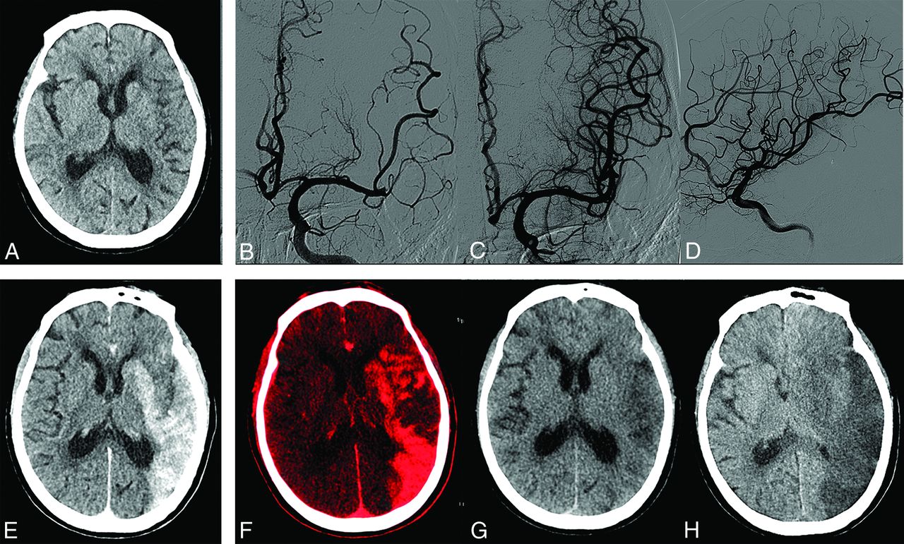
  • FIG 2.
    • Download figure
    • Open in new tab
    • Download powerpoint
    FIG 2.

    Examples of ROIs drawn on axial, coronal, and sagittal iodine overlay maps.
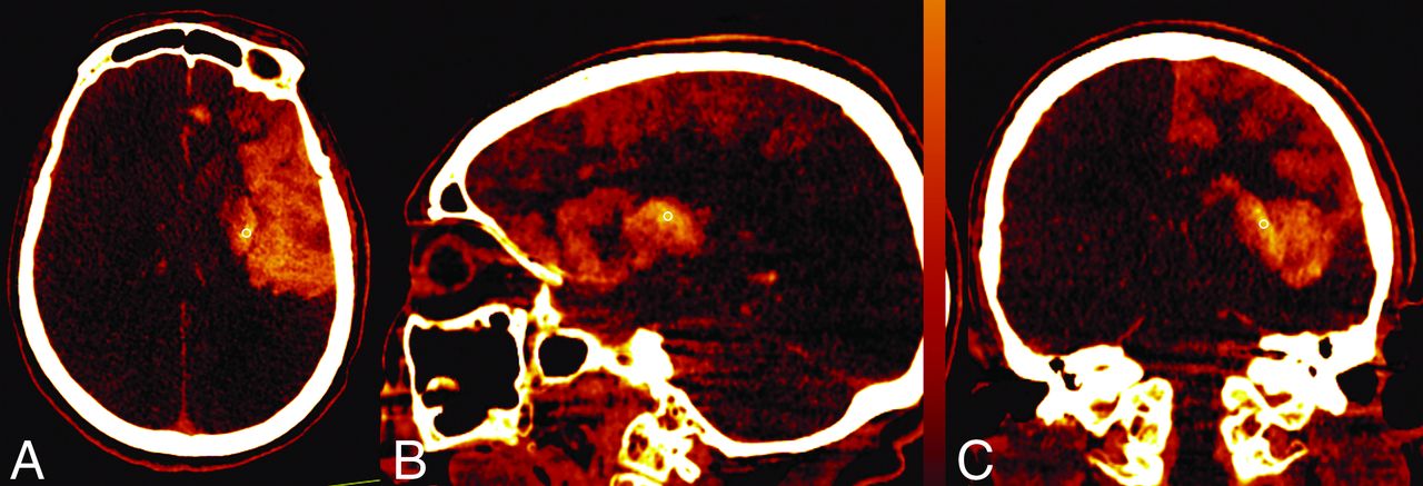
  • FIG 3.
    • Download figure
    • Open in new tab
    • Download powerpoint
    FIG 3.

    A 75-year-old woman with an ASPECTS of 7 on preprocedural CT (A). B, C, and D, Left M2 middle trunk occlusion with modified TICI 3 recanalization. E, Simulated conventional 120 kV(peak) images show hyperattenuation involving the left basal ganglia and temporal, frontal, and parietal lobes. F, Iodine overlay maps show contrast staining, with the AIIC (2.4 mg I/mL) and NICR (151.9%), respectively. G, Virtual noncontrast CT reveals no hemorrhage signs. H, Contrast staining resolved on 36-hour follow-up CT. Her 90-day mRS score was 4.
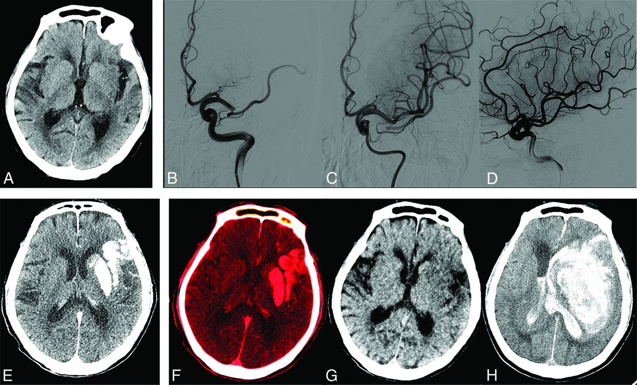
  • FIG 4.
    • Download figure
    • Open in new tab
    • Download powerpoint
    FIG 4.

    An 80-year-old man with an ASPECTS of 7 on admission CT (A). B, C, and D, Left M1 occlusion with modified TICI 3 recanalization. E, Simulated conventional 120 kV(p) images show contrast staining involving the left basal ganglia and temporal lobe. F, Iodine overlay maps show contrast staining, with the AIIC (4.9 mg I/mL) and the NICR (317.3%), respectively. G, Virtual noncontrast CT shows no hemorrhage signs in the area with hyperattenuation in simulated conventional 120 kV(p) images. H, Parenchymal hematoma type 2 develops with a marked midline shift on 24-hour follow-up CT. The patient died 6 days after recanalization (mRS score of 6).
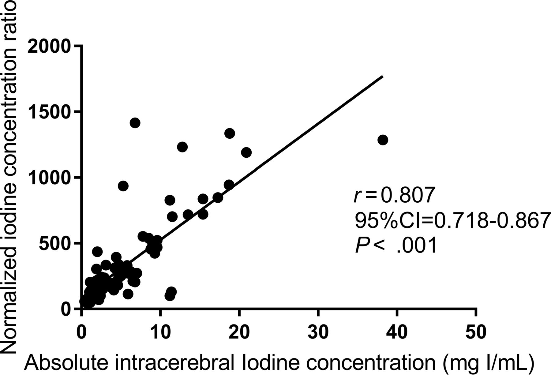
  • FIG 5.
    • Download figure
    • Open in new tab
    • Download powerpoint
    FIG 5.

    The NICR shows a strong correlation with the absolute intracerebral iodine concentration.

Tables

  • Figures
    • View popup
    Table 1:

    Scanning parameters of the noncontrast CT and noncontrast DECT

    Single-Source CTDual-Energy CT
    ScannerSomatom Emotion 16Somatom FORCE
    ManufacturerSiemensSiemens
    Scanning modeSpiralSpiral
    Scanning directionCaudal-cranialCaudal-cranial
    kV(p) (kV)11080/Sn 150
    Reference mAs240310/207
    Collimation (mm)16 × 0.6192 × 0.6
    Section thickness (mm)51
    Rotation time (sec)0.61
    Pitch0.650.7
    CARE Dose 4DaOnOn
    Reconstruction (A + B)
     KernelH41s medium+Hr40
     Iterative algorithmNAADMIRE 2
    • Note:—ADMIRE indicates advanced modeled iterative reconstruction; NA, not applicable.

    • ↵a Siemens.

    • View popup
    Table 2:

    Univariate analysis of patients’ baseline characteristics in cohorts with iodine staining without and with ICH (n = 95)

    Patient CharacteristicsNo Hemorrhage (n = 58)Hemorrhage (n = 37)P Value
    Age (mean) (SD) (yr)68 (12.1)67.7 (11.6).897a
    Male sex (No.) (%)28 (48.3%)19 (51.3%).770b
    History of hypertension (No.) (%)28 (48.3%)18 (48.6%).972b
    History of diabetes mellitus (No.) (%)13 (22.4%)17 (45.9%).016b
    History of atrial fibrillation (No.) (%)28 (48.3%)12 (32.4%).105b
    MT duration (median) (IQR) (min)70 (55–95)60 (46–90).310c
    Time of symptom onset to puncture (median) (IQR) (min)200 (144–313)230 (178–343).169c
    ASPECTS (median) (IQR)8 (7–9)8 (7–9).348c
    Baseline NIHSS (mean) (SD)16 (6)19 (7).067a
    rLMC (mean) (SD)11.8 (4.3)9.4 (4.6).01a
    Platelet count (median) (IQR) (109/L)133.8 (96.7–175.4)130 (100.3–167.9).694c
    hs-CRP (median) (IQR) (mg I/L)8.4 (1.2–45.3)6.9 (0.5–41.2).904c
    Occlusion site
     ICA occlusion (No.) (%)15 (25%)13 (31.5%.334b
     M1 trunk occlusion (No.) (%)30 (51.7%)18 (48.6%).770b
     M2 trunk occlusion (No.) (%)13 (22.4%)6 (16.2%).461b
    Thrombectomy devices
     Solitaired13 (22.4%)8 (21.6%).983b
     Sofiae+Aperiof13 (22.4%)10 (27%).609b
     Aperio20 (34.5%)14 (37.8%).739b
     Sofia12 (20.7%)5 (13.5%).374b
    Attempts of aspiration or stent retriever (median) (IQR)1 (1–1)1 (1-1).840c
    Antiplatelet therapy (No.) (%)39 (67.2)25 (67.6).974b
    Thrombolytic therapy (No.) (%)12 (20.7)7 (18.9).833b
    Anticoagulant therapy (No.) (%)12 (20.7)6 (16.2).587b
    Statin treatment (No.) (%)29 (50)24 (64.9).155b
    • Note:—rLMC indicates regional leptomeningeal collateral score; hs-CRP, hypersensitive c-reactive protein.

    • ↵a One-way ANOVA.

    • ↵b Pearson χ2 test.

    • ↵c Mann-Whitney test.

    • ↵d Covidien.

    • ↵e Microvention.

    • ↵f Acandis.

    • View popup
    Table 3:

    DECT parameters in groups without and with subsequent ICH

    DECT ParametersFollow-up CT
    No ICHICHP Value
    AIIC (median) (IQR) (mg I/mL)2.0 (1.7–4.5)5.9 (2.7–11.4)<.001
    NICR (median) (IQR) (%),144.5 (105.8–221.0)330.6 (181.8–703.3)<.001
    90-Day mRS (4–6) (%)30.2 (29/96)57.1 (24/42).003
    • View popup
    Table 4:

    AIIC and NICR parameters in terms of the hemorrhage classification on the follow-up CT

    AIIC (Median) (IQR) (mg I/mL)P ValueNICR (Median) (IQR) (%)P Value
    HI (HI1+HI2)4.2 (2.3–9.9).258252.8 (186.8–611.2).465
    PH (PH1+PH2)8.3 (2.8–11.4)429.8 (182.1–716.9)
    HI +PH13.7 (2.1–8.9).024248.2 (164.6–468.7).08
    PH29.3 (4.9–13.3)455.3 (218.4–801.3)
    • Note:—HI indicates hemorrhagic infarction; PH, parenchymal hematoma.

PreviousNext
Back to top

In this issue

American Journal of Neuroradiology: 43 (11)
American Journal of Neuroradiology
Vol. 43, Issue 11
1 Nov 2022
  • Table of Contents
  • Index by author
  • Complete Issue (PDF)
Advertisement
Print
Download PDF
Email Article

Thank you for your interest in spreading the word on American Journal of Neuroradiology.

NOTE: We only request your email address so that the person you are recommending the page to knows that you wanted them to see it, and that it is not junk mail. We do not capture any email address.

Enter multiple addresses on separate lines or separate them with commas.
Quantitative Intracerebral Iodine Extravasation in Risk Stratification for Intracranial Hemorrhage in Patients with Acute Ischemic Stroke
(Your Name) has sent you a message from American Journal of Neuroradiology
(Your Name) thought you would like to see the American Journal of Neuroradiology web site.
CAPTCHA
This question is for testing whether or not you are a human visitor and to prevent automated spam submissions.
Cite this article
C. Ma, D. Xu, Q. Hui, X. Gao, M. Peng
Quantitative Intracerebral Iodine Extravasation in Risk Stratification for Intracranial Hemorrhage in Patients with Acute Ischemic Stroke
American Journal of Neuroradiology Nov 2022, 43 (11) 1589-1596; DOI: 10.3174/ajnr.A7671

Citation Manager Formats

  • BibTeX
  • Bookends
  • EasyBib
  • EndNote (tagged)
  • EndNote 8 (xml)
  • Medlars
  • Mendeley
  • Papers
  • RefWorks Tagged
  • Ref Manager
  • RIS
  • Zotero
0 Responses
Respond to this article
Share
Bookmark this article
Iodine Extravasation in Intracranial Hemorrhage
C. Ma, D. Xu, Q. Hui, X. Gao, M. Peng
American Journal of Neuroradiology Nov 2022, 43 (11) 1589-1596; DOI: 10.3174/ajnr.A7671
del.icio.us logo Twitter logo Facebook logo Mendeley logo
  • Tweet Widget
  • Facebook Like
  • Google Plus One
Purchase

Jump to section

  • Article
    • Abstract
    • ABBREVIATIONS:
    • MATERIALS AND METHODS
    • RESULTS
    • DISCUSSION
    • CONCLUSIONS
    • Footnotes
    • References
  • Figures & Data
  • Info & Metrics
  • Responses
  • References
  • PDF

Related Articles

  • No related articles found.
  • PubMed
  • Google Scholar

Cited By...

  • Determinants and Clinical Relevance of Iodine Contrast Extravasation after Endovascular Thrombectomy: A Dual-Energy CT Study
  • Crossref (5)
  • Google Scholar

This article has been cited by the following articles in journals that are participating in Crossref Cited-by Linking.

  • Prognostic value of contrast staining on dual-energy CT after endovascular therapy in acute ischemic stroke: a meta-analysis
    Fan Yang, Yi Zeng, Fei Zhu, Xiaoyan Hu
    BMC Neurology 2023 23 1
  • Determinants and Clinical Relevance of Iodine Contrast Extravasation after Endovascular Thrombectomy: A Dual-Energy CT Study
    Clémence Hoche, Alba Henderson, Héloïse Ifergan, Marie Gaudron, Christophe Magni, Igor Maldonado, Jean-Philippe Cottier, Marco Pasi, Grégoire Boulouis, Clara Cohen
    American Journal of Neuroradiology 2024 45 1
  • Machine learning, clinical-radiomics approach with HIM for hemorrhagic transformation prediction after thrombectomy and treatment
    Sheng Hu, Junyu Liu, Jiayi Hong, Yuting Chen, Ziwen Wang, Jibo Hu, Shiying Gai, Xiaochao Yu, Jingjing Fu
    Frontiers in Neurology 2025 16
  • Post-endovascular treatment, blood-brain barrier disruption, predicts patient outcomes better than pre-treatment status
    Huazheng Zhai, Yao Li, Ruiqi Jia, Jun Cao, Qiang Wei, Weimin Yang, Jingye Wang
    Neurological Sciences 2024 45 9
  • Quantitative CT imaging: where are we, and what is missing?
    Kevin J Treb, Ahmed O El Sadaney, Andrea Ferrero, Francis I Baffour, Shuai Leng, Lifeng Yu, Prabhakar Shantha Rajiah, Joel G Fletcher, Cynthia H McCollough, Kishore Rajendran
    British Journal of Radiology 2025

More in this TOC Section

  • Diagnostic Neuroradiology of Monoclonal Antibodies
  • ML for Glioma Molecular Subtype Prediction
  • Segmentation of Brain Metastases with BLAST
Show more Adult Brain

Similar Articles

Advertisement

Indexed Content

  • Current Issue
  • Accepted Manuscripts
  • Article Preview
  • Past Issues
  • Editorials
  • Editors Choice
  • Fellow Journal Club
  • Letters to the Editor

Cases

  • Case Collection
  • Archive - Case of the Week
  • Archive - Case of the Month
  • Archive - Classic Case

Special Collections

  • Special Collections

Resources

  • News and Updates
  • Turn around Times
  • Submit a Manuscript
  • Author Policies
  • Manuscript Submission Guidelines
  • Evidence-Based Medicine Level Guide
  • Publishing Checklists
  • Graphical Abstract Preparation
  • Imaging Protocol Submission
  • Submit a Case
  • Become a Reviewer/Academy of Reviewers
  • Get Peer Review Credit from Publons

Multimedia

  • AJNR Podcast
  • AJNR SCANtastic
  • Video Articles

About Us

  • About AJNR
  • Editorial Board
  • Not an AJNR Subscriber? Join Now
  • Alerts
  • Feedback
  • Advertise with us
  • Librarian Resources
  • Permissions
  • Terms and Conditions

American Society of Neuroradiology

  • Not an ASNR Member? Join Now

© 2025 by the American Society of Neuroradiology All rights, including for text and data mining, AI training, and similar technologies, are reserved.
Print ISSN: 0195-6108 Online ISSN: 1936-959X

Powered by HighWire