
Article Figures & Data
Figures
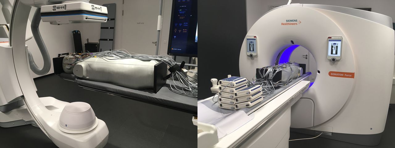
- FIG 1.
The anthropomorphic ATOM phantom used for effective dose measurement. The phantom in an experimental setup for 3D acquisition is equipped with MOSFET dosimeters on an Artis icono biplane angiography system (left) and on the Somatom Force CT scanner (right).
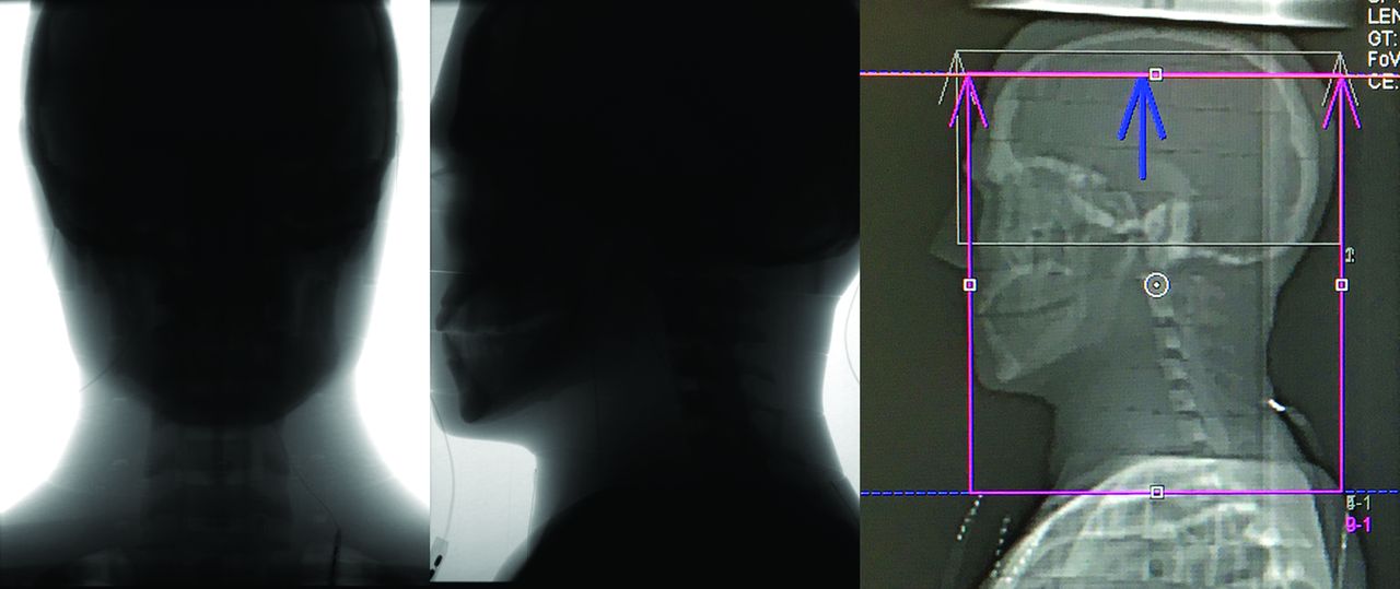
- FIG 2.
Lateral view of the investigated head area of the phantom for uncollimated measurement (left) and collimated measurement (middle) of the 60s DCT head perfusion protocol on the Artis icono and for perfusion measurement on the Somatom Force (right).
- FIG 3.
The position of the investigated head area of the phantom for carotid measurement on the Artis icono in frontal (left) and lateral (middle) views and on the Somatom Force in the lateral view (right).
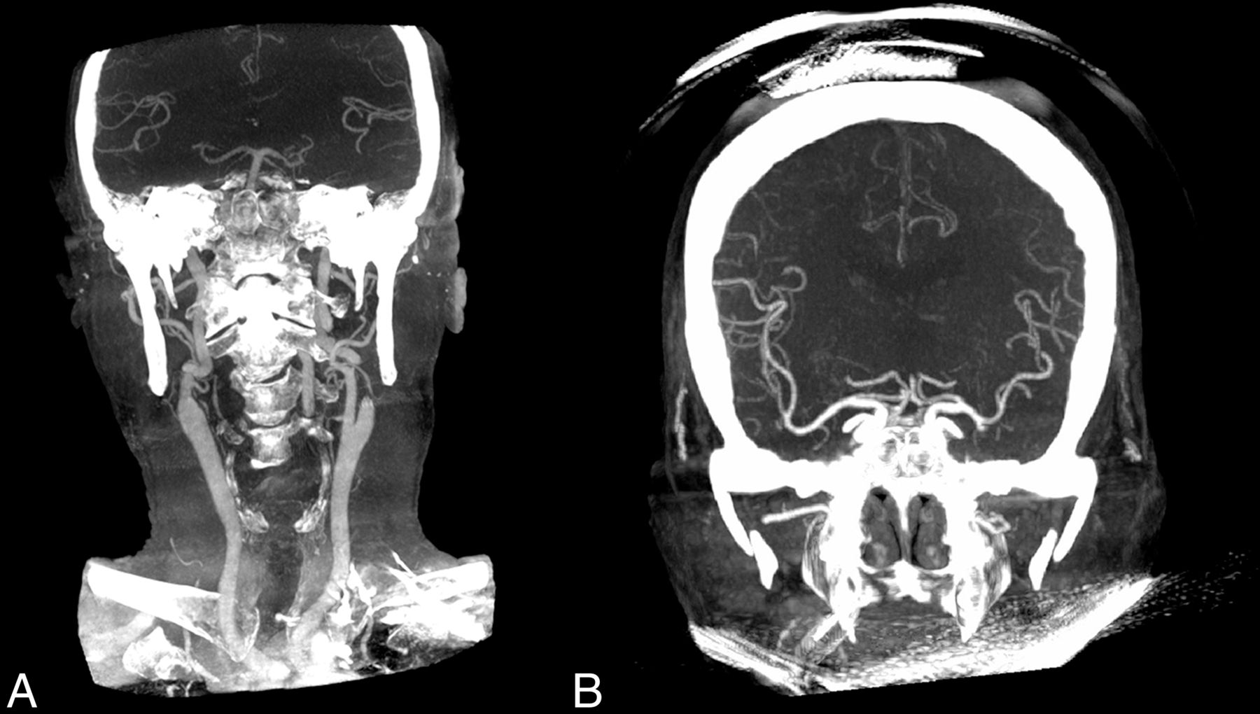
- FIG 4.
Comparison of the coverage area of portrait FDCT-A (A) and landscape FDCT-A (B) (based on reconstructed coronal MIPs).


Tables
- Table 1:
Technical parameters of investigated 3D imaging protocols (brain perfusion, portrait angiography with head and neck angiography), measured eye lens dose (for left, right eye and mean), and effective dose for the anthropomorphic ATOM male phantom on Artis icono
3D Imaging Protocol Parameters 60s DCT Head Perfusion (10 Rotations) 4s DCT Head Portrait (1 Rotation) Reconstructed volume size (diameter × height) (cm) 24 × 18.5 (uncollimated) 24 × 15a 18.5 × 24 (uncollimated) Tube voltage (nominal) (kV) 70 70 90 Dose/frame (nominal) (nGy/f) 360 360 1200 Rotation range 200° 200° 200° Angulation step (df) 0.8 0.8 0.8 Eye lens dose (mean) (mGY) 65 and 69 (SD, 67) 54 and 58 (SD, 56) 7.7 and 8.3 (SD, 8) Estimated effective dose (mSv) 4.52 2.88 0.91 Note:—60s DCT indicates 60s Dyna-CT; 4s DCT, 4s Dyna CT.
↵a Collimation was defined on the basis of usual clinical workflow at the University Hospital Basel.
- Table 2:
Technical parameters of investigated protocols (brain perfusion and head and neck angiography), measured eye lens dose (for left, right eye and mean), and effective dose for the anthropomorphic ATOM male phantom on Somatom Force
3D Imaging Protocol Parameters NeuroVPCT_Prolonged, DynMulti4D NeuroVPCT_Prolonged, Head Angio Scan coverage (cm) 11.4 24 Tube voltage (kV) 70 90 Scan duration (sec) 60 NA Number of cycles @ 1.5 seconds cycle time 30 NA CTDIvol (mGy) 144.2 19,9 DLP (mGy × cm) 2169.5 550 Eye lens dose (mean) (mGy) 119 and 125 (SD, 122) 11.8 and 11.7 (SD, 11.8) Estimated effective dose (mSv) 2.17 1.35 Note:—CTDIvol indicates volume CT dose index; DLP, dose-length product; NA, not applicable.
- Table 3:
Organ dose in milligrays for selected organs and tissues of investigated 3D imaging protocols on the Artis icono
Organ 60s DCT Head Perfusion (10 Rotations, Uncollimated) 60s DCT Head Perfusion (10 Rotations, Collimated) 4s DCT Head Portrait(1 Rotation) Brain 100.8 84.7 11.8 Salivary glands 56.2 7.4 11.4 Thyroid 3.5 1.8 6.1 Lung 1.3 0.8 0.2 Red bone marrow 17.4 12.1 2.4 Esophagus 1.1 0.7 0.8 - Table 4:
Organ dose in milligrays for selected organs and tissues of investigated 3D imaging protocols on the Somatom force
Organ NeuroVPCT_Prolonged, DynMulti4D NeuroVPCT_Prolonged, Head Angio Brain 71.6 7.8 Salivary glands 6.4 8 Thyroid 1.3 17.2 Lung 0.4 1.3 Red bone marrow 9.2 1.3 Esophagus 0.4 2.0


