Advertisement

Main menu

  • Home
  • Content
    • Current Issue
    • Accepted Manuscripts
    • Article Preview
    • Past Issue Archive
    • AJNR Case Collection
    • Case of the Week Archive
    • Classic Case Archive
    • Case of the Month Archive
  • Special Collections
    • Spinal CSF Leak Articles (Jan 2020-June 2024)
    • 2024 AJNR Journal Awards
    • Most Impactful AJNR Articles
  • Multimedia
    • AJNR Podcast
    • AJNR Scantastics
    • Video Articles
  • For Authors
    • Submit a Manuscript
    • Author Policies
    • Fast publishing of Accepted Manuscripts
    • Graphical Abstract Preparation
    • Manuscript Submission Guidelines
    • Imaging Protocol Submission
    • Submit a Case for the Case Collection
  • About Us
    • About AJNR
    • Editorial Board
  • More
    • Become a Reviewer/Academy of Reviewers
    • Subscribers
    • Permissions
    • Alerts
    • Feedback
    • Advertisers
    • ASNR Home
  • Other Publications
    • ajnr

User menu

  • Alerts
  • Log in

Search

  • Advanced search
American Journal of Neuroradiology

American Journal of Neuroradiology

ASHNR American Society of Functional Neuroradiology ASHNR American Society of Pediatric Neuroradiology ASSR
  • Alerts
  • Log in

Advanced Search

  • Home
  • Content
    • Current Issue
    • Accepted Manuscripts
    • Article Preview
    • Past Issue Archive
    • AJNR Case Collection
    • Case of the Week Archive
    • Classic Case Archive
    • Case of the Month Archive
  • Special Collections
    • Spinal CSF Leak Articles (Jan 2020-June 2024)
    • 2024 AJNR Journal Awards
    • Most Impactful AJNR Articles
  • Multimedia
    • AJNR Podcast
    • AJNR Scantastics
    • Video Articles
  • For Authors
    • Submit a Manuscript
    • Author Policies
    • Fast publishing of Accepted Manuscripts
    • Graphical Abstract Preparation
    • Manuscript Submission Guidelines
    • Imaging Protocol Submission
    • Submit a Case for the Case Collection
  • About Us
    • About AJNR
    • Editorial Board
  • More
    • Become a Reviewer/Academy of Reviewers
    • Subscribers
    • Permissions
    • Alerts
    • Feedback
    • Advertisers
    • ASNR Home
  • Follow AJNR on Twitter
  • Visit AJNR on Facebook
  • Follow AJNR on Instagram
  • Join AJNR on LinkedIn
  • RSS Feeds

Welcome to the new AJNR, Updated Hall of Fame, and more. Read the full announcements.


AJNR is seeking candidates for the position of Associate Section Editor, AJNR Case Collection. Read the full announcement.


Research ArticleExtracranial Vascular

Impact Analysis of Different CT Configurations of Carotid Artery Plaque Calcifications on Cerebrovascular Events

L. Saba, H. Chen, R. Cau, G.D. Rubeis, G. Zhu, F. Pisu, B. Jang, G. Lanzino, J.S. Suri, Y. Qi and M. Wintermark
American Journal of Neuroradiology February 2022, 43 (2) 272-279; DOI: https://doi.org/10.3174/ajnr.A7401
L. Saba
aFrom the Department of Radiology (L.S., R.C., F.P.), Azienda Ospedaliero Universitaria di Cagliari, Cagliari, Italy
  • Find this author on Google Scholar
  • Find this author on PubMed
  • Search for this author on this site
  • ORCID record for L. Saba
H. Chen
bDepartment of Radiology (H.C., G.Z., B.J., M.W.), Division of Neuroradiology, Stanford University Hospital, Stanford, California
  • Find this author on Google Scholar
  • Find this author on PubMed
  • Search for this author on this site
  • ORCID record for H. Chen
R. Cau
aFrom the Department of Radiology (L.S., R.C., F.P.), Azienda Ospedaliero Universitaria di Cagliari, Cagliari, Italy
  • Find this author on Google Scholar
  • Find this author on PubMed
  • Search for this author on this site
  • ORCID record for R. Cau
G.D. Rubeis
cDepartment of Radiology, UOC of Diagnostic and Interventional Neuroradiology (G.D.R.), San Camillo-Forlanini Hospital, Rome, Italy
  • Find this author on Google Scholar
  • Find this author on PubMed
  • Search for this author on this site
  • ORCID record for G.D. Rubeis
G. Zhu
bDepartment of Radiology (H.C., G.Z., B.J., M.W.), Division of Neuroradiology, Stanford University Hospital, Stanford, California
  • Find this author on Google Scholar
  • Find this author on PubMed
  • Search for this author on this site
  • ORCID record for G. Zhu
F. Pisu
aFrom the Department of Radiology (L.S., R.C., F.P.), Azienda Ospedaliero Universitaria di Cagliari, Cagliari, Italy
  • Find this author on Google Scholar
  • Find this author on PubMed
  • Search for this author on this site
  • ORCID record for F. Pisu
B. Jang
bDepartment of Radiology (H.C., G.Z., B.J., M.W.), Division of Neuroradiology, Stanford University Hospital, Stanford, California
  • Find this author on Google Scholar
  • Find this author on PubMed
  • Search for this author on this site
  • ORCID record for B. Jang
G. Lanzino
dDepartment of Neurologic Surgery (G.L.), Mayo Clinic, Rochester, Minnesota
  • Find this author on Google Scholar
  • Find this author on PubMed
  • Search for this author on this site
  • ORCID record for G. Lanzino
J.S. Suri
eStroke Monitoring and Diagnostic Division (J.S.S.), AtheroPoint, Roseville, California
  • Find this author on Google Scholar
  • Find this author on PubMed
  • Search for this author on this site
  • ORCID record for J.S. Suri
Y. Qi
fDepartment of Xuanwu Hospital (Y.Q.), Capital Medical University, Beijing, Chin
  • Find this author on Google Scholar
  • Find this author on PubMed
  • Search for this author on this site
  • ORCID record for Y. Qi
M. Wintermark
bDepartment of Radiology (H.C., G.Z., B.J., M.W.), Division of Neuroradiology, Stanford University Hospital, Stanford, California
  • Find this author on Google Scholar
  • Find this author on PubMed
  • Search for this author on this site
  • ORCID record for M. Wintermark
  • Article
  • Figures & Data
  • Info & Metrics
  • Responses
  • References
  • PDF
Loading

Abstract

BACKGROUND AND PURPOSE: CT is considered the standard reference both for quantification and characterization of carotid artery calcifications. Our aim was to investigate the relationship among different types of calcium configurations detected with CT within the plaque with a novel classification and to investigate the prevalence of cerebrovascular events.

MATERIALS AND METHODS: Seven hundred ninety patients (men = 332; mean age, 69.7 [SD, 13] years; 508 symptomatic for cerebrovascular symptoms and 282 asymptomatic) who underwent computed tomography of the carotid arteries were retrospectively included in this institutional review board–approved study. The plaque was classified into 6 types according to the different types of calcium configurations as the following: type 1, complete absence of calcification within the plaque; type 2, intimal or superficial calcifications; type 3, deep or bulky calcifications; type 4, adventitial calcifications with internal soft plaque of <2 mm thickness; type 5, mixed patterns with intimal and bulky calcifications; and type 6, positive rim sign.

RESULTS: The highest prevalence of cerebrovascular events was observed for type 6, for which 89 of the 99 cases were symptomatic. Type 6 plaque had the highest degree of correlation with TIA, stroke, symptoms, and ipsilateral infarct for both sides with a higher prevalence in younger patients. The frequency of symptoms observed by configuration type significantly differed between right and left plaques, with symptoms observed more frequently in type 6 calcification on the right side (50/53; 94%) than on the left side (39/46; 85%, P < .001).

CONCLUSIONS: We propose a novel carotid artery plaque configuration classification that is associated with the prevalence of cerebrovascular events. If confirmed in longitudinal analysis, this classification could be used to stratify the risk of occurrence of ischemic events.

ABBREVIATION:

IPH
intraplaque hemorrhage

Ischemic stroke is one of the leading causes of mortality and disability worldwide.1 Several studies have demonstrated that atherosclerosis involving the carotid arteries is responsible for a significant portion of acute ischemic strokes (up to 25%).2,3 In the past 20 years, the role of carotid plaque imaging in risk stratification of ischemic stroke has evolved considerably. Several recently published articles suggest that the mere degree of stenosis, considered the only criterion for risk stratification at a time when advanced imaging was not available, is not the best parameter to predict the stability and vulnerability of the plaque and its potential for causing cerebrovascular events.2,4,5 Therefore, there has been great emphasis on identifying additional imaging features that can be used to select the best therapeutic approach.

A number of parameters have been identified that are associated with an increased risk of plaque rupture, including intraplaque haemorrhage,6,7 lipid-rich necrotic core,8 rupture of the fibrous cap, and contrast plaque enhancement.9 Other features such as a thick fibrous cap have been linked to a reduced risk of stroke. The role of calcium, present in 50%–60% of carotid plaques, is controversial,10 and the relationship between the amount and topographic distribution of vascular wall calcifications and plaque vulnerability have not been evaluated. Some studies have found that carotid plaque calcification is associated with plaque stability and may be considered a protective plaque feature,11⇓⇓-14 while others have found that it could represent an independent marker of ischemic symptoms.15 The significance of calcifications and their distribution in carotid artery plaques are controversial,15⇓-17 and it is unclear whether the level of carotid calcification should be considered an element of plaque stability or instability. Eisenmenger et al18 proposed a model that includes the positive rim sign, maximum soft-plaque thickness, the NASCET stenosis, and ulceration to predict intraplaque hemorrhages (IPHs) on computed tomography (CT). A positive rim sign was defined as adventitial calcification (<2 mm thick) with internal soft plaque (>2 mm thickness). This model had excellent IPH prediction with an area under the curve of 0.94.18

Calcium in a carotid artery plaque can be identified with different imaging techniques. Among these, CT is considered the standard reference both for quantification and characterization of carotid artery calcifications.19⇓-21 Our hypothesis is that it is possible to classify the CTA patterns of calcifications in the carotid plaque by identifying subgroups that can be more frequently associated with the presence of symptomatic cerebrovascular events. We have, therefore, developed a novel classification based on the different calcium configurations.

In this study, we identified various carotid artery plaque subgroups on the basis of their morphology as visualized on CTA and assessed their clinical significance in a large cohort of symptomatic and asymptomatic patients.

MATERIALS AND METHODS

Study Design and Patient Population

Institutional review board approval was obtained, and informed consent was waived because of the retrospective nature of the study. This 2-center retrospective study was performed in 2 tertiary centers (Azienda Ospedaliera Universitaria di Cagliari [site A] and Stanford University Hospital [site B] between March 2013 and May 2020. All examinations were performed according to established standard-of-care CTA protocols. Patient characteristics including age, sex, clinical symptoms, medical history, and clinical outcomes were recorded.

Patients 18 years of age or older with bilateral carotid artery plaques (thickness of ≥1.5 mm, according to the Mannheim intima-media thickness consensus) were included.22 Excluded were subjects younger than 18 years of age and those in whom the CTA was performed for reasons other than suspected atherosclerotic disease (ie, dissection) or other etiologies for ischemic stroke such as a cardiac embolic source, embolism from the thoracic aorta, and vertebrobasilar artery disease. The degree of stenosis was measured according to the NASCET criteria.23 We also excluded patients with other neurologic conditions such as brain neoplasms, demyelinating disease, and so forth.

According to a standardized protocol, CTA of the carotid artery was obtained in asymptomatic patients when carotid sonography showed significant stenosis (>50% measured according to the NASCET criteria) or features of plaque vulnerability (ulcerations, irregular surface) and when carotid sonography could not adequately assess the degree of stenosis and plaque characteristics because of anatomic conditions. Moreover, all subjects presenting with acute cerebrovascular events underwent CTA of the carotid arteries at the time of their acute CT of the head.

Definition of Symptomatic

Patients were classified by the treating neurologist using the Trial of Org 10172 in Acute Stroke Treatment (TOAST) criteria.24 Consequently, a patient was considered symptomatic after experiencing a TIA or ischemic stroke in either cerebral hemisphere. TIA was considered a brief (<24 hours) episode of neurologic dysfunction such as hemiparesis, hemisensory loss, dysarthria, dysphasia, or monocular blindness. If the episode of neurologic dysfunction exceeded 24 hours, it was classified as a stroke. A lacunar stroke was defined as a stroke causing one of the traditional clinical lacunar syndromes without evidence of cerebral cortical dysfunction. Such patients also had normal CT/MR imaging findings or a relevant brain stem or subcortical hemispheric lesion with a diameter of <0.5 cm.25 A patient was considered symptomatic within a time window of up to 3 months after symptom onset. For our model of analysis, we considered both carotid arteries in each patient: The symptomatic carotid was the one ipsilateral to the symptoms and the asymptomatic carotid was the contralateral one.

CT Technique

Due to the range of time for data collection and the bicenter approach, CTA of the carotid arteries was performed with multiple scanner technologies according to a standardized protocol. As a general approach, patients were placed in the supine position with the head tilted back to prevent dental artifacts. The coverage was from the aortic arch to the carotid siphon in a caudocranial direction.

Plaque Calcification Configuration

Carotid plaques were classified into 6 subgroups based on calcium configurations (Fig 1):

  • Type 1: complete absence of calcification within the plaque

  • Type 2: intimal or superficial calcifications

  • Type 3: deep or bulky calcifications

  • Type 4: adventitial calcifications with internal soft plaque of <2-mm-thickness; negative rim sign

  • Type 5: mixed patterns with intimal and bulky calcifications

  • Type 6: positive rim sign.

FIG 1.
  • Download figure
  • Open in new tab
  • Download powerpoint
FIG 1.

Scheme showing the different types of configurations with the corresponding CTA findings.

A superficial calcification was defined as a calcified nodule located at the intimal-lumen interface or close to the lumen, whereas a deep calcification was defined as a calcified nodule located at the media/adventitia border or close to the adventitia. The presence of both deep and superficial calcifications was regarded as a mixed category.26 Bulky calcification was defined as calcification measuring >2 mm thick without associated thin adventitial calcification.18

The classification was performed by 2 radiologists; in the case of conflicts between the readers, the decision was made by a third senior reader expert in vascular imaging. Interobserver and intraobserver (within a 2-month timeframe) agreement was recorded. Interactive window/level settings were usually set at W850:L300.27 This approach was adopted because calcifications may show variable edge and halo blurring depending on the window level, which could change the size (expressed in millimeters) of the calcification; therefore, we opted for this static approach.

Statistical Analysis

Continuous variables were presented as mean (SD) or median (with interquartile range) values. Categoric variables were described as frequency rates and percentages. Comparisons of continuous data were performed using the independent samples t test or Mann-Whitney U test. Shapiro-Wilk tests were used to check continuous variables for normal distribution. Categoric variables were compared using a χ2 test or Fisher exact test as appropriate. Comparisons between groups were performed using a 1-way ANOVA for continuous variables with normal distributions; the Kruskal-Wallis test was used for continuous variables with non-normal distributions. A Tukey multiple comparison test was performed to determine statistically significant changes between each group. Inter-/intraobserver agreement was measured using a weighted Cohen κ analysis. A P value < .05 was considered statistically significant. The Pearson ρ was used to correlate types of plaque with traditional risk factors, infarct, stroke, symptoms, and degree of stenosis. All statistical analyses were performed using SPSS Statistics, Version 25 (IBM).

RESULTS

Baseline Characteristics

The study included 790 patients (men = 42%, women = 58%; mean age, 69.7 [SD, 13] years; age range, 23–102 years) with bilateral plaques, 508 symptomatic and 282 asymptomatic. Clinical and demographic characteristics are summarized in Table 1. The mean degree of stenosis was 61% in the symptomatic patients and 30% in the asymptomatic ones. Plaque on the right side was 39%; and on the left side, 41%.

View this table:
  • View inline
  • View popup
Table 1:

Demographic and risk factors  

Plaque Calcification Type and Symptoms

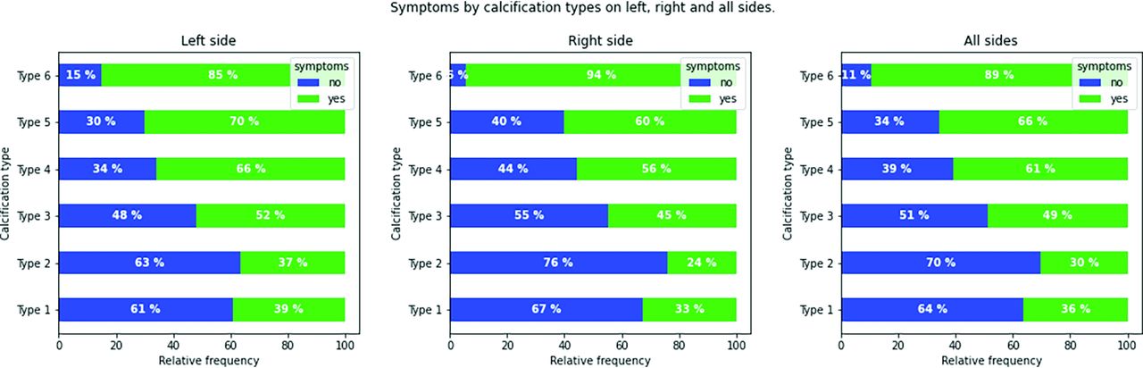
The crosstabulation among the types of calcifications is given in Table 2. The frequency of symptoms observed by configuration type significantly differed between right and left plaques, with symptoms observed more frequently in type 6 calcification on the right side (50/53; 94%) than on the left side (39/46, 85%; P < .001).

View this table:
  • View inline
  • View popup
Table 2:

Crosstabulation between calcified plaque on the right and left sides

We found that the absence of calcification on one side is frequently associated with absence of calcification on the contralateral side. However, the weighted overall Cohen κ value was poor, with a value of 0.297. We also tested the inter- and intraobserver concordance; the values are summarized in the Online Supplemental Data.

In the analysis of the correlations among the types of calcifications and symptomatic status (by considering 1580 sides), the highest prevalence of symptomatic plaques was observed for type 6, in which 89 of the 99 plaques were symptomatic. A complete summary of the findings is shown in Fig 2.

FIG 2.
  • Download figure
  • Open in new tab
  • Download powerpoint
FIG 2.

Bar plots in symptomatic and asymptomatic subjects according to the plaque configuration for the total plaque count (A), the right side (B), and left side (C).

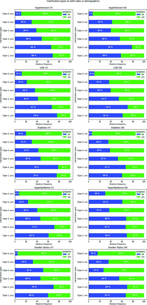
We also checked the impact of side by analyzing the association between the right/left carotid artery plaque configurations and the presence of symptoms (right stroke, left stroke/absence of stroke for asymptomatic patients); the results are summarized in Fig 2. The Pearson χ2 test showed a statistically significant difference for different types of configurations for both right and left plaques (P < .001). Figure 3 shows the relationship among different plaque configurations on the right/left side versus the measured risk factors.

FIG 3.
  • Download figure
  • Open in new tab
  • Download powerpoint
FIG 3.

Bar plots in symptomatic and asymptomatic subjects according to the plaque configuration for the total on the right (upper row) and left (lower row) sides for cardiovascular risk factors. CAD indicates coronary artery disease; Y, yes; N, no.

There was an increasing correlation from type 1 to type 6 plaque configurations with symptoms, stroke, ipsilateral infarct, TIA, and NASCET degree of stenosis for both sides. In particular, the highest degree of correlation was found for ipsilateral infarct, ranging from −0.067 for type 1 to 0.21 for type 6 on right side and from −0.11 for type 1 to 0.21 for type 6 on left side (see Fig 4 for further details). No other traditional risk factors for cerebrovascular diseases reached the same degree of correlation (Fig 4). Moreover, as observed in the Online Supplemental Data, by plotting the types of calcifications with age and degree of stenosis, type 6 plaque configuration was more common in younger patients, with a bimodal distribution for stenosis degree (mild and severe) on both sides.

FIG 4.
  • Download figure
  • Open in new tab
  • Download powerpoint
FIG 4.

The Pearson ρ correlogram shows the correlation test among the different demographic variables and the calcification types. Each box represents a correlation test, the blue values represent negative correlations, and the red values represent positive associations. CAD indicates coronary artery disease; L, left; R, right.

DISCUSSION

In the present study, we grouped the different types of calcium configurations we encountered in our daily clinical practice by proposing a novel classification system to verify whether some configurations are more likely to be associated with cerebrovascular events.

The analysis of the association between the types of calcifications and the presence of symptoms (by considering 1580 sides) showed that the highest risk of cerebrovascular events is seen for type 6, in which 89 of the 99 patients were symptomatic. These results were also confirmed by the correlation analysis, in which type 6 had the highest degree of correlation with symptoms, stroke, TIA, and ipsilateral infarct. Our results also confirm that the presence of a positive rim sign is significantly associated with cerebrovascular events, but from the analysis of the other ORs, it is quite obvious that the different types of calcium configurations play a different role, with an increasing OR according to the analyzed class. Moreover, after analyzing the relative frequency of plaque configurations among different ages and degrees of stenosis,28,29 type 6 was more frequent in younger patients, with a bimodal distribution with regard to the NASCET degree of stenosis (mild and severe).

The type 1 class identifies those carotid artery plaques without calcification within the plaque, and somewhat surprisingly, we found that plaques without any calcium have an OR of 0.5, which suggests that such plaques are not associated with an increased frequency of cerebrovascular events.

When one introduces a new classification, it is important to test the inter-/intraobserver agreement, and we found that the configurations are quite good and reproducible. The overall concordance is good, with a better intraobserver agreement than interobserver agreement. In particular, the type 1 category has excellent values of reproducibility; also, types 4 and 6 have optimal values. Types 2 and 3 have suboptimal values. However, a further evolution to improve the reproducibility could be to apply machine learning algorithms,30 to move from a subjective assessment to a more objective and reproducible type classification.

As expected, we found that the mean degree of stenosis was 64% in symptomatic patients and 35% in asymptomatic ones by confirming that the degree of stenosis is an important parameter, even if this could be considered an indirect parameter influenced by the volume of plaque and the different remodeling pathways.

We have also checked the impact by side, by analyzing the association between the right/left carotid artery plaque configurations and the presence of symptoms (right stroke/left stroke/absence of stroke for asymptomatic patients), and a statistically significant difference for different types of configurations for both right and left plaques was found. Figure 3 shows the relationship between the different plaque configurations on the right/left sides versus the measured risk factors. Moreover, we found that the absence of calcification on one side is not associated with the absence of calcification on the contralateral side: The Cohen κ value was poor (0.297).

To date, there are no grading systems describing carotid artery calcium configurations and distributions. However, there are studies that have demonstrated how some types of configurations can influence the risk of ipsilateral cerebrovascular events. Eisenmenger et al18 showed that the positive rim sign is predictive of an increased stroke risk because it is associated with the presence of IPH within the plaque. Recently, Yang et al26 demonstrated that superficial calcifications were associated with an increased risk of IPH and subsequent risk of cerebrovascular events. Xu et al31 reported a similar association in 2010 using MR imaging. This could be explained by exploring the pathophysiology of the IPH, in which the leakage from the vasa vasorum could produce a blood collection with subsequent space occupation and shift and compression of the adjacent structures with superimposition of the calcification. However, the problem could be also more complex: Van den Bouwhuijsen et al,32 in 2015, found that a larger carotid calcification volume at the level of the bifurcation was related to the presence of IPH. This is an interesting finding because this association did not seem to be driven simply by plaque size or atherosclerotic burden because it did not alter after an adjustment was made for wall thickness. The association was even more pronounced in persons with a low degree of carotid stenosis. They also found that larger calcification volumes were associated with a lower prevalence of lipid core, which was more pronounced at a higher degree of stenosis (>30%).

The findings of this study do not suggest that some types of calcifications are vulnerable but that these are associated with other parameters related to the vulnerability. This concept is clear by comparing types 4 and 6 in which the calcium morphology is exactly the same, but the difference is due to the thickness of the soft-tissue component (<2 and >2 mm for types 4 and 6, respectively). Our results confirm that the role of calcium is complex. It is implicated in different metabolic processes, which are sometimes conflicting and overlapping.17 One important point is that with CT, we can explore a partial element of this complexity, the geometric configuration, and we have found that this could potentially represent a biomarker.

The potential utility of this novel classification relies on the opportunity to classify carotid artery plaque according to the calcium configuration and characteristics, and CT is an optimal technique for this task because it provides excellent visualization of calcification due to the marked x-ray attenuation properties of calcium.33 Further prospective studies would be necessary to test the potential impact of these configurations on the occurrence of cerebrovascular events.34

Our study has limitations. The first one is that due to the retrospective nature of our analysis, the 2 cohorts we have analyzed were not similar due to the statistically significant difference in the hyperlipidemia prevalence. This could have introduced a bias because hyperlipidemia could play a role in the atherogenic process by stimulating some specific pathways. In particular, medial calcification is not associated with atherosclerosis, and it is not typically linked to dyslipidemia, whereas this association is more robust for intimal calcifications35 because hyperlipidemia leads to accumulation of lipoproteins in the subendothelial space of cardiovascular tissues, which leads to formation of mildly oxidized phospholipids, which are known bioactive factors in vascular cell calcification.36,37 Second, this is a retrospective cross-sectional study in which we have not explored the relationship between the types of carotid plaque configurations and the occurrence of cerebrovascular events but we have assessed the relationship between the calcium configuration and the presence of events.

Our data need confirmation in a prospective, longitudinal cohort to confirm our hypothesis and validate our classification; thus, our results should be considered preliminary. Therefore, although suggesting that some types of calcium configurations within the plaque can be markers of instability, we cannot distinguish whether there is a causative relationship between the 2 or if the calcification is a mere epiphenomenon of the concomitant atherosclerotic process. Nevertheless, our results suggest that imaging can explore the complexity of calcium configurations and offer an option of stratification and that calcium configurations may be of value as an indirect marker of plaque instability.

CONCLUSIONS

We suggest a carotid artery plaque configuration classification that is associated with the presence of cerebrovascular events. If confirmed in longitudinal analysis, this classification could be used to stratify the risk of occurrence of ischemic events.

Footnotes

  • Disclosure forms provided by the authors are available with the full text and PDF of this article at www.ajnr.org.

References

  1. 1.↵
    1. Gorelick PB
    . The global burden of stroke: persistent and disabling. Lancet Neurol 2019;18:417–18 doi:10.1016/S1474-4422(19)30030-4 pmid:30871943
    CrossRefPubMed
  2. 2.↵
    1. Saba L,
    2. Saam T,
    3. Jäger HR, et al
    . Imaging biomarkers of vulnerable carotid plaques for stroke risk prediction and their potential clinical implications. Lancet Neurol 2019;18:559–72 doi:10.1016/S1474-4422(19)30035-3 pmid:30954372
    CrossRefPubMed
  3. 3.↵
    1. Kamel H,
    2. Merkler AE,
    3. Iadecola C, et al
    . Tailoring the approach to embolic stroke of undetermined source. JAMA Neurol 2019;76:855–57 doi:10.1001/jamaneurol.2019.0591 pmid:30958521
    CrossRefPubMed
  4. 4.↵
    1. Wasserman BA,
    2. Wityk RJ,
    3. Trout HH, et al
    . Low-grade carotid stenosis: looking beyond the lumen with MRI. Stroke 2005;36:2504–13 doi:10.1161/01.STR.0000185726.83152.00 pmid:16239630
    Abstract/FREE Full Text
  5. 5.↵
    1. Naghavi M,
    2. Libby P,
    3. Falk E, et al
    . From vulnerable plaque to vulnerable patient: a call for new definitions and risk assessment strategies, Part II. Circulation 2003;108:1772–78 doi:10.1161/01.CIR.0000087481.55887.C9 pmid:14557340
    Abstract/FREE Full Text
  6. 6.↵
    1. Schindler A,
    2. Schinner R,
    3. Altaf N, et al
    . Prediction of stroke risk by detection of hemorrhage in carotid plaques: meta-analysis of individual patient data. JACC Cardiovasc Imaging 2020;13:395–406 doi:10.1016/j.jcmg.2019.03.028 pmid:31202755
    Abstract/FREE Full Text
  7. 7.↵
    1. Saba L,
    2. Micheletti G,
    3. Brinjikji W, et al
    . Carotid intraplaque-hemorrhage volume and its association with cerebrovascular events. AJNR Am J Neuroradiol 2019;40:1731–37 doi:10.3174/ajnr.A6189 pmid:31558503
    Abstract/FREE Full Text
  8. 8.↵
    1. Underhill HR,
    2. Hatsukami TS,
    3. Fayad ZA, et al
    . MRI of carotid atherosclerosis: clinical implications and future directions. Nat Rev Cardiol 2010;7:165–73 doi:10.1038/nrcardio.2009.246 pmid:20101259
    CrossRefPubMed
  9. 9.↵
    1. Saba L,
    2. Lai ML,
    3. Montisci R, et al
    . Association between carotid plaque enhancement shown by multidetector CT angiography and histologically validated microvessel density. Eur Radiol 2012;22:2237–45 doi:10.1007/s00330-012-2467-5 pmid:22572988
    CrossRefPubMed
  10. 10.↵
    1. Baradaran H,
    2. Al-Dasuqi K,
    3. Knight-Greenfield A, et al
    . Association between carotid plaque features on CTA and cerebrovascular ischemia: a systematic review and meta-analysis. AJNR Am J Neuroradiol 2017;38:2321–26 doi:10.3174/ajnr.A5436 pmid:29074638
    Abstract/FREE Full Text
  11. 11.↵
    1. Kwee RM
    . Systematic review on the association between calcification in carotid plaques and clinical ischemic symptoms. J Vasc Surg 2010;51:1015–25 doi:10.1016/j.jvs.2009.08.072 pmid:20045615
    CrossRefPubMed
  12. 12.↵
    1. Nandalur KR,
    2. Hardie AD,
    3. Raghavan P, et al
    . Composition of the stable carotid plaque: Insights from a multidetector computed tomography study of plaque volume. Stroke 2007;38:935–40 doi:10.1161/01.STR.0000257995.74834.92 pmid:17272781
    Abstract/FREE Full Text
  13. 13.↵
    1. Saba L,
    2. Montisci R,
    3. Sanfilippo R, et al
    . Multidetector row CT of the brain and carotid artery: a correlative analysis. Clin Radiol 2009;64:767–78 doi:10.1016/j.crad.2009.03.009 pmid:19589415
    CrossRefPubMed
  14. 14.↵
    1. Barrett HE,
    2. Van der Heiden K,
    3. Farrell E, et al
    . Calcifications in atherosclerotic plaques and impact on plaque biomechanics. J Biomech 2019;87:1–12 doi:10.1016/j.jbiomech.2019.03.005 pmid:30904335
    CrossRefPubMed
  15. 15.↵
    1. Nandalur KR,
    2. Baskurt E,
    3. Hagspiel KD, et al
    . Carotid artery calcification on CT may independently predict stroke risk. AJR Am J Roentgenol 2006;186:547–52 doi:10.2214/AJR.04.1216 pmid:16423966
    CrossRefPubMed
  16. 16.↵
    1. Moradi M,
    2. Mehdi M,
    3. Mahdavi B, et al
    . The relation of calcium volume score and stenosis of carotid artery. J Stroke Cerebrovasc Dis 2020;29:104493 doi:10.1016/j.jstrokecerebrovasdis.2019.104493 pmid:31734123
    CrossRefPubMed
  17. 17.↵
    1. Saba L,
    2. Nardi V,
    3. Cau R, et al
    . Carotid artery plaque calcifications: lessons from histopathology to diagnostic imaging. Stroke 2022;53:290–97 doi:10.1161/STROKEAHA.121.035692 pmid:34753301
    CrossRefPubMed
  18. 18.↵
    1. Eisenmenger LB,
    2. Aldred BW,
    3. Kim SE, et al
    . Prediction of carotid intraplaque hemorrhage using adventitial calcification and plaque thickness on CTA. AJNR Am J Neuroradiol 2016;37:1496–503 doi:10.3174/ajnr.A4765 pmid:27102316
    Abstract/FREE Full Text
  19. 19.↵
    1. Saba L,
    2. Lanzino G,
    3. Lucatelli P, et al
    . Carotid plaque CTA analysis in symptomatic subjects with bilateral intraplaque hemorrhage: a preliminary analysis. AJNR Am J Neuroradiol 2019;40:1538–45 doi:10.3174/ajnr.A6160 pmid:31395662
    Abstract/FREE Full Text
  20. 20.↵
    1. Saba L,
    2. Yuan C,
    3. Hatsukami TS, et al
    . Carotid Artery Wall Imaging: Perspective and Guidelines from the ASNR Vessel Wall Imaging Study Group and Expert Consensus Recommendations of the American Society of Neuroradiology. AJNR Am J Neuroradiol 2018;39:E9–31 doi:10.3174/ajnr.A5488 pmid:29326139
    Abstract/FREE Full Text
  21. 21.↵
    1. Baradaran H,
    2. Ng CR,
    3. Gupta A, et al
    . Extracranial internal carotid artery calcium volume measurement using computer tomography. Int Angiol 2017;36:445–61 doi:10.23736/S0392-9590.17.03811-1 pmid:28541017
    CrossRefPubMed
  22. 22.↵
    1. Touboul PJ,
    2. Hennerici MG,
    3. Meairs S, et al
    . Mannheim Carotid Intima-Media Thickness and Plaque Consensus (2004–2006–2011). Cerebrovasc Dis 2012;34:290–96 doi:10.1159/000343145 pmid:23128470
    CrossRefPubMed
  23. 23.↵
    1. Saba L,
    2. Mallarini G
    . Comparison between quantification methods of carotid artery stenosis and computed tomographic angiography. J Comput Assist Tomogr 2010;34:421–30 doi:10.1097/RCT.0b013e3181d0f640] pmid:20498548
    CrossRefPubMed
  24. 24.↵
    1. McArdle PF,
    2. Kittner SJ,
    3. Ay H, et al
    ; NINDS SiGN Study. Agreement between TOAST and CCS ischemic stroke classification: the NINDS SiGN Study. Neurology 2014;83:1653–60 doi:10.1212/WNL.0000000000000942 pmid:25261504
    Abstract/FREE Full Text
  25. 25.↵
    1. Benson JC,
    2. Payabvash S,
    3. Mortazavi S, et al
    . CT perfusion in acute lacunar stroke: detection capabilities based on infarct location. AJNR Am J Neuroradiol 2016;37:2239–44 doi:10.3174/ajnr.A4904 pmid:27538902
    Abstract/FREE Full Text
  26. 26.↵
    1. Yang J,
    2. Pan X,
    3. Zhang B, et al
    . Superficial and multiple calcifications and ulceration associate with intraplaque hemorrhage in the carotid atherosclerotic plaque. Eur Radiol 2018;28:4968–77 doi:10.1007/s00330-018-5535-7 pmid:29876705
    CrossRefPubMed
  27. 27.↵
    1. Saba L,
    2. Mallarin G
    . Window settings for the study of calcified carotid plaques with multidetector CT angiography. AJNR Am J Neuroradiol 2009;30:1445–50 doi:10.3174/ajnr.A1509 pmid:19299487
    Abstract/FREE Full Text
  28. 28.↵
    1. Howard DPJ,
    2. Gaziano L,
    3. Rothwell PM
    . Risk of stroke in relation to degree of asymptomatic carotid stenosis: a population-based cohort study, systematic review, and meta-analysis. Lancet Neurol 2021;20:193–202 doi:10.1016/S1474-4422(20)30484-1 pmid:33609477
    CrossRefPubMed
  29. 29.↵
    1. Kelly-Hayes M
    . Influence of age and health behaviors on stroke risk: lessons from longitudinal studies. J Am Geriatr Soc 2010;58:(Suppl 2):S325–28 doi:10.1111/j.1532-5415.2010.02915.x] pmid:21029062
    CrossRefPubMed
  30. 30.↵
    1. Saba L,
    2. Biswas M,
    3. Kuppili V, et al
    . The present and future of deep learning in radiology. Eur J Radiol 2019;114:14–24 doi:10.1016/j.ejrad.2019.02.038 pmid:31005165
    CrossRefPubMed
  31. 31.↵
    1. Xu X,
    2. Ju H,
    3. Cai J, et al
    . High-resolution MR study of the relationship between superficial calcification and the stability of carotid atherosclerotic plaque. Int J Cardiovasc Imaging 2010;26:143–50 doi:10.1007/s10554-009-9578-3 pmid:20052617
    CrossRefPubMed
  32. 32.↵
    1. van den Bouwhuijsen QJ,
    2. Bos D,
    3. Ikram MA, et al
    . Coexistence of calcification, intraplaque hemorrhage and lipid core within the asymptomatic atherosclerotic carotid plaque: the Rotterdam Study. Cerebrovasc Dis 2015;39:319–24 doi:10.1159/000381138 pmid:25966822
    CrossRefPubMed
  33. 33.↵
    1. Mccollough CH,
    2. Boedeker K,
    3. Cody D, et al
    . Principles and applications of multienergy CT: report of AAPM Task Group 291. Med Phys 2020;47:e881–912 doi:10.1002/mp.14157 pmid:32215937
    CrossRefPubMed
  34. 34.↵
    1. Saba L,
    2. Brinjikji W,
    3. Spence JD, et al
    . Roadmap consensus on carotid artery plaque imaging and impact on therapy strategies and guidelines: an international, multispecialty, expert review and position statement. AJNR Am J Neuroradiol 2021;42:1566–75 doi:10.3174/ajnr.A7223 pmid:34326105
    Abstract/FREE Full Text
  35. 35.↵
    1. Akers EJ,
    2. Nicholls SJ,
    3. Bartolo BD
    . Plaque calcification. Arterioscler Thromb Vasc Biol 2019;39:1902–10 doi:10.1161/ATVBAHA.119.311574 pmid:31462089
    CrossRefPubMed
  36. 36.↵
    1. Tintut Y,
    2. Hsu JJ,
    3. Demer LL
    . Lipoproteins in cardiovascular calcification: potential targets and challenges. Front Cardiovasc Med 2018;5:172 doi:10.3389/fcvm.2018.00172 pmid:30533416
    CrossRefPubMed
  37. 37.↵
    1. Demer LL,
    2. Tintut Y
    . Vascular calcification: pathobiology of a multifaceted disease. Circulation 2008;117:2938–48 doi:10.1161/CIRCULATIONAHA.107.743161 pmid:18519861
    FREE Full Text
  • Received July 14, 2021.
  • Accepted after revision November 15, 2021.
  • © 2022 by American Journal of Neuroradiology
PreviousNext
Back to top

In this issue

American Journal of Neuroradiology: 43 (2)
American Journal of Neuroradiology
Vol. 43, Issue 2
1 Feb 2022
  • Table of Contents
  • Index by author
  • Complete Issue (PDF)
Advertisement
Print
Download PDF
Email Article

Thank you for your interest in spreading the word on American Journal of Neuroradiology.

NOTE: We only request your email address so that the person you are recommending the page to knows that you wanted them to see it, and that it is not junk mail. We do not capture any email address.

Enter multiple addresses on separate lines or separate them with commas.
Impact Analysis of Different CT Configurations of Carotid Artery Plaque Calcifications on Cerebrovascular Events
(Your Name) has sent you a message from American Journal of Neuroradiology
(Your Name) thought you would like to see the American Journal of Neuroradiology web site.
CAPTCHA
This question is for testing whether or not you are a human visitor and to prevent automated spam submissions.
Cite this article
L. Saba, H. Chen, R. Cau, G.D. Rubeis, G. Zhu, F. Pisu, B. Jang, G. Lanzino, J.S. Suri, Y. Qi, M. Wintermark
Impact Analysis of Different CT Configurations of Carotid Artery Plaque Calcifications on Cerebrovascular Events
American Journal of Neuroradiology Feb 2022, 43 (2) 272-279; DOI: 10.3174/ajnr.A7401

Citation Manager Formats

  • BibTeX
  • Bookends
  • EasyBib
  • EndNote (tagged)
  • EndNote 8 (xml)
  • Medlars
  • Mendeley
  • Papers
  • RefWorks Tagged
  • Ref Manager
  • RIS
  • Zotero
0 Responses
Respond to this article
Share
Bookmark this article
Impact Analysis of Different CT Configurations of Carotid Artery Plaque Calcifications on Cerebrovascular Events
L. Saba, H. Chen, R. Cau, G.D. Rubeis, G. Zhu, F. Pisu, B. Jang, G. Lanzino, J.S. Suri, Y. Qi, M. Wintermark
American Journal of Neuroradiology Feb 2022, 43 (2) 272-279; DOI: 10.3174/ajnr.A7401
del.icio.us logo Twitter logo Facebook logo Mendeley logo
  • Tweet Widget
  • Facebook Like
  • Google Plus One
Purchase

Jump to section

  • Article
    • Abstract
    • ABBREVIATION:
    • MATERIALS AND METHODS
    • RESULTS
    • DISCUSSION
    • CONCLUSIONS
    • Footnotes
    • References
  • Figures & Data
  • Info & Metrics
  • Responses
  • References
  • PDF

Related Articles

  • No related articles found.
  • PubMed
  • Google Scholar

Cited By...

  • MR Imaging of Carotid Artery Atherosclerosis: Updated Evidence on High-Risk Plaque Features and Emerging Trends
  • Crossref (24)
  • Google Scholar

This article has been cited by the following articles in journals that are participating in Crossref Cited-by Linking.

  • International Union of Angiology (IUA) consensus paper on imaging strategies in atherosclerotic carotid artery imaging: From basic strategies to advanced approaches
    Luca Saba, Pier Luigi Antignani, Ajay Gupta, Riccardo Cau, Kosmas I. Paraskevas, Pavel Poredos, Bruce A. Wasserman, Hooman Kamel, Efthymios D. Avgerinos, Rodrigo Salgado, Federico Caobelli, Leonardo Aluigi, Luis Savastano, Martin Brown, Tom Hatsukami, Emad Hussein, Jasjit S. Suri, Armado Mansilha, Max Wintermark, Daniel Staub, Jose Fernandes Montequin, Ruben Tomas Toro Rodriguez, Niranjan Balu, Jan Pitha, M. Eline Kooi, Brajesh K. Lal, J. David Spence, Giuseppe Lanzino, Hugh Stephen Marcus, Marcello Mancini, Seemant Chaturvedi, Ales Blinc
    Atherosclerosis 2022 354
  • Dual-Source Photon-Counting Computed Tomography—Part I: Clinical Overview of Cardiac CT and Coronary CT Angiography Applications
    Filippo Cademartiri, Antonella Meloni, Laura Pistoia, Giulia Degiorgi, Alberto Clemente, Carmelo De Gori, Vincenzo Positano, Simona Celi, Sergio Berti, Michele Emdin, Daniele Panetta, Luca Menichetti, Bruna Punzo, Carlo Cavaliere, Eduardo Bossone, Luca Saba, Riccardo Cau, Ludovico La Grutta, Erica Maffei
    Journal of Clinical Medicine 2023 12 11
  • Embolic strokes of undetermined source: a clinical consensus statement of the ESC Council on Stroke, the European Association of Cardiovascular Imaging and the European Heart Rhythm Association of the ESC
    George Ntaios, Helmut Baumgartner, Wolfram Doehner, Erwan Donal, Thor Edvardsen, Jeff S Healey, Bernard Iung, Hooman Kamel, Scott E Kasner, Eleni Korompoki, Babak B Navi, Christian Pristipino, Luca Saba, Renate B Schnabel, Emma Svennberg, Gregory Y H Lip
    European Heart Journal 2024 45 19
  • MR Imaging of Carotid Artery Atherosclerosis: Updated Evidence on High-Risk Plaque Features and Emerging Trends
    J.C. Benson, L. Saba, G. Bathla, W. Brinjikji, V. Nardi, G. Lanzino
    American Journal of Neuroradiology 2023 44 8
  • Cardiovascular Applications of Photon-Counting CT Technology: A Revolutionary New Diagnostic Step
    Antonella Meloni, Filippo Cademartiri, Vicenzo Positano, Simona Celi, Sergio Berti, Alberto Clemente, Ludovico La Grutta, Luca Saba, Eduardo Bossone, Carlo Cavaliere, Bruna Punzo, Erica Maffei
    Journal of Cardiovascular Development and Disease 2023 10 9
  • Carotid stenosis and cryptogenic stroke
    Luca Saba, Riccardo Cau, Giacomo Spinato, Jasjit S. Suri, Marta Melis, Gianluca De Rubeis, Pierluigi Antignani, Ajay Gupta
    Journal of Vascular Surgery 2024 79 5
  • Pearls and Pitfalls of Carotid Artery Imaging
    Riccardo Cau, Ajay Gupta, Marianne Eline Kooi, Luca Saba
    Radiologic Clinics of North America 2023 61 3
  • Quantifying Carotid Stenosis: History, Current Applications, Limitations, and Potential: How Imaging Is Changing the Scenario
    Luca Saba, Roberta Scicolone, Elias Johansson, Valentina Nardi, Giuseppe Lanzino, Stavros K. Kakkos, Gianluca Pontone, Andrea D. Annoni, Kosmas I. Paraskevas, Allan J. Fox
    Life 2024 14 1
  • Photon-Counting Computed Tomography in Atherosclerotic Plaque Characterization
    Riccardo Cau, Luca Saba, Antonella Balestrieri, Antonella Meloni, Lorenzo Mannelli, Ludovico La Grutta, Eduardo Bossone, Cesare Mantini, Carola Politi, Jasjit S. Suri, Carlo Cavaliere, Bruna Punzo, Erica Maffei, Filippo Cademartiri
    Diagnostics 2024 14 11
  • Lessons from autopsy: Topographical variability of atherosclerosis plaques
    Gavino Faa, Riccardo Cau, Alberto Ravarino, Andrea Canino, Peter Van Eyken, Matteo Fraschini, Jasjit S Suri, Luca Saba
    Journal of Public Health Research 2024 13 2

More in this TOC Section

  • Proximal Vertebral Artery Variants and Embryology
  • High-Risk Plaque Features in Carotid MRI
  • Nonstenotic Carotid Plaques and Stroke Review
Show more Extracranial Vascular

Similar Articles

Advertisement

Indexed Content

  • Current Issue
  • Accepted Manuscripts
  • Article Preview
  • Past Issues
  • Editorials
  • Editors Choice
  • Fellow Journal Club
  • Letters to the Editor

Cases

  • Case Collection
  • Archive - Case of the Week
  • Archive - Case of the Month
  • Archive - Classic Case

Special Collections

  • Special Collections

Resources

  • News and Updates
  • Turn around Times
  • Submit a Manuscript
  • Author Policies
  • Manuscript Submission Guidelines
  • Evidence-Based Medicine Level Guide
  • Publishing Checklists
  • Graphical Abstract Preparation
  • Imaging Protocol Submission
  • Submit a Case
  • Become a Reviewer/Academy of Reviewers
  • Get Peer Review Credit from Publons

Multimedia

  • AJNR Podcast
  • AJNR SCANtastic
  • Video Articles

About Us

  • About AJNR
  • Editorial Board
  • Not an AJNR Subscriber? Join Now
  • Alerts
  • Feedback
  • Advertise with us
  • Librarian Resources
  • Permissions
  • Terms and Conditions

American Society of Neuroradiology

  • Not an ASNR Member? Join Now

© 2025 by the American Society of Neuroradiology All rights, including for text and data mining, AI training, and similar technologies, are reserved.
Print ISSN: 0195-6108 Online ISSN: 1936-959X

Powered by HighWire