
Article Figures & Data
Figures
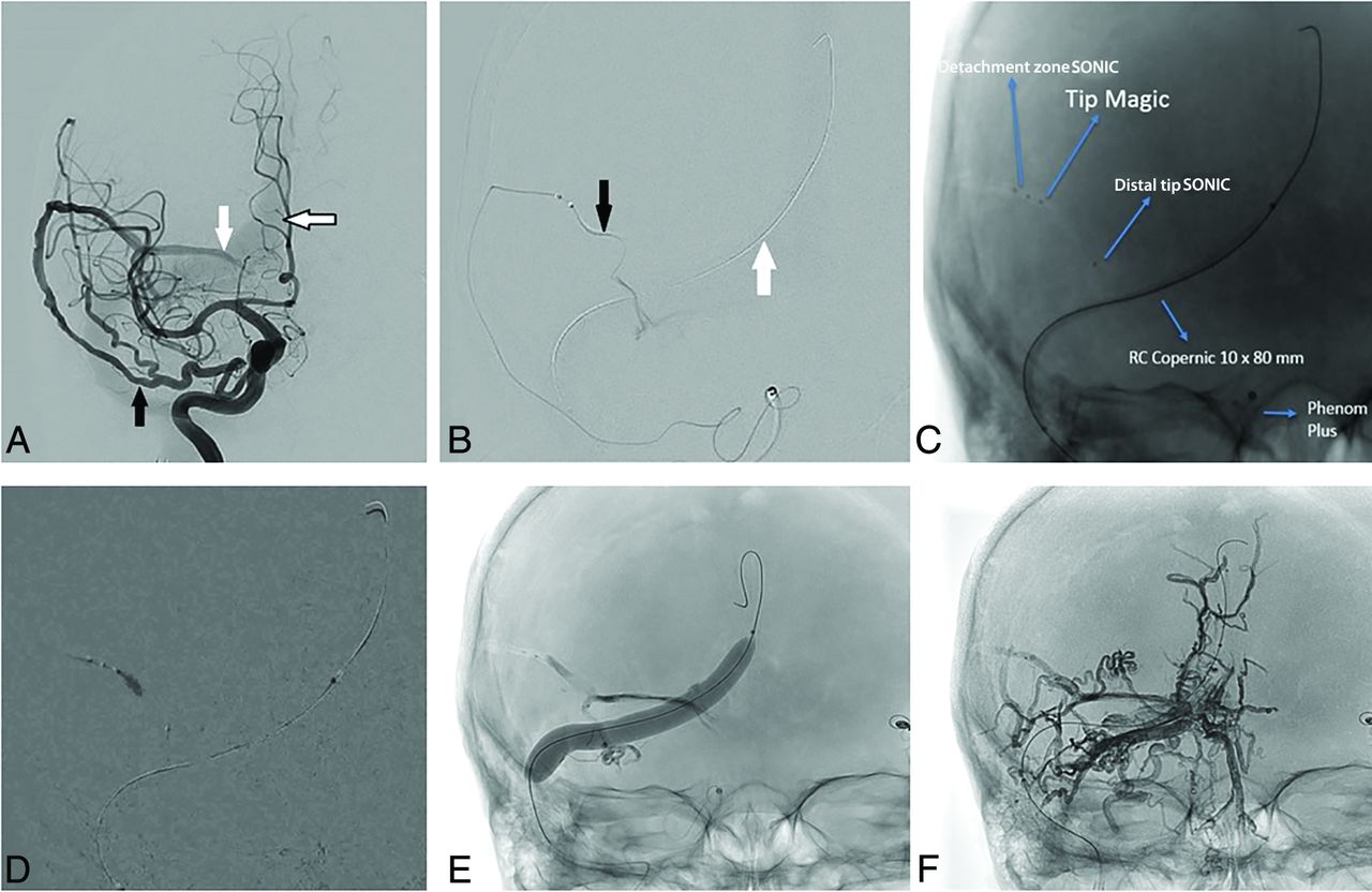
- FIG 1.
Transarterial embolization of a transverse sinus AVF with sinus balloon protection. A, Anterior-posterior projection of a right ICA angiogram demonstrates a right transverse sinus dural AVF supplied by a recurrent meningeal artery (black arrow), which originates from the ophthalmic artery. It drains into the right transverse sinus (white arrow), where there is early venous filling. Sinus reflux is present (black border arrow). B, A detachable-tip microcatheter (SONIC; Balt) has been advanced through this artery, and its tip has been placed distally for a microcatheter DSA. The detachable segment is marked by the black arrow. The noninflated sinus-protection balloon (Copernic RC 10 × 80 mm; Balt) has been placed in the right transverse sinus and torcula (white arrow). C, Spot anterior-posterior film before embolization shows 2 microcatheters adjacent to each other for a modified pressure cooker technique. Arrows and labels indicate the proximal detachment point of the SONIC microcatheter, the distal tip of the flow-directed second microcatheter (Magic; Balt), the distal tip of the SONIC microcatheter, and the sinus-protection balloon (not yet inflated). D, Anterior-posterior subtracted fluoroscopic image demonstrates a glue-Lipiodol (Guerbet) mixture being injected from the distal tip of the second microcatheter (Magic) and forming a cast over the detachable segment of the SONIC microcatheter (pressure cooker technique). E, Spot anterior-posterior film demonstrates the inflated sinus-protection balloon, the glue cast, and an inverted-Y shaped early EVOH cast forming in the early stages of the embolization. F, Spot anterior-posterior film following completion of embolization and deflation of the balloon shows an extensive EVOH cast surrounding the fistulous point, forming a tunnel around the sinus, and extending into multiple adjacent arterial feeders (predominantly from occipital artery feeders).
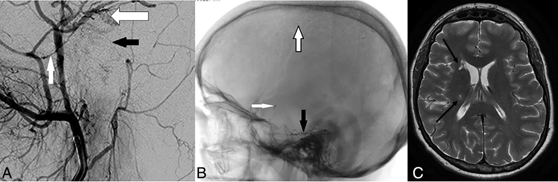
- FIG 2.
Transarterial glue embolization of a petrous dural AVF, complicated by brain parenchymal infarction. A, Lateral projection of an external carotid artery angiogram (magnified to show small vessels) demonstrates the petrous branch of the middle meningeal artery (white arrow) and the stylomastoid branch of the combined occipital/posterior auricular artery (black arrow) forming an anastomotic arch over the petrous temporal bone. This is the facial nerve arterial arcade and is the vasa nervosum to the geniculate ganglion and the intraosseous segments of the facial nerve. Adjacent to this is a complex focus of abnormal dural vessels (black border arrow) from a petrous dural AVF. Note the proximity of the AVF to the vasa nervosum. Thus, the more distal feeder of the middle meningeal artery was used for embolization. B, Following transarterial glue embolization, a spot lateral projection demonstrates a glue-Lipiodol cast filling the AVF (black arrow) but also droplets of glue in the intracranial ICA (white arrow) and distal cerebral convexity branches (black border arrow). These were due to transmission of glue across EC-IC anastomoses, most likely from middle meningeal artery branches into the lateral tentorial branch of the meningohypophyseal trunk. The patient woke with a temporary contralateral hemiparesis, which slowly resolved during several weeks. C, Axial TSE T2 image from follow-up MR imaging demonstrates small foci of gliosis in the right caudate head and posterior limb of the right internal capsule (black arrows), resulting from the prior embolic glue-related infarcts.
- FIG 3.
DSA and transarterial embolization using EVOH of a right-sided Borden III/Cognard IV petrous dural AVF. All images are in the lateral projection. A, Right external carotid artery injection demonstrates arterial supply to the AVF from the squamous temporal branch of the right middle meningeal artery (black-border arrow), from an enlarged petrous branch of the middle meningeal artery (white arrow), and from an enlarged stylomastoid artery (black arrow). Note the facial nerve arterial arcade formed by the latter 2 vessels. B, A microcatheter is present in the squamous branch of the middle meningeal artery (black-border arrow). Microcatheter-injection DSA from a distal point in the vessel demonstrates the fistulous point (white arrow) entering an ectatic petrosal varix. Note that the microcatheter tip is >20 mm from the origin of the petrous branch of the middle meningeal artery as seen in A, allowing reflux of EVOH back to the proximal marker and detachment of the 15-mm-length tip without penetration into the facial nerve arcade. C, Spot film following EVOH embolization via the squamous branch of the middle meningeal artery demonstrates the EVOH cast (black arrow) across the fistulous point and into the venous sac. The proximal marker of the detachable tip is visible (white arrow) and is well distal to the origin of the petrous branch of the middle meningeal artery as seen in A. D, Magnified right common carotid artery injection following embolization demonstrates the subtracted EVOH cast (black-border arrow) with no remnant filling the AVF. The petrous branch of the middle meningeal artery (white arrows) and the stylomastoid artery (black arrow) remains patent after treatment. The patient had preserved facial nerve function postprocedure. Reproduced - from Bhatia et al.15


