
Article Figures & Data
Figures
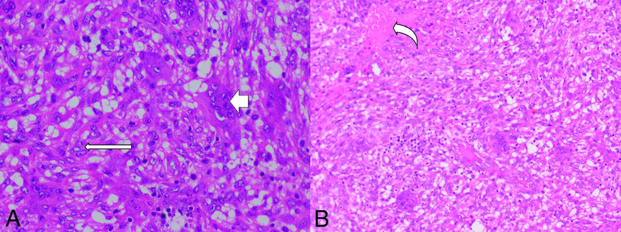
- FIG 1.
A, H&E, original magnification ×200. Cellular lesion composed of sheets of oval- to spindle-shaped cells admixed with many multinucleated giant cells (short arrow), the nuclei of which resemble stromal cells (long arrow). Mitotic activity is inconspicuous. B, H&E original magnification ×100. Hemorrhagic foci (curved arrow) and mild lymphocytic infiltrates in the surrounding stroma.
- FIG 2.
GCG of the mandible. Orthopantomogram shows an expansile, lytic central mandibular lesion with lobulated margins (asterisk). Note the resorption of the roots of the central incisors (arrow).
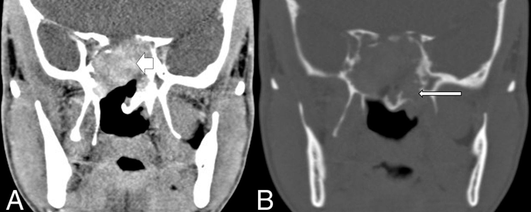
- FIG 3.
GCG of the sphenoid bone. A, Contrast-enhanced CT of the skull base shows a lobulated, enhancing mass in the sphenoid sinus (short arrow). B, On the bone window, the mass expands the sphenoid bone and there are multiple foci of a cortical breach (long arrow) Note the absence of matrix mineralization in the lesion.
- FIG 4.
MR imaging features of GCG. A, Heterogeneous intermediate-to-high signal intensity (short arrow) on the T1-weighted sagittal image. B, Mixed intermediate-to-hyperintense (white asterisk) and hypointense (long arrow) solid components on T2-weighted axial image in the sphenoid bone and clival GCG.
- FIG 5.
GCG of the orbit with secondary ABC changes appearing as a multiloculated lesion with multiple fluid-fluid levels (arrow) of differential signal intensity on T1-weighted (A) and T2-weighted (B) axial images.
- FIG 6.
Multiloculated GCG of the left temporal bone with fluid-fluid levels (short arrow, A) on axial T2-weighted and layers of hypointense signal (long arrow, B) on a susceptibility-weighted imaging.
- FIG 7.
Patterns of enhancement in GCG (on postgadolinium T1WI). A, Solid, sphenoidal GCG shows heterogeneous enhancement (short arrow). B, Multiloculated right-orbital GCG with enhancing walls (long arrow).
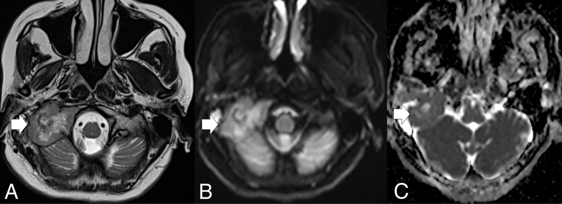
- FIG 8.
GCG of the right petrous bone. The lesion (arrow) is heterogeneous with peripheral, intermediate-signal-intensity soft tissue and central cystic area on (A) T2-weighted. DWI (B) and ADC map (C) of the lesion.


Tables
GCG GCT ABC Brown Tumor Epithelioid stroma Spindled stroma, oval-to-elongated nuclei Plump epithelioid with oval nuclei Spindled stroma, oval-to-elongated nuclei Spindled stroma, oval-to-elongated nuclei with fibrous stroma dividing it into lobules Giant cells 12 Nuclei in clusters around hemorrhagic foci >12 Uniformly distributed nuclei Smaller giant cells, in clusters, woven bone present, less hemorrhage Few giant cells Immunohistochemistry (p63 positivity) Yes Yes No No Gene mutation H3f3a mutation No Yes No No USP6 rearrangement No No Yes No Male Female Total Orbit 1 0 1 Mandible 3 2 5 Maxilla 3 5 8 Temporal 1 2 3 Sphenoid/clivus 2 1 3


Jump to section
Related Articles
Cited By...
- No citing articles found.