Advertisement

Main menu

  • Home
  • Content
    • Current Issue
    • Accepted Manuscripts
    • Article Preview
    • Past Issue Archive
    • AJNR Case Collection
    • Case of the Week Archive
    • Classic Case Archive
    • Case of the Month Archive
  • Special Collections
    • Spinal CSF Leak Articles (Jan 2020-June 2024)
    • 2024 AJNR Journal Awards
    • Most Impactful AJNR Articles
  • Multimedia
    • AJNR Podcast
    • AJNR Scantastics
    • Video Articles
  • For Authors
    • Submit a Manuscript
    • Author Policies
    • Fast publishing of Accepted Manuscripts
    • Graphical Abstract Preparation
    • Manuscript Submission Guidelines
    • Imaging Protocol Submission
    • Submit a Case for the Case Collection
  • About Us
    • About AJNR
    • Editorial Board
  • More
    • Become a Reviewer/Academy of Reviewers
    • Subscribers
    • Permissions
    • Alerts
    • Feedback
    • Advertisers
    • ASNR Home
  • Other Publications
    • ajnr

User menu

  • Alerts
  • Log in

Search

  • Advanced search
American Journal of Neuroradiology

American Journal of Neuroradiology

ASHNR American Society of Functional Neuroradiology ASHNR American Society of Pediatric Neuroradiology ASSR
  • Alerts
  • Log in

Advanced Search

  • Home
  • Content
    • Current Issue
    • Accepted Manuscripts
    • Article Preview
    • Past Issue Archive
    • AJNR Case Collection
    • Case of the Week Archive
    • Classic Case Archive
    • Case of the Month Archive
  • Special Collections
    • Spinal CSF Leak Articles (Jan 2020-June 2024)
    • 2024 AJNR Journal Awards
    • Most Impactful AJNR Articles
  • Multimedia
    • AJNR Podcast
    • AJNR Scantastics
    • Video Articles
  • For Authors
    • Submit a Manuscript
    • Author Policies
    • Fast publishing of Accepted Manuscripts
    • Graphical Abstract Preparation
    • Manuscript Submission Guidelines
    • Imaging Protocol Submission
    • Submit a Case for the Case Collection
  • About Us
    • About AJNR
    • Editorial Board
  • More
    • Become a Reviewer/Academy of Reviewers
    • Subscribers
    • Permissions
    • Alerts
    • Feedback
    • Advertisers
    • ASNR Home
  • Follow AJNR on Twitter
  • Visit AJNR on Facebook
  • Follow AJNR on Instagram
  • Join AJNR on LinkedIn
  • RSS Feeds

Welcome to the new AJNR, Updated Hall of Fame, and more. Read the full announcements.


AJNR is seeking candidates for the position of Associate Section Editor, AJNR Case Collection. Read the full announcement.


Research ArticleNeurovascular/Stroke Imaging

Prevalence and Characteristics of Intracranial Aneurysms in Hereditary Hemorrhagic Telangiectasia

How-Chung Cheng, Marie E. Faughnan, Karel G. terBrugge, Hon-Man Liu, Timo Krings and the Brain Vascular Malformation Consortium Hereditary Hemorrhagic Telangiectasia Investigator Group
American Journal of Neuroradiology December 2023, 44 (12) 1367-1372; DOI: https://doi.org/10.3174/ajnr.A8058
How-Chung Cheng
aFrom the Division of Neuroradiology (H.-C.C., K.G.t., T.K.), Joint Department of Medical Imaging, Toronto Western Hospital, University Health Network, Toronto, Ontario, Canada
bDivision of Neurosurgery (H.-C.C.), Department of Surgery, Fu Jen Catholic University Hospital, Fu Jen Catholic University, New Taipei City, Taiwan
  • Find this author on Google Scholar
  • Find this author on PubMed
  • Search for this author on this site
  • ORCID record for How-Chung Cheng
Marie E. Faughnan
cToronto HHT Centre (M.E.F.), St. Michael's Hospital and Li Ka Shing Knowledge Institute, Toronto, Ontario, Canada
dDivision of Respirology (M.E.F.), Department of Medicine, University of Toronto, Toronto, Ontario, Canada
  • Find this author on Google Scholar
  • Find this author on PubMed
  • Search for this author on this site
  • ORCID record for Marie E. Faughnan
Karel G. terBrugge
aFrom the Division of Neuroradiology (H.-C.C., K.G.t., T.K.), Joint Department of Medical Imaging, Toronto Western Hospital, University Health Network, Toronto, Ontario, Canada
  • Find this author on Google Scholar
  • Find this author on PubMed
  • Search for this author on this site
Hon-Man Liu
eDepartment of Medical Imaging (H.-M.L.), Fu Jen Catholic University Hospital, Fu Jen Catholic University, New Taipei City, Taiwan
  • Find this author on Google Scholar
  • Find this author on PubMed
  • Search for this author on this site
  • ORCID record for Hon-Man Liu
Timo Krings
aFrom the Division of Neuroradiology (H.-C.C., K.G.t., T.K.), Joint Department of Medical Imaging, Toronto Western Hospital, University Health Network, Toronto, Ontario, Canada
  • Find this author on Google Scholar
  • Find this author on PubMed
  • Search for this author on this site
  • ORCID record for Timo Krings
  • Article
  • Figures & Data
  • Info & Metrics
  • Responses
  • References
  • PDF
Loading

Abstract

BACKGROUND AND PURPOSE: The association between hereditary hemorrhagic telangiectasia and intracranial aneurysms remains controversial. This study evaluated the prevalence and characteristics of intracranial aneurysms in patients with hereditary hemorrhagic telangiectasia with brain vascular malformations.

MATERIALS AND METHODS: Between 2007 and 2021, patients enrolled in the Brain Vascular Malformation Consortium with definite hereditary hemorrhagic telangiectasia, the presence of brain vascular malformations, and available angiographic studies of the brain were retrospectively reviewed. Angiographic features of intracranial aneurysms and their relationship to coexisting brain vascular malformations were analyzed. We also examined the association between baseline clinical features and the presence of intracranial aneurysms.

RESULTS: One hundred eighty patients were included. A total of 14 intracranial aneurysms were found in 9 (5%) patients, and 4 intracranial aneurysms were considered flow-related aneurysms. Patients with intracranial aneurysms were significantly older than patients without intracranial aneurysms (mean, 48.1 [SD, 18.2] years versus 33.5 [SD, 21.0] years; P = .042). If we excluded flow-related intracranial aneurysms, the prevalence of intracranial aneurysms was 3.3%. All intracranial aneurysms were in the anterior circulation, were unruptured, and had an average maximal diameter of 3.9 (SD, 1.5) mm. No intracranial aneurysms were found in pediatric patients with hereditary hemorrhagic telangiectasia. No statistically significant correlation was observed among other baseline demographics, hereditary hemorrhagic telangiectasia features, and the presence of intracranial aneurysms.

CONCLUSIONS: The prevalence of intracranial aneurysms in this large cohort study is comparable with that in the general population and might be increased slightly due to hemodynamic factors associated with shunting brain vascular malformations.

ABBREVIATIONS:

HHT
hereditary hemorrhagic telangiectasia
IA
intracranial aneurysm
VM
vascular malformation

Hereditary hemorrhagic telangiectasia (HHT) or Osler-Weber-Rendu syndrome is an autosomal dominant disorder characterized by mucocutaneous telangiectasias and vascular malformations (VMs) in organs, including the liver, gastrointestinal tract, lungs, brain, and spinal cord.1,2 It is caused by mutations in one of several genes involved in the transforming growth factor-β/bone morphogenetic protein signaling pathway. Loss-of-function mutations in HHT genes have been shown to impair endothelial cell proliferation and migration during angiogenesis and vascular remodeling.1,3,4 Approximately 96% of individuals with a definite clinical diagnosis of HHT have a mutation in the endoglin (ENG) gene or activin A receptor type II-like kinase 1 (ACVRL1) gene,5 and about 3% of patients have a mutation in the SMAD4 gene. The diagnosis of HHT is made clinically using the Curaçao criteria, which include spontaneous and recurrent nose bleeds, multiple mucocutaneous telangiectasias (of the lips, oral cavity, nose, or fingers), visceral VMs (of the liver, lungs, or CNS), and a first-degree relative with HHT. Patients are diagnosed with “definite HHT” when 3 of these 4 criteria are met, whereas those who fulfill 2 criteria are considered to have “possible HHT.”6 For asymptomatic individuals from a family with known HHT, the International HHT Guidelines recommend genetic testing for diagnosis.5

Patients with HHT present with a wide variety of cerebrovascular diseases. Between 10% and 20% of patients have brain VMs, classified in previous studies as pial AVFs, nidus-type AVMs, or capillary vascular malformations.7⇓⇓⇓-11 Other nonshunting vascular lesions such as cavernous malformations, developmental venous anomalies, and intracranial aneurysms (IAs) were also found in patients with HHT.8,11,12

Although earlier studies reported the prevalence of IAs in the HHT population,8,11,13,14 no studies have addressed the characteristics of these aneurysms and their relationship to AVMs. The present study aimed to determine the prevalence of IAs in the HHT population, identify angiographic features of IAs, and examine the correlation between IAs and brain VMs in patients with HHT. Notably, a recent study found a strikingly high 14.5% prevalence of IAs in an HHT cohort, contrary to previous studies.14 Current screening recommendations may need refinement if this higher prevalence of IAs is confirmed in our data set.

MATERIALS AND METHODS

Study Population

We analyzed patient data from the HHT project of the Brain Vascular Malformation Consortium. Patients with either a confirmed genetic diagnosis or a definite clinical diagnosis of HHT, based on the presence of at least 3 of the Curaçao criteria,6 were enrolled in the Brain Vascular Malformation Consortium HHT project as previously reported.10,15 The Brain Vascular Malformation Consortium HHT cohort aimed for a 25% recruitment target for patients with brain VMs.15 From January 2007 to December 2021, we included all recruited patients with brain VMs and available angiographic studies (ie, CTA, MRA, and/or DSA). All patients provided written, informed consent. The study was approved by each participating center's institutional review board.

Data Collection

For each patient, we collected data including age at enrollment, sex, clinical presentations, family history, and genetic testing results (ENG, ACVRL1, SMAD4) when available. Radiologic data included biplanar 6-vessel selective cerebral angiography, contrast-enhanced MRA or TOF-MRA using 1.5T or 3T scanners, and CTA. All images were retrospectively reviewed by 2 of the authors (H.-C.C. and T.K.) in consensus. Images were assessed for the presence of IAs and shunting lesions such as AVMs and AVFs, as classified in previous studies.10,16 The aneurysm location, the number of aneurysms per patient, their geometric measurements, and their relationship to the intracranial shunting lesions were recorded. We measured aneurysm neck width, dome-to-neck ratio, and aspect ratio on the PACS. Aneurysms with a neck diameter of ≥4 mm or a dome-to-neck ratio of <2 were classified as wide-neck.17,18 A Spetzler-Martin grade was calculated for each AVM when the patient had a concurrent IA.19 IAs were considered unrelated aneurysms when they presented on vessels that were not AVM feeders, whereas those arising from vessels contributing to perfusion of the nidus were termed flow-related aneurysms.20 Flow-related aneurysms were further classified as proximal when located at the circle of Willis up to the primary bifurcation, while those more distally located were considered distal.

Statistical Tests

We determined the prevalence of IAs by the number of patients having at least 1 IA divided by the total number of included patients with HHT. We tested for differences in age, sex, the presence of Curaçao criteria, and the presence of AVMs/AVFs between patients with IAs and those without. Continuous variables were compared using the Student t test, and categoric variables were compared using the Fisher exact test. We considered P values < .05 as statistically significant. All analyses used SPSS (IBM).

RESULTS

One hundred eighty patients with HHT and brain VMs as well as available angiographic studies were included in the study. The average age at enrollment was 34.2 (SD, 21.1) years, with 54 (30%) subjects younger than 18 years of age. Female patients accounted for 61.7% of the population. One hundred thirty-one patients had positive findings on genetic testing: One hundred nine (83.2%) had an ENG mutation, 21 (16.0%) had an ACVRL1 mutation, and 1 (0.7%) had a SMAD4 mutation. Basic demographics and clinical features of the study population are summarized in Table 1.

View this table:
  • View inline
  • View popup
Table 1:

Basic demographics of 180 subjects with HHT and brain VMs

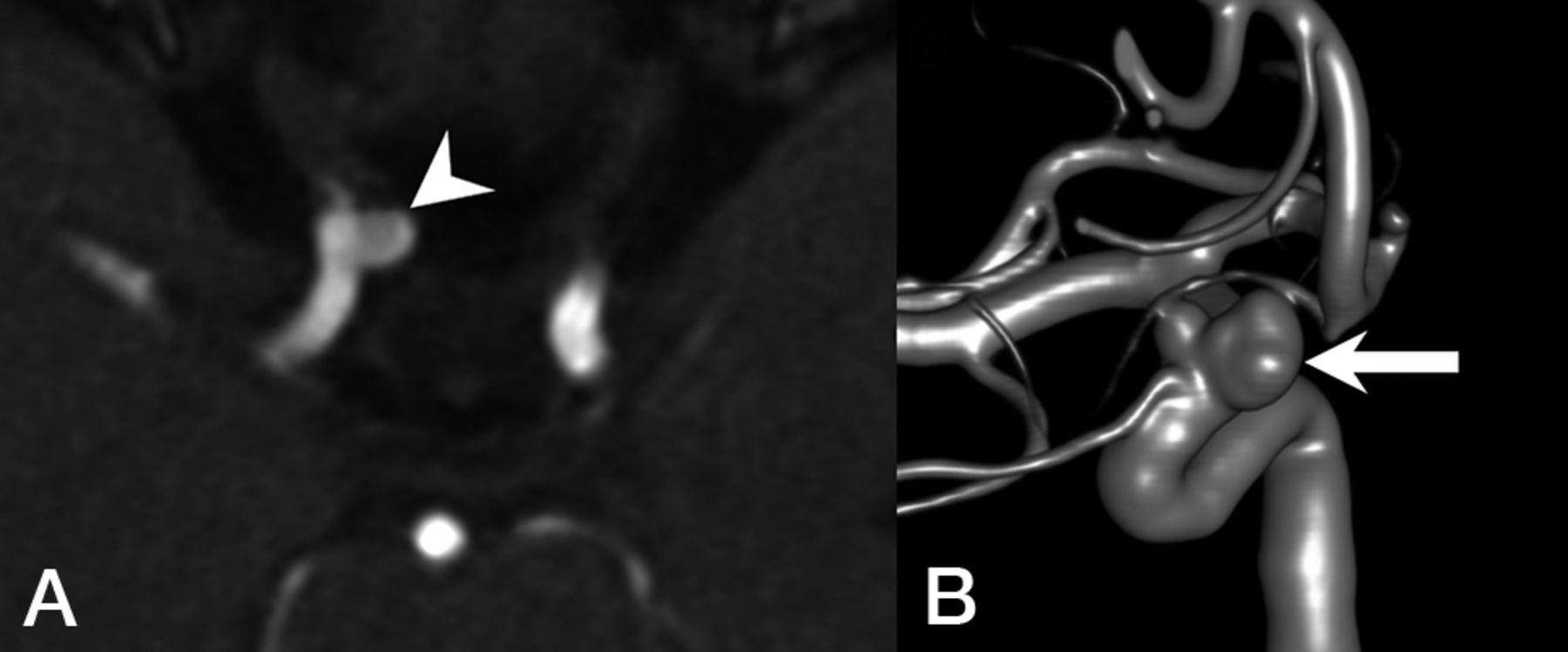
Fourteen IAs were noted in 9 (5%) patients, and 3 (33%) patients had multiple aneurysms. No pediatric patients with HHT in this cohort had an IA. All IAs were unruptured and were found in the anterior circulation, most commonly located in the ICA (64%), followed by the anterior cerebral artery (21%). The average neck size and maximal aneurysm diameter were 3.7 (SD, 1.3) mm and 3.9 (SD, 1.5) mm, respectively. The average aspect ratio was 0.92 (SD, 0.30). All aneurysms had a dome-to-neck ratio of <2. A representative example of IAs is shown in Fig 1.

Fig 1.
  • Download figure
  • Open in new tab
  • Download powerpoint
Fig 1.

Representative images of a patient with HHT with an IA unrelated to AVMs. A, MRA demonstrates an ophthalmic segment aneurysm of the right ICA (arrowhead) projecting medially. B, 3D reconstruction of a DSA shows a bilobed, wide-neck, right ophthalmic segment aneurysm of the right ICA (arrow).

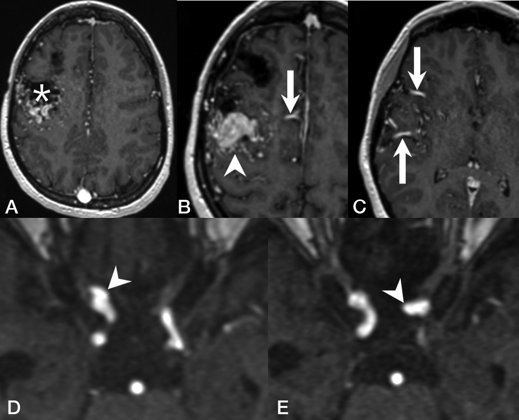
Of the 9 patients with IAs, 6 had nidus-type brain AVMs, 1 had an AVF, and 2 had a capillary vascular malformation. Ten (71%) of the 14 IAs were considered unrelated to the VMs, whereas 4 (29%) were considered flow-related aneurysms arising from a feeding artery of a shunting VM. One patient with a right frontal Spetzler-Martin grade III AVM and a nidus size of 4 cm had 1 aneurysm on each of the ICAs (Fig 2). Another patient with a left frontal AVF and a large venous pouch had an aneurysm on the ipsilateral anterior cerebral artery. The last patient had 2 Spetzler-Martin grade I AVMs in the same hemisphere as the aneurysm. The characteristics of IAs and angiographic features are summarized in the Online Supplemental Data. Patients with IAs were significantly older compared with patients without IAs (mean, 48.1 [SD, 18.2] years versus 33.5 1 [SD, 21.0] years; P = .042). No statistically significant association was found between patients with or without IAs regarding sex, the presence of the Curaçao criteria, or the presence of concurrent shunting brain VMs (AVMs and/or AVFs) (Table 2).

Fig 2.
  • Download figure
  • Open in new tab
  • Download powerpoint
Fig 2.

Representative images of a patient with HHT with flow-related cerebral aneurysms. T1 contrast-enhanced MR images demonstrate a Spetzler-Martin grade III right frontal AVM with a nidus size of 4 cm (A, asterisk) and venous varices (B, arrowhead), supplied by several dilated arterial feeders from the right anterior cerebral arteries (B, arrow) and the right middle cerebral arteries (C, arrow). MRA demonstrates a clinoid segment aneurysm of the right ICA (D, arrowhead) and another aneurysm arising from the ophthalmic segment of the left ICA (E, arrowhead).

View this table:
  • View inline
  • View popup
Table 2:

Bivariate analysis between baseline demographics and IAs

DISCUSSION

The association between HHT and IAs remains controversial. In a report on neurovascular manifestations of patients with HHT, Brinjikji et al11 demonstrated a 2.1%. prevalence of IAs. Woodall et al8 evaluated 230 patients with HHT and found a 2.4% prevalence of IAs. Ring et al13 looked for arterial aneurysms in all locations in an HHT cohort and reported a 4.3% IA prevalence. A very recent study from Perez Akly et al14 found 33 (14.5%) patients with HHTs with IAs, >3-fold higher than the prevalence rate of 3.2% in the general population,21 thus potentially changing the clinical practice regarding active screening for IAs in patients with HHT. This remarkable discrepancy and the potential impact of encountering a high prevalence of IAs in patients with HHT prompted the present study.

We report a prevalence of IAs of 5% in this largest series of patients with HHT with brain VMs. In the subgroup of patients not having flow-related IAs, the prevalence of IAs was 3.3%, very similar to that of the general population. Although we included specifically patients with HHT with brain VMs, our observation is in line with the reported prevalences by Brinjikji et al,11 Woodall et al,8 and Ring et al.13 In addition, our study cohort shares similar prevalences of epistaxis,8,11,14 mucosal telangiectasias,1,11,22 and pulmonary AVMs1,11,23,24 with previous reports, and, therefore, is likely generalizable to unselected patients with HHT. Because children can develop complications of HHT,25 we believe that it is necessary to include the pediatric population in this epidemiologic study to better understand the prevalence and natural history of IAs and, more important, to determine whether HHT is associated with an increased risk of IAs. Moreover, we are convinced that by including all patients with available angiographic studies of all types (ie, CTA, MRA, and/or DSA), our observed prevalence of IAs more accurately reflects the prevalence of IAs in the HHT population. According to our findings and those of the latter 3 studies,8,11,13 the prevalence of IAs in HHT is similar to that in the general population, with a small additional prevalence of IAs in patients with HHT with coexisting shunting brain VMs.

This observation is in keeping with pathogenetic concepts of HHT in several ways. First, it is postulated that in HHT, the targets of the dysfunction are the venules instead of the arteries.16 Early histopathologic studies demonstrated that the clinically detectable lesions of HHT start with focal dilation of postcapillary venules, which enlarge and connect with arterioles, eventually forming direct arteriovenous shuntings as capillaries disappear.1,26 More recent animal studies also suggest that the ENG/ACVRL1 signaling pathway mediates the endothelial cell shear stress response and is essential for flow-induced endothelial cell migration against the direction of blood flow, starting from growing veins, ie, flow-migration coupling. Loss of ENG or ACVRL1 expression in venous and capillary endothelial cells is sufficient to cause AVMs as a result of impaired flow-migration coupling, whereas ENG or ACVRL1 deletion in arterial endothelial cells does not lead to AVM formation.4,27⇓⇓-30 Histopathologically, HHT is unlikely to be associated with a higher prevalence of IAs. Second, although several reports have associated the interruption of the transforming growth factor-β signaling pathway with a higher prevalence of visceral aneurysms in patients with HHT due to its important role in extracellular matrix remodeling,13,31 there is no genetic evidence to support applying the same hypothesis to IAs. ENG, one of the most commonly found mutated genes in patients with HHT, has repeatedly been reported to have no association with IAs.32,33

In a study about sequencing transforming growth factor-β pathway genes in familial IA cases, Santiago-Sim et al34 demonstrated that mutations in transforming growth factor-β receptor genes are not a major cause of IAs. Furthermore, a recently published genome-wide association study also demonstrated no evidence of polygenic overlap between other aortic aneurysms and IAs,35 while another meta-analysis of genome-wide association study on genetic risk factors for IAs found no linkage between transforming growth factor-β-related genes and the development of IAs.36 Finally, unlike some other clinical features of HHT that become apparent in early childhood and evolve, no IAs were found in the pediatric population of our cohort. In addition, the mean age of patients with IAs in this study was 48 years, comparable with the mean age of 47–55 years described in non-HHT populations with IAs.37,38 If IAs and VMs share the same pathogenesis, there would presumably be more evidence of IAs in younger patients with HHT.

IAs in this study were most commonly found in the ICA and were small with a mean maximal diameter of 3.9 mm. Similar to findings in the previous studies,8,11,14 no patients in this cohort presented with SAH, which may hint at a benign nature of IAs in HHT. Although there were a few case reports describing ruptured IAs in patients with HHT,39,40 most were flow-related aneurysms associated with a shunting brain VM.

In this study, we found no association between IAs and shunting brain VMs. Because approximately 71% of included patients with HHT had at least 1 shunting brain VM, it is possible that we could not detect any significant difference in brain VM prevalence between patients with and without IAs, given this high prevalence of brain VMs in our cohort. Notably, merely 29% of the IAs in this study were related to the shunting brain VMs, and only 2% of the AVMs were found to have flow-related aneurysms, as opposed to 18% described previously by a meta-analysis on the natural history of brain AVMs and concurrent IAs in the non-HHT population.41 One possible explanation is thart most of the AVMs found in patients with HHT are <3 cm;10,11,42 therefore, the hemodynamic factors related to the shunting in the AVM nidus, such as higher shear stress on the vessel wall, are less prominent among patients with HHT.

In conclusion, the prevalence of IAs in patients with HHT with brain VMs is similar to that in the general population. In addition, we observed that IAs in patients with HHTs are seen in adults, but not children, and they are typically small with a low likelihood of hemorrhagic presentation. Thus, our observations do not support a recommendation for routine screening of patients with HHT specifically for IAs.

This study has limitations. First, it has some inherent selection bias because we included only patients with brain VMs. Although the genetic linkage between IAs and brain VMs, if any, remains undetermined, the prevalence of IAs in our study was in line with that reported in the literature. Second, genetic information was not available for one-third of the included individuals; therefore, there was an insufficient sample size to assess the association between IAs and genes mutated. Also, our study cohort included few patients with ACVRL1 or SMAD4 mutations, unsurprisingly, given the known association between brain VMs and ENG mutation, among patients with HHT. Additional studies will be required to clarify the association between these genes and IAs. Finally, the limited sample size of IAs may reduce the power of the study to detect statistically significant correlations such as the association between IAs and shunting brain VMs, but there was no evidence of even a trend association.

CONCLUSIONS

From this large cohort of patients with HHT and brain VMs, we demonstrate that the prevalence of IAs in HHT is similar to that of the general population and is not clearly associated with the presence of shunting brain VMs. Further studies are required to explore the mechanistic linkage between HHT and IA formation and the natural history of IAs in the HHT population.

ACKNOWLEDGMENTS

We would like to thank Professor Shiao-Chi Wu of National Yang Ming Chiao Tung University for statistical analysis.

Brain Vascular Malformation Consortium Hereditary Hemorrhagic Telangiectasia Investigator Group:

Mary E. Atherton, Murali M. Chakinala, Marianne S. Clancy, Marie E. Faughnan, James R. Gossage, Adrienne M. Hammill, Katharine Henderson, Steven Hetts, Peter Hountras, Vivek Iyer, Raj S. Kasthuri, Helen Kim, Timo Krings, Michael T. Lawton, Doris Lin, Johannes Jurgen Mager, Douglas A. Marchuk, Justin P. McWilliams, Jamie McDonald, Ludmila Pawlikowska, Jeffrey Pollak, Felix Ratjen, Karen Swanson, Dilini Vethanayagam, Shantel Weinsheimer, Andrew J White, Pearce Wilcox.

Footnotes

  • The Brain Vascular Malformation Consortium is part of the Rare Diseases Clinical Research Network, which is funded by the National Institutes of Health and led by the National Center for Advancing Translational Sciences through its Office of Rare Diseases Research. The Brain Vascular Malformation Consortium is funded under grant No. U54NS065705 as a collaboration between the National Center for Advancing Translational Sciences and the National Institute of Neurological Disorders and Stroke. All Rare Diseases Clinical Research Network consortia are supported by the network's Data Management and Coordinating Center (U2CTR002818). Funding support for the Data Management and Coordinating Center is provided by National Center for Advancing Translational Sciences and the National Institute of Neurological Disorders and Stroke.

  • M.E.F. was also supported by the Li Ka Shing Knowledge Institute. This work was funded, in part, by the Holt-Hornsby and Andreae Fund and University Medical Imaging Toronto.

  • Disclosure forms provided by the authors are available with the full text and PDF of this article at www.ajnr.org.

References

  1. 1.↵
    1. Shovlin CL
    . Hereditary haemorrhagic telangiectasia: pathophysiology, diagnosis and treatment. Blood Rev 2010;24:203–19 doi:10.1016/j.blre.2010.07.001 pmid:20870325
    CrossRefPubMed
  2. 2.↵
    1. McDonald J,
    2. Bayrak-Toydemir P,
    3. Pyeritz RE
    . Hereditary hemorrhagic telangiectasia: an overview of diagnosis, management, and pathogenesis. Genet Med 2011;13:607–16 doi:10.1097/GIM.0b013e3182136d32 pmid:21546842
    CrossRefPubMed
  3. 3.↵
    1. McDonald J,
    2. Wooderchak-Donahue W,
    3. VanSant Webb C, et al
    . Hereditary hemorrhagic telangiectasia: genetics and molecular diagnostics in a new era. Front Genet 2015;6:1 doi:10.3389/fgene.2015.00001 pmid:25674101
    CrossRefPubMed
  4. 4.↵
    1. Jin Y,
    2. Muhl L,
    3. Burmakin M, et al
    . Endoglin prevents vascular malformation by regulating flow-induced cell migration and specification through VEGFR2 signaling. Nat Cell Biol 2017;19:639–52 doi:10.1038/ncb3534 pmid:28530660
    CrossRefPubMed
  5. 5.↵
    1. Faughnan ME,
    2. Mager JJ,
    3. Hetts SW, et al
    . Second international guidelines for the diagnosis and management of hereditary hemorrhagic telangiectasia. Ann Intern Med 2020;173:989–1001 doi:10.7326/M20-1443 pmid:32894695
    CrossRefPubMed
  6. 6.↵
    1. Shovlin CL,
    2. Guttmacher AE,
    3. Buscarini E, et al
    . Diagnostic criteria for hereditary hemorrhagic telangiectasia (Rendu-Osler-Weber syndrome). Am J Med Genet 2000;91:66–67 doi:10.1002/(SICI)1096-8628(20000306)91:1<66::AID-AJMG12>3.0.CO;2-P pmid:10751092
    CrossRefPubMed
  7. 7.↵
    1. Nishida T,
    2. Faughnan ME,
    3. Krings T, et al
    . Brain arteriovenous malformations associated with hereditary hemorrhagic telangiectasia: gene-phenotype correlations. Am J Med Genet A 2012;158A:2829–34 doi:10.1002/ajmg.a.35622 pmid:22991266
    CrossRefPubMed
  8. 8.↵
    1. Woodall MN,
    2. McGettigan M,
    3. Figueroa R, et al
    . Cerebral vascular malformations in hereditary hemorrhagic telangiectasia. J Neurosurg 2014;120:87–92 doi:10.3171/2013.10.JNS122402 pmid:24236651
    CrossRefPubMed
  9. 9.↵
    1. Brinjikji W,
    2. Iyer VN,
    3. Sorenson T, et al
    . Cerebrovascular manifestations of hereditary hemorrhagic telangiectasia. Stroke 2015;46:3329–37 doi:10.1161/STROKEAHA.115.010984 pmid:26405205
    FREE Full Text
  10. 10.↵
    1. Krings T,
    2. Kim H,
    3. Power S, et al
    ; Brain Vascular Malformation Consortium HHT Investigator Group. Neurovascular manifestations in hereditary hemorrhagic telangiectasia: imaging features and genotype-phenotype correlations. AJNR Am J Neuroradiol 2015;36:863–70 doi:10.3174/ajnr.A4210 pmid:25572952
    Abstract/FREE Full Text
  11. 11.↵
    1. Brinjikji W,
    2. Iyer VN,
    3. Yamaki V, et al
    . Neurovascular manifestations of hereditary hemorrhagic telangiectasia: a consecutive series of 376 patients during 15 years. AJNR Am J Neuroradiol 2016;37:1479–86 doi:10.3174/ajnr.A4762 pmid:27012295
    Abstract/FREE Full Text
  12. 12.↵
    1. Gaetani E,
    2. Peppucci E,
    3. Agostini F, et al
    . Distribution of cerebrovascular phenotypes according to variants of the ENG and ACVRL1 genes in subjects with hereditary hemorrhagic telangiectasia. J Clin Med 2022;11:2685 doi:10.3390/jcm11102685 pmid:35628811
    CrossRefPubMed
  13. 13.↵
    1. Ring NY,
    2. Latif MA,
    3. Hafezi-Nejad N, et al
    . Prevalence of and factors associated with arterial aneurysms in patients with hereditary hemorrhagic telangiectasia: 17-year retrospective series of 418 patients. J Vasc Interv Radiol 2021;32:1661–69 doi:10.1016/j.jvir.2021.08.018 pmid:34478850
    CrossRefPubMed
  14. 14.↵
    1. Perez Akly MS,
    2. Vazquez C,
    3. Besada CH, et al
    . Prevalence of intracranial aneurysms in hereditary hemorrhagic telangiectasia: report from a single reference center. AJNR Am J Neuroradiol 2022;43:844–49 doi:10.3174/ajnr.A7505 pmid:35589139
    Abstract/FREE Full Text
  15. 15.↵
    1. Akers AL,
    2. Ball KL,
    3. Clancy M, et al
    . Brain vascular malformation consortium: overview, progress and future directions. J Rare Disord 2013;1:5 pmid:25221778
    PubMed
  16. 16.↵
    1. Krings T,
    2. Ozanne A,
    3. Chng SM, et al
    . Neurovascular phenotypes in hereditary haemorrhagic telangiectasia patients according to age. Review of 50 consecutive patients aged 1 day-60 years. Neuroradiology 2005;47:711–20 doi:10.1007/s00234-005-1390-8 pmid:16136265
    CrossRefPubMed
  17. 17.↵
    1. Fernandez Zubillaga A,
    2. Guglielmi G,
    3. Viñuela F, et al
    . Endovascular occlusion of intracranial aneurysms with electrically detachable coils: correlation of aneurysm neck size and treatment results. AJNR Am J Neuroradiol 1994;15:815–20 pmid:8059647
    Abstract/FREE Full Text
  18. 18.↵
    1. Debrun GM,
    2. Aletich VA,
    3. Kehrli P, et al
    . Aneurysm geometry: an important criterion in selecting patients for Guglielmi detachable coiling. Neurol Med Chir (Tokyo) 1998;38(Suppl):1–20 doi:10.2176/nmc.38.suppl_1 pmid:10234971
    CrossRefPubMed
  19. 19.↵
    1. Spetzler RF,
    2. Martin NA
    . A proposed grading system for arteriovenous malformations. J Neurosurg 1986;65:476–78 doi:10.3171/jns.1986.65.4.0476 pmid:3760956
    CrossRefPubMed
  20. 20.↵
    1. Rammos SK,
    2. Gardenghi B,
    3. Bortolotti C, et al
    . Aneurysms associated with brain arteriovenous malformations. AJNR Am J Neuroradiol 2016;37:1966–67 doi:10.3174/ajnr.A4869 pmid:27339951
    Abstract/FREE Full Text
  21. 21.↵
    1. Thompson BG,
    2. Brown RD Jr.,
    3. Amin-Hanjani S, et al
    ; American Stroke Association. Guidelines for the management of patients with unruptured intracranial aneurysms: a Guideline for Healthcare Professionals from the American Heart Association/American Stroke Association. Stroke 2015;46:2368–400 doi:10.1161/STR.0000000000000070 pmid:26089327
    Abstract/FREE Full Text
  22. 22.↵
    1. Hyldahl SJ,
    2. El-Jaji MQ,
    3. Schuster A, et al
    . Skin and mucosal telangiectatic lesions in hereditary hemorrhagic telangiectasia patients. Int J Dermatol 2022;61:1497–505 doi:10.1111/ijd.16320 pmid:35792874
    CrossRefPubMed
  23. 23.↵
    1. Jaskolka J,
    2. Wu L,
    3. Chan RP, et al
    . Imaging of hereditary hemorrhagic telangiectasia. AJR Am J Roentgenol 2004;183:307–14 doi:10.2214/ajr.183.2.1830307 pmid:15269017
    CrossRefPubMed
  24. 24.↵
    1. Fish A,
    2. Henderson K,
    3. Moushey A, et al
    . Incidence of spontaneous pulmonary AVM rupture in HHT patients. J Clin Med 2021;10:4714 doi:10.3390/jcm10204714 pmid:34682838
    CrossRefPubMed
  25. 25.↵
    1. Kilian A,
    2. Latino GA,
    3. White AJ, et al
    ; the Brain Vascular Malformation Consortium HHT Investigator Group. Genotype-phenotype correlations in children with HHT. J Clin Med 2020;9:2714 doi:10.3390/jcm9092714 pmid:32842615
    CrossRefPubMed
  26. 26.↵
    1. Braverman IM,
    2. Keh A,
    3. Jacobson BS
    . Ultrastructure and three-dimensional organization of the telangiectases of hereditary hemorrhagic telangiectasia. J Invest Dermatol 1990;95:422–27 doi:10.1111/1523-1747.ep12555569 pmid:2212727
    CrossRefPubMed
  27. 27.↵
    1. Crist AM,
    2. Lee AR,
    3. Patel NR, et al
    . Vascular deficiency of Smad4 causes arteriovenous malformations: a mouse model of hereditary hemorrhagic telangiectasia. Angiogenesis 2018;21:363–80 doi:10.1007/s10456-018-9602-0 pmid:29460088
    CrossRefPubMed
  28. 28.↵
    1. Bernabeu C,
    2. Bayrak-Toydemir P,
    3. McDonald J, et al
    . Potential second-hits in hereditary hemorrhagic telangiectasia. J Clin Med 2020;9:3571 doi:10.3390/jcm9113571 pmid:33167572
    CrossRefPubMed
  29. 29.↵
    1. Singh E,
    2. Redgrave RE,
    3. Phillips HM, et al
    . Arterial endoglin does not protect against arteriovenous malformations. Angiogenesis 2020;23:559–66 doi:10.1007/s10456-020-09731-z pmid:32506200
    CrossRefPubMed
  30. 30.↵
    1. Park H,
    2. Furtado J,
    3. Poulet M, et al
    . Defective flow-migration coupling causes arteriovenous malformations in hereditary hemorrhagic telangiectasia. Circulation 2021;144:805–22 doi:10.1161/CIRCULATIONAHA.120.053047 pmid:34182767
    CrossRefPubMed
  31. 31.↵
    1. Moulinet T,
    2. Mohamed S,
    3. Deibener-Kaminsky J, et al
    . High prevalence of arterial aneurysms in hereditary hemorrhagic telangiectasia. Int J Cardiol 2014;176:1414–16 doi:10.1016/j.ijcard.2014.08.016 pmid:25150474
    CrossRefPubMed
  32. 32.↵
    1. Krex D,
    2. Ziegler A,
    3. Schackert HK, et al
    . Lack of association between endoglin intron 7 insertion polymorphism and intracranial aneurysms in a white population: evidence of racial/ethnic differences. Stroke 2001;32:2689–94 doi:10.1161/hs1101.098660 pmid:11692035
    Abstract/FREE Full Text
  33. 33.↵
    1. Onda H,
    2. Kasuya H,
    3. Yoneyama T, et al
    . Endoglin is not a major susceptibility gene for intracranial aneurysm among Japanese. Stroke 2003;34:1640–44 doi:10.1161/01.STR.0000075770.70554.99 pmid:12775886
    Abstract/FREE Full Text
  34. 34.↵
    1. Santiago-Sim T,
    2. Mathew-Joseph S,
    3. Pannu H, et al
    . Sequencing of TGF-beta pathway genes in familial cases of intracranial aneurysm. Stroke 2009;40:1604–11 doi:10.1161/STROKEAHA.108.540245 pmid:19299629
    CrossRefPubMed
  35. 35.↵
    1. van 't Hof FN,
    2. Ruigrok YM,
    3. Lee CH, et al
    ; Aneurysm Consortium; Vascular Research Consortium of New Zealand. Shared genetic risk factors of intracranial, abdominal, and thoracic aneurysms. J Am Heart Assoc 2016;5:e002603 doi:10.1161/JAHA.115.002603 pmid:27418160
    Abstract/FREE Full Text
  36. 36.↵
    1. Alg VS,
    2. Sofat R,
    3. Houlden H, et al
    . Genetic risk factors for intracranial aneurysms: a meta-analysis in more than 116,000 individuals. Neurology 2013;80:2154–65 doi:10.1212/WNL.0b013e318295d751 pmid:23733552
    Abstract/FREE Full Text
  37. 37.↵
    1. Weir B,
    2. Disney L,
    3. Karrison T
    . Sizes of ruptured and unruptured aneurysms in relation to their sites and the ages of patients. J Neurosurg 2002;96:64–70 doi:10.3171/jns.2002.96.1.0064 pmid:11794606
    CrossRefPubMed
  38. 38.↵
    1. Backes D,
    2. Vergouwen MD,
    3. Tiel Groenestege AT, et al
    . PHASES score for prediction of intracranial aneurysm growth. Stroke 2015;46:1221–26 doi:10.1161/STROKEAHA.114.008198 pmid:25757900
    Abstract/FREE Full Text
  39. 39.↵
    1. Helmchen C,
    2. Nahser HC,
    3. Yousry T, et al
    . Therapy of cerebral aneurysms and arteriovenous vascular malformations in hereditary hemorrhagic telangiectasia (Rendu-Osler-Weber disease) [in German]. Nervenarzt 1995;66:124–28 pmid:7715751
    PubMed
  40. 40.↵
    1. Ishii D,
    2. Takechi A,
    3. Seyama G, et al
    . Ruptured large basilar artery aneurysm associated with an arteriovenous malformation in hereditary hemorrhagic telangiectasia. Neurol Med Chir (Tokyo) 2012;52:502–05 doi:10.2176/nmc.52.502 pmid:22850500
    CrossRefPubMed
  41. 41.↵
    1. Gross BA,
    2. Du R
    . Natural history of cerebral arteriovenous malformations: a meta-analysis. J Neurosurg 2013;118:437–43 doi:10.3171/2012.10.JNS121280 pmid:23198804
    CrossRefPubMed
  42. 42.↵
    1. Brinjikji W,
    2. Iyer VN,
    3. Wood CP, et al
    . Prevalence and characteristics of brain arteriovenous malformations in hereditary hemorrhagic telangiectasia: a systematic review and meta-analysis. J Neurosurg 2017;127:302–10 doi:10.3171/2016.7.JNS16847 pmid:27767404
    CrossRefPubMed
  • Received July 24, 2023.
  • Accepted after revision October 6, 2023.
  • © 2023 by American Journal of Neuroradiology
PreviousNext
Back to top

In this issue

American Journal of Neuroradiology: 44 (12)
American Journal of Neuroradiology
Vol. 44, Issue 12
1 Dec 2023
  • Table of Contents
  • Index by author
  • Complete Issue (PDF)
Advertisement
Print
Download PDF
Email Article

Thank you for your interest in spreading the word on American Journal of Neuroradiology.

NOTE: We only request your email address so that the person you are recommending the page to knows that you wanted them to see it, and that it is not junk mail. We do not capture any email address.

Enter multiple addresses on separate lines or separate them with commas.
Prevalence and Characteristics of Intracranial Aneurysms in Hereditary Hemorrhagic Telangiectasia
(Your Name) has sent you a message from American Journal of Neuroradiology
(Your Name) thought you would like to see the American Journal of Neuroradiology web site.
CAPTCHA
This question is for testing whether or not you are a human visitor and to prevent automated spam submissions.
Cite this article
How-Chung Cheng, Marie E. Faughnan, Karel G. terBrugge, Hon-Man Liu, Timo Krings, the Brain Vascular Malformation Consortium Hereditary Hemorrhagic Telangiectasia Investigator Group
Prevalence and Characteristics of Intracranial Aneurysms in Hereditary Hemorrhagic Telangiectasia
American Journal of Neuroradiology Dec 2023, 44 (12) 1367-1372; DOI: 10.3174/ajnr.A8058

Citation Manager Formats

  • BibTeX
  • Bookends
  • EasyBib
  • EndNote (tagged)
  • EndNote 8 (xml)
  • Medlars
  • Mendeley
  • Papers
  • RefWorks Tagged
  • Ref Manager
  • RIS
  • Zotero
0 Responses
Respond to this article
Share
Bookmark this article
Intracranial Aneurysms in HHT
How-Chung Cheng, Marie E. Faughnan, Karel G. terBrugge, Hon-Man Liu, Timo Krings, the Brain Vascular Malformation Consortium Hereditary Hemorrhagic Telangiectasia Investigator Group
American Journal of Neuroradiology Dec 2023, 44 (12) 1367-1372; DOI: 10.3174/ajnr.A8058
del.icio.us logo Twitter logo Facebook logo Mendeley logo
  • Tweet Widget
  • Facebook Like
  • Google Plus One
Purchase

Jump to section

  • Article
    • Abstract
    • ABBREVIATIONS:
    • MATERIALS AND METHODS
    • RESULTS
    • DISCUSSION
    • CONCLUSIONS
    • ACKNOWLEDGMENTS
    • Footnotes
    • References
  • Figures & Data
  • Info & Metrics
  • Responses
  • References
  • PDF

Related Articles

  • No related articles found.
  • PubMed
  • Google Scholar

Cited By...

  • No citing articles found.
  • Crossref (2)
  • Google Scholar

This article has been cited by the following articles in journals that are participating in Crossref Cited-by Linking.

  • Advanced Technologies in Vascular Neurosurgery
    Abhijith R. Bathini, Maged Ghoche, Seyed Farzad Maroufi, Brandon A. Nguyen, Maria José Pachón-Londoño, Ataollah Shahbandi, Devi P. Patra, Bernard R. Bendok
    2025
  • Increasing Endoglin Deletion in Endothelial Cells Exacerbates the Severity of Brain Arteriovenous Malformation in Mouse
    Zahra Shabani, Leandro Barbosa Do Prado, Rui Zhang, Wan Zhu, Sonali S. Shaligram, Alka Yadav, Calvin Wang, Hua Su
    Biomedicines 2024 12 8

More in this TOC Section

  • Automated Detection of Vessel Occlusions
  • HIR in Unsuccessfully Reperfused AIS-LVO Patients
  • Outcomes After EV Treatment of Cerebral Vasospasm
Show more Neurovascular/Stroke Imaging

Similar Articles

Advertisement

Indexed Content

  • Current Issue
  • Accepted Manuscripts
  • Article Preview
  • Past Issues
  • Editorials
  • Editors Choice
  • Fellow Journal Club
  • Letters to the Editor

Cases

  • Case Collection
  • Archive - Case of the Week
  • Archive - Case of the Month
  • Archive - Classic Case

Special Collections

  • Special Collections

Resources

  • News and Updates
  • Turn around Times
  • Submit a Manuscript
  • Author Policies
  • Manuscript Submission Guidelines
  • Evidence-Based Medicine Level Guide
  • Publishing Checklists
  • Graphical Abstract Preparation
  • Imaging Protocol Submission
  • Submit a Case
  • Become a Reviewer/Academy of Reviewers
  • Get Peer Review Credit from Publons

Multimedia

  • AJNR Podcast
  • AJNR SCANtastic
  • Video Articles

About Us

  • About AJNR
  • Editorial Board
  • Not an AJNR Subscriber? Join Now
  • Alerts
  • Feedback
  • Advertise with us
  • Librarian Resources
  • Permissions
  • Terms and Conditions

American Society of Neuroradiology

  • Not an ASNR Member? Join Now

© 2025 by the American Society of Neuroradiology All rights, including for text and data mining, AI training, and similar technologies, are reserved.
Print ISSN: 0195-6108 Online ISSN: 1936-959X

Powered by HighWire