
Article Figures & Data
Figures
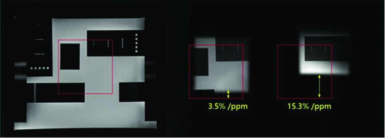
- FIG 1.
A comparison of CSDE with HISE and PRESS in a phantom containing 100% water. The red square indicates the FOV in the center of the phantom, while the middle image is the result of HISE, and the right image is the result of PRESS.
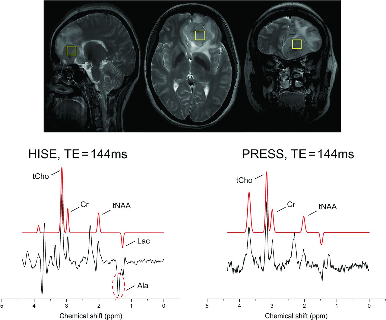
- FIG 2.
A 43-year-old woman with a mass located in the left frontal lobe, suggesting a meningioma. It was an atypical meningioma (World Health Organization grade II) with invasion into the brain parenchyma. The HISE technique observed relatively strong Ala signal, while the PRESS technique could not differentiate between the Ala and Lac peaks. Both HISE and PRESS did not detect Lip signals. The yellow box in anatomical images represented the region of the volume of the SVS scan.
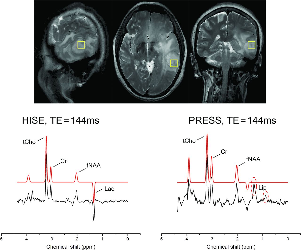
- FIG 3.
A 67 -year-old woman with a mass in the left temporal lobe with surrounding edema. On the basis of multimodal MR imaging, a high-grade glioma was suspected. Clinical correlation was recommended. The glioma was classified as a glioblastoma (World Health Organization grade IV, IDH1 wild-type). In the HISE, a Lac signal was detected. In PRESS, due to chemical shift displacement effects, a Lip signal originating from the scalp was detected, and the Lac signal was covered by the Lip signal. The yellow box in anatomical images represented the region of the volume of the SVS scan.
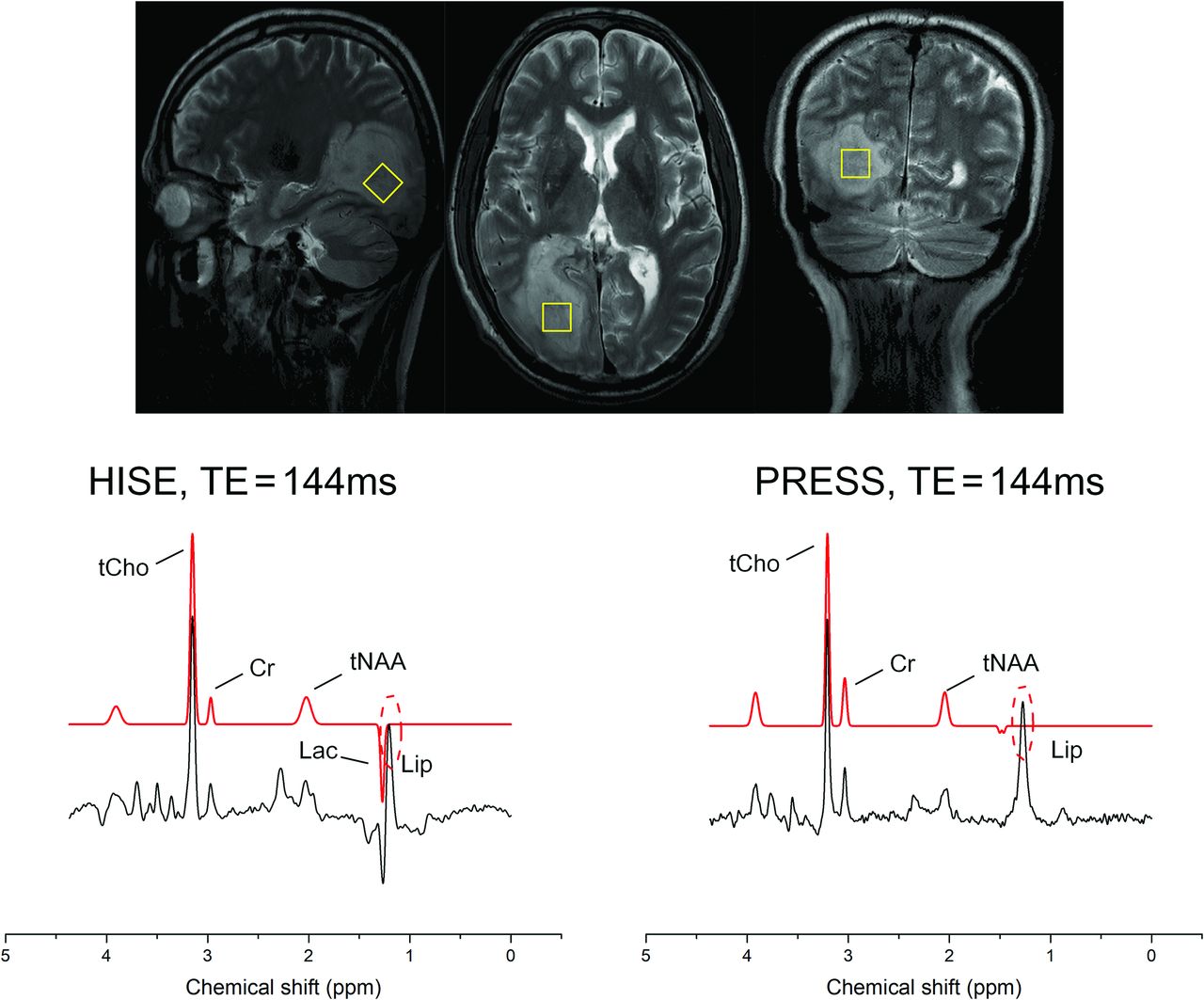
- FIG 4.
A 52-year-old man with a right occipital lobe space-occupying lesion, which was classified as a brain metastasis (originating from lung cancer). In the HISE, both Lac and Lip signals were detected. In PRESS, only a Lip signal was detected, and the Lac signal was covered by the Lip signal. The yellow box in anatomical images represented the region of the volume of the SVS scan.
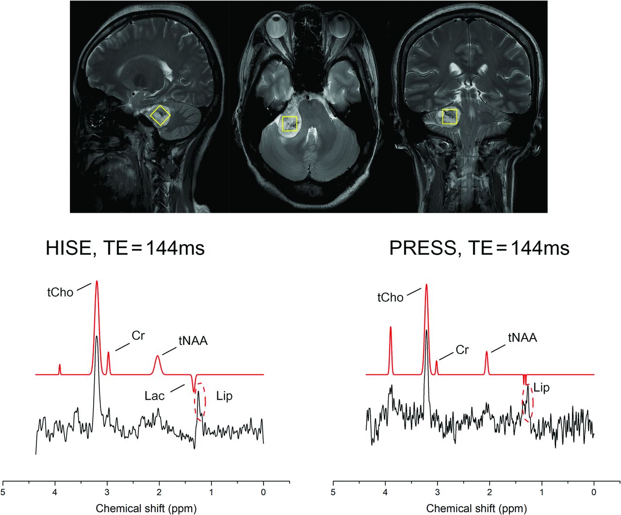
- FIG 5.
A 71-year-old woman with a space-occupying lesion in the right cerebellopontine angle, confirmed to be an acoustic neuroma after surgery. In the HISE, a small Lac signal was detected, In PRESS, the Lac signal was also covered by the Lip signal. The yellow box in anatomical images represented the region of the volume of the SVS scan.


Tables
Parameters HISE PRESS TR 2500 ms 2500 ms TE 144 ms 144 ms Voxel size 15 ×15 ×15 mm3 15 ×15 ×15 mm3 Bandwidth 1000 kHz 1000 kHz Averages 100 100 Spectral sampling 1024 1024 Flip angle 90° 90° Phase-cycling schemes 2 blocks 4 blocks Water-suppressed bandwidth 90 kHz 90 kHz Acquisition time 4 min 19 sec 4 min 14 sec Note:—min indicates minute; sec, second.
- Table 2:
A comparison of average values (mean [SD]) of FWHM and SNR of prominent metabolites for the HISE and PRESS SVS scans among all casesa
Metabolite FWHM P Value SNR P Value HISE PRESS HISE PRESS tNAA 10.92 (SD, 2.59) 14.12 (SD, 3.62) .001b 14.61 (SD, 7.66) 9.53 (SD, 5.48) .001b Cr 10.87 (SD, 3.31) 11.75 (SD, 4.72) .455 20.02 (SD, 14.06) 9.44 (SD, 6.12) .000c tCho 12.07 (SD, 2.74) 12.74 (SD, 3.45) .215 63.23 (SD, 43.32) 29.72 (SD, 19.19) .000c - Table 3:
The detection of specific metabolites (Lac, Ala, and Lip) at TE = 144 ms for the HISE and PRESS SVS scans, respectivelya
No. Sex Age (yr) Pathologic Diagnosis HISE PRESS Lac Ala Lip Lac Ala Lip Case 1 F 71 Acoustic neuroma + − + − − + Case 2 M 47 Acoustic neuroma + − − − − − Case 3 M 52 Metastases (lung origin) + ? + − − + Case 4 M 58 Metastases (gastrointestinal origin) + − + − − + Case 5 M 61 Non-Hodgkin lymphoma + − + − − + Case 6 M 66 Meningioma, WHO I + + − − − − Case 7 F 52 Meningioma, WHO I + + − − − − Case 8 F 67 Meningioma, WHO I + ? − + − − Case 9 F 43 Meningioma, WHO II + + − − + − Case 10 M 52 Meningioma, WHO I + ? + − − + Case 11 F 34 Meningioma, WHO I + + − − ? − Case 12 F 56 Meningioma, WHO I − − + − − + Case 13 F 50 Meningioma, WHO I + + − − − − Case 14 F 58 Meningioma, WHO I − + + − ? + Case 15 F 42 Oligodendroglioma, WHO III + − − − − − Case 16 F 49 Oligodendroglioma, WHO II + − − − − − Case 17 F 35 GBM, WHO IV + − − − − + Case 18 M 45 GBM, WHO IV + ? + − − + Case 19 F 64 GBM, WHO IV + ? + + − + Case 20 F 67 GBM, WHO IV + − − − − + Case 21 F 70 GBM, WHO IV + ? + − − + Case 22 M 66 GBM, WHO IV + − − + − + Case 23 F 69 GBM, WHO IV + − + + − + Note:—F indicates female; M, male; WHO, World Health Organization; GBM, glioblastoma.
↵a The plus sign represents the detection of strong signal peaks; the question mark represents the presence of faint or inconspicuous signal peaks; and the minus sign represents the almost undetectable signal peaks.


Jump to section
Related Articles
Cited By...
- No citing articles found.