Advertisement

Main menu

  • Home
  • Content
    • Current Issue
    • Accepted Manuscripts
    • Article Preview
    • Past Issue Archive
    • AJNR Case Collection
    • Case of the Week Archive
    • Classic Case Archive
    • Case of the Month Archive
  • Special Collections
    • Spinal CSF Leak Articles (Jan 2020-June 2024)
    • 2024 AJNR Journal Awards
    • Most Impactful AJNR Articles
  • Multimedia
    • AJNR Podcast
    • AJNR Scantastics
    • Video Articles
  • For Authors
    • Submit a Manuscript
    • Author Policies
    • Fast publishing of Accepted Manuscripts
    • Graphical Abstract Preparation
    • Manuscript Submission Guidelines
    • Imaging Protocol Submission
    • Submit a Case for the Case Collection
  • About Us
    • About AJNR
    • Editorial Board
  • More
    • Become a Reviewer/Academy of Reviewers
    • Subscribers
    • Permissions
    • Alerts
    • Feedback
    • Advertisers
    • ASNR Home
  • Other Publications
    • ajnr

User menu

  • Alerts
  • Log in

Search

  • Advanced search
American Journal of Neuroradiology

American Journal of Neuroradiology

ASHNR American Society of Functional Neuroradiology ASHNR American Society of Pediatric Neuroradiology ASSR
  • Alerts
  • Log in

Advanced Search

  • Home
  • Content
    • Current Issue
    • Accepted Manuscripts
    • Article Preview
    • Past Issue Archive
    • AJNR Case Collection
    • Case of the Week Archive
    • Classic Case Archive
    • Case of the Month Archive
  • Special Collections
    • Spinal CSF Leak Articles (Jan 2020-June 2024)
    • 2024 AJNR Journal Awards
    • Most Impactful AJNR Articles
  • Multimedia
    • AJNR Podcast
    • AJNR Scantastics
    • Video Articles
  • For Authors
    • Submit a Manuscript
    • Author Policies
    • Fast publishing of Accepted Manuscripts
    • Graphical Abstract Preparation
    • Manuscript Submission Guidelines
    • Imaging Protocol Submission
    • Submit a Case for the Case Collection
  • About Us
    • About AJNR
    • Editorial Board
  • More
    • Become a Reviewer/Academy of Reviewers
    • Subscribers
    • Permissions
    • Alerts
    • Feedback
    • Advertisers
    • ASNR Home
  • Follow AJNR on Twitter
  • Visit AJNR on Facebook
  • Follow AJNR on Instagram
  • Join AJNR on LinkedIn
  • RSS Feeds

Welcome to the new AJNR, Updated Hall of Fame, and more. Read the full announcements.


AJNR is seeking candidates for the position of Associate Section Editor, AJNR Case Collection. Read the full announcement.


Research ArticlePediatric Neuroimaging

Prognostic Significance of Central Skull Base Remodeling in Chiari II Malformation

John T. Freiling, Nilesh K. Desai, Stephen F. Kralik, William E. Whitehead and Thierry A.G.M. Huisman
American Journal of Neuroradiology April 2024, 45 (4) 488-493; DOI: https://doi.org/10.3174/ajnr.A8142
John T. Freiling
aFrom the Edward B. Singleton Department of Radiology (J.T.F., N.K.D., S.F.K., T.A.G.M.H.), Texas Children’s Hospital & Baylor College of Medicine, Houston, Texas
  • Find this author on Google Scholar
  • Find this author on PubMed
  • Search for this author on this site
  • ORCID record for John T. Freiling
Nilesh K. Desai
aFrom the Edward B. Singleton Department of Radiology (J.T.F., N.K.D., S.F.K., T.A.G.M.H.), Texas Children’s Hospital & Baylor College of Medicine, Houston, Texas
  • Find this author on Google Scholar
  • Find this author on PubMed
  • Search for this author on this site
  • ORCID record for Nilesh K. Desai
Stephen F. Kralik
aFrom the Edward B. Singleton Department of Radiology (J.T.F., N.K.D., S.F.K., T.A.G.M.H.), Texas Children’s Hospital & Baylor College of Medicine, Houston, Texas
  • Find this author on Google Scholar
  • Find this author on PubMed
  • Search for this author on this site
  • ORCID record for Stephen F. Kralik
William E. Whitehead
bDepartment of Neurosurgery (W.E.W.), Texas Children’s Hospital & Baylor College of Medicine, Houston, Texas
  • Find this author on Google Scholar
  • Find this author on PubMed
  • Search for this author on this site
Thierry A.G.M. Huisman
aFrom the Edward B. Singleton Department of Radiology (J.T.F., N.K.D., S.F.K., T.A.G.M.H.), Texas Children’s Hospital & Baylor College of Medicine, Houston, Texas
  • Find this author on Google Scholar
  • Find this author on PubMed
  • Search for this author on this site
  • ORCID record for Thierry A.G.M. Huisman
  • Article
  • Figures & Data
  • Info & Metrics
  • Responses
  • References
  • PDF
Loading

Article Figures & Data

Figures

  • Tables
  • FIG 1.
    • Download figure
    • Open in new tab
    • Download powerpoint
    FIG 1.

    Comparison of normal and remodeled basiocciput and posterior atlanto-occipital membranes. A and B, Sagittal CT images demonstrate the normal appearance of the basiocciput (arrow in A) and outward convexity of the basiocciput (arrow in B). C and D, Sagittal T2-weighted images demonstrate the normal appearance of the PAOM (arrow in C) and outward convexity of the PAOM (arrow in D). Image insets demonstrate larger FOV sagittal images and the location of corresponding zoomed-in FOV.
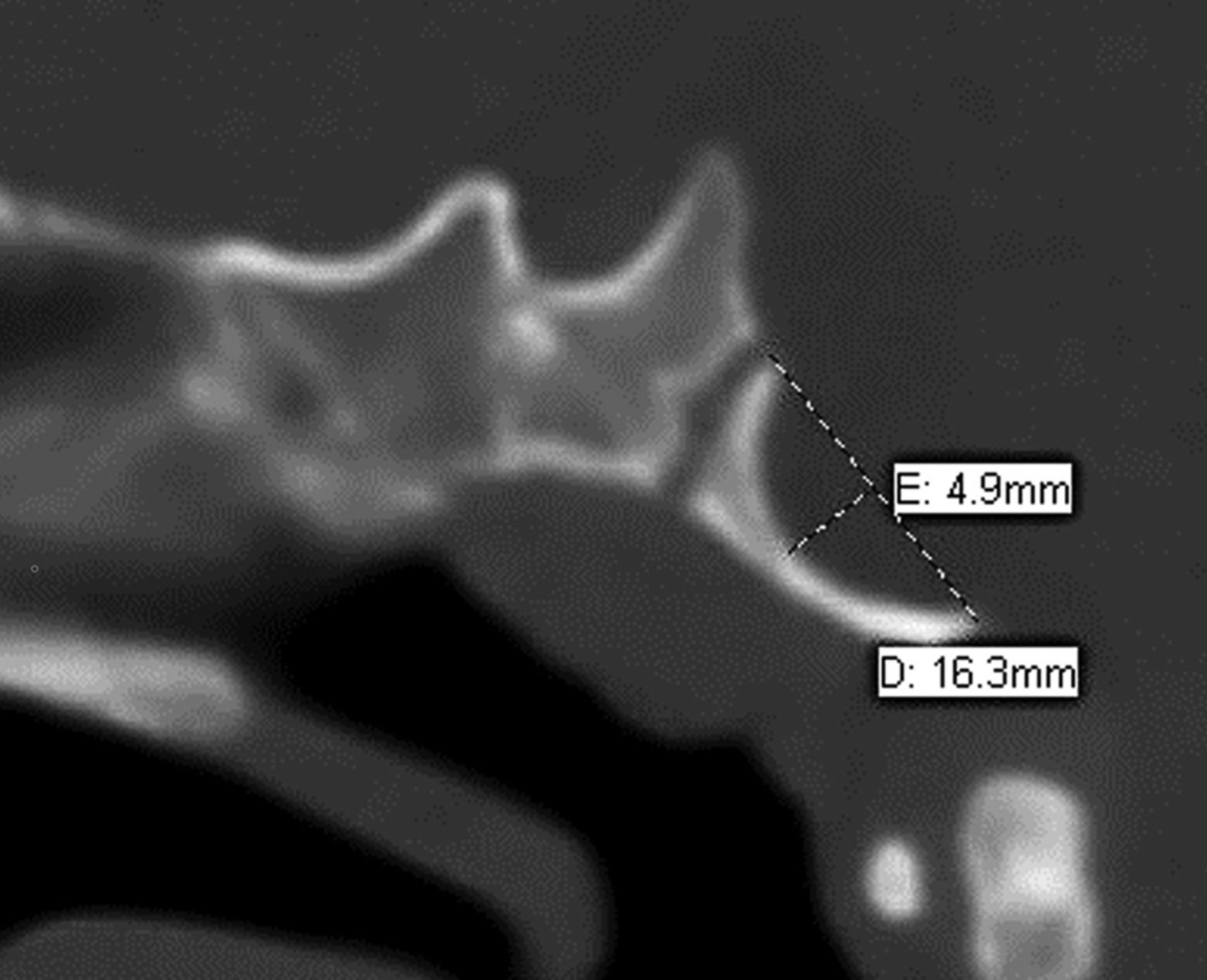
  • FIG 2.
    • Download figure
    • Open in new tab
    • Download powerpoint
    FIG 2.

    Sagittal CT of the skull base demonstrating outward convexity of the basiocciput in a 10-month-old infant with CM2. Basiocciput length is measured from the basion to the dorsal spheno-occipital synchondrosis. Convexity is measured as the maximum distance orthogonal to the length line.
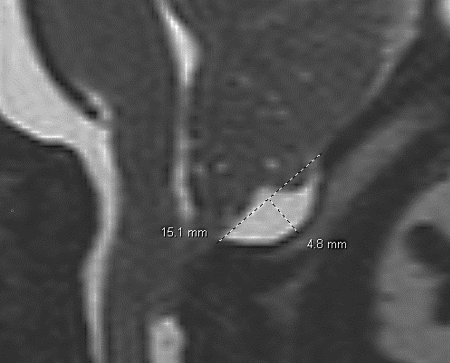
  • FIG 3.
    • Download figure
    • Open in new tab
    • Download powerpoint
    FIG 3.

    Sagittal T2-weighted image of the posterior craniocervical junction in a 1-month-old infant with CM2. C1-opisthion length is measured from the posterior C1 arch to the opisthion. Posterior atlanto-occipital membrane convexity is measured as the maximum distance orthogonal to the length line.
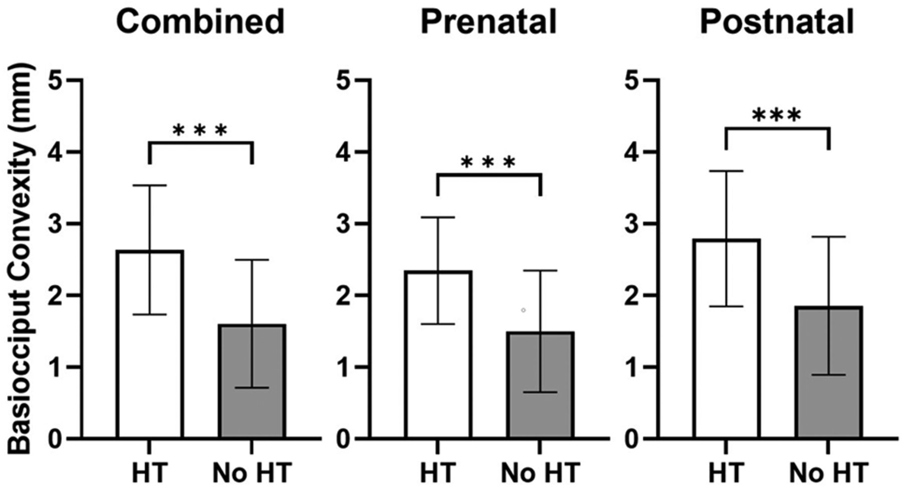
  • FIG 4.
    • Download figure
    • Open in new tab
    • Download powerpoint
    FIG 4.

    Basiocciput convexity is increased in patients requiring HT. Basiocciput convexity (millimeters) was measured in all patients requiring HT and not requiring HT. Patients were then stratified by prenatal or postnatal repair of the ONTD and basiocciput convexities and were compared. Bars represent the mean (SD) with triple asterisks P < .001 representing statistical significance as determined by an unpaired Student t test.
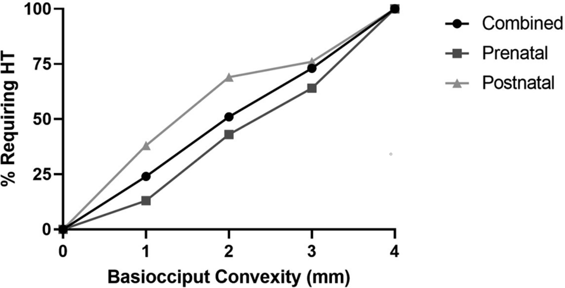
  • FIG 5.
    • Download figure
    • Open in new tab
    • Download powerpoint
    FIG 5.

    An increase in basiocciput convexity was associated with a higher percentage of patients requiring HT. The percentage of patients requiring HT at varying basiocciput convexities (1–4+ mm) was determined in the combined prenatal and postnatal cohorts.

Tables

  • Figures
    • View popup
    Table 1:

    CT and MR imaging data of patients who underwent either prenatal or postnatal surgical repair of an ONTD, according to whether they required HTa

     HTNo HTP Value
    Combined prenatal and postnatal    
     Mean basiocciput convexity (mm)2.6 (SD, 0.9)1.6 (SD, 0.9)<.001
     Mean basiocciput length (mm)15.2 (SD, 1.8)17.3 (SD, 2.2)<.001
     Mean basiocciput convexity/length ratio0.17 (SD, 0.06)0.1 (SD, 0.06)<.001
     Mean convexity of PAOM (mm)2.6 (SD, 1.6)1.9 (SD, 1.4).003
     Mean lateral ventricle width (mm)35.1 (SD, 26.2)29.4 (SD, 9.9).10
     Mean FOD (mm)134.8 (SD, 17.2)152.2 (SD, 15.5)<.001
     Mean PFD (mm)51.4 (SD, 9.2)65.9 (SD, 9.6)<.001
     Mean FOD/PFD ratio2.7 (SD, 0.4)2.3 (SD, 0.2)<.001
    Prenatal only   
     Mean basiocciput convexity (mm)2.3 (SD, 0.7)1.5 (SD, 0.8)<.001
     Mean basiocciput length (mm)15.6 (SD, 2.1)17.7 (SD, 2.2)<.001
     Mean basiocciput convexity/length ratio0.15 (SD, 0.05)0.08 (SD, 0.05)<.001
     Mean convexity of PAOM (mm)2.6 (SD, 1.9)1.9 (SD, 1.4).09
     Mean lateral ventricle width (mm)35 (SD, 10.9)28.8 (SD, 9.9).02
     Mean FOD (mm)140.5 (SD, 19.0)154.2 (SD, 12.6)<.001
     Mean PFD (mm)56 (SD, 10.4)67.3 (SD, 8.0<.001
     Mean FOD/PFD ratio2.6 (SD, 0.3)2.3 (SD, 0.2)<.001
    Postnatal only   
     Mean basiocciput convexity (mm)2.8 (SD, 0.9)1.9 (SD, 1.0)<.001
     Mean basiocciput length (mm)15 (SD, 1.6)16.5 (SD, 2.0).002
     Mean basiocciput convexity/length ratio0.19 (SD, 0.06)0.12 (SD, 0.07)<.001
     Mean convexity of PAOM (mm)2.7 (SD, 1.5)1.8 (SD, 1.5).02
     Mean lateral ventricle width (mm)35.1 (SD, 31.2)30.7 (SD, 10.0).53
     Mean FOD (mm)131.6 (SD, 15.5)148 (SD, 20.0)<.001
     Mean PFD (mm)48.9 (SD, 7.5)62.8 (SD, 11.8)<.001
     Mean FOD/PFD ratio2.7 (SD, 0.4)2.4 (SD, 0.2)<.001
    • ↵a Data are presented as mean (SD). P values were determined by an unpaired Student t test.

    • View popup
    Table 2:

    CT and MR imaging data of all patients regardless of whether they received HT, according to whether they received prenatal or postnatal surgical repair of an ONTDa

     All patientsPrenatalPostnatalP Value
    Mean basiocciput convexity (mm)1.8 (SD, 0.9)2.5 (SD, 1.0)<.001
    Mean basiocciput length (mm)16.9 (SD, 2.4)15.4 (SD, 1.9)<.001
    Mean basiocciput convexity/length ratio0.11 (SD, 0.06)0.17 (SD, 0.07)<.001
    Mean convexity of PAOM (mm)2.2 (SD, 1.6)2.4 (SD, 1.5).35
    Mean lateral ventricle width (mm)31.1 (SD, 10.6)33.8 (SD, 27.0).44
    Mean FOD (mm)149.1 (SD, 16.6)136.6 (SD, 18.4)<.001
    Mean PFD (mm)63.1 (SD, 10.5)53.1 (SD, 11.1)<.001
    Mean FOD/PFD ratio2.4 (SD, 0.3)2.6 (SD, 0.4)<.001
    • ↵a Data are presented as mean (SD). P values were determined by an unpaired Student t test.

PreviousNext
Back to top

In this issue

American Journal of Neuroradiology: 45 (4)
American Journal of Neuroradiology
Vol. 45, Issue 4
1 Apr 2024
  • Table of Contents
  • Index by author
  • Complete Issue (PDF)
Advertisement
Print
Download PDF
Email Article

Thank you for your interest in spreading the word on American Journal of Neuroradiology.

NOTE: We only request your email address so that the person you are recommending the page to knows that you wanted them to see it, and that it is not junk mail. We do not capture any email address.

Enter multiple addresses on separate lines or separate them with commas.
Prognostic Significance of Central Skull Base Remodeling in Chiari II Malformation
(Your Name) has sent you a message from American Journal of Neuroradiology
(Your Name) thought you would like to see the American Journal of Neuroradiology web site.
CAPTCHA
This question is for testing whether or not you are a human visitor and to prevent automated spam submissions.
Cite this article
John T. Freiling, Nilesh K. Desai, Stephen F. Kralik, William E. Whitehead, Thierry A.G.M. Huisman
Prognostic Significance of Central Skull Base Remodeling in Chiari II Malformation
American Journal of Neuroradiology Apr 2024, 45 (4) 488-493; DOI: 10.3174/ajnr.A8142

Citation Manager Formats

  • BibTeX
  • Bookends
  • EasyBib
  • EndNote (tagged)
  • EndNote 8 (xml)
  • Medlars
  • Mendeley
  • Papers
  • RefWorks Tagged
  • Ref Manager
  • RIS
  • Zotero
0 Responses
Respond to this article
Share
Bookmark this article
Central Skull Base Remodeling in Chiari II
John T. Freiling, Nilesh K. Desai, Stephen F. Kralik, William E. Whitehead, Thierry A.G.M. Huisman
American Journal of Neuroradiology Apr 2024, 45 (4) 488-493; DOI: 10.3174/ajnr.A8142
del.icio.us logo Twitter logo Facebook logo Mendeley logo
  • Tweet Widget
  • Facebook Like
  • Google Plus One
Purchase

Jump to section

  • Article
    • Abstract
    • ABBREVIATIONS:
    • MATERIALS AND METHODS
    • RESULTS
    • DISCUSSION
    • CONCLUSIONS
    • Footnotes
    • References
  • Figures & Data
  • Info & Metrics
  • Responses
  • References
  • PDF

Related Articles

  • No related articles found.
  • PubMed
  • Google Scholar

Cited By...

  • No citing articles found.
  • Crossref
  • Google Scholar

This article has not yet been cited by articles in journals that are participating in Crossref Cited-by Linking.

More in this TOC Section

  • Neuroimaging Delineation and Progression of SLSMD
  • New CNS Embryonal Tumor Insights via MRI Analysis
  • Susceptibility Mapping in Newborn Brain Development
Show more PEDIATRIC NEUROIMAGING

Similar Articles

Advertisement

Indexed Content

  • Current Issue
  • Accepted Manuscripts
  • Article Preview
  • Past Issues
  • Editorials
  • Editors Choice
  • Fellow Journal Club
  • Letters to the Editor

Cases

  • Case Collection
  • Archive - Case of the Week
  • Archive - Case of the Month
  • Archive - Classic Case

Special Collections

  • Special Collections

Resources

  • News and Updates
  • Turn around Times
  • Submit a Manuscript
  • Author Policies
  • Manuscript Submission Guidelines
  • Evidence-Based Medicine Level Guide
  • Publishing Checklists
  • Graphical Abstract Preparation
  • Imaging Protocol Submission
  • Submit a Case
  • Become a Reviewer/Academy of Reviewers
  • Get Peer Review Credit from Publons

Multimedia

  • AJNR Podcast
  • AJNR SCANtastic
  • Video Articles

About Us

  • About AJNR
  • Editorial Board
  • Not an AJNR Subscriber? Join Now
  • Alerts
  • Feedback
  • Advertise with us
  • Librarian Resources
  • Permissions
  • Terms and Conditions

American Society of Neuroradiology

  • Not an ASNR Member? Join Now

© 2025 by the American Society of Neuroradiology All rights, including for text and data mining, AI training, and similar technologies, are reserved.
Print ISSN: 0195-6108 Online ISSN: 1936-959X

Powered by HighWire