Advertisement

Main menu

  • Home
  • Content
    • Current Issue
    • Accepted Manuscripts
    • Article Preview
    • Past Issue Archive
    • AJNR Case Collection
    • Case of the Week Archive
    • Classic Case Archive
    • Case of the Month Archive
  • Special Collections
    • Spinal CSF Leak Articles (Jan 2020-June 2024)
    • 2024 AJNR Journal Awards
    • Most Impactful AJNR Articles
  • Multimedia
    • AJNR Podcast
    • AJNR Scantastics
    • Video Articles
  • For Authors
    • Submit a Manuscript
    • Author Policies
    • Fast publishing of Accepted Manuscripts
    • Graphical Abstract Preparation
    • Manuscript Submission Guidelines
    • Imaging Protocol Submission
    • Submit a Case for the Case Collection
  • About Us
    • About AJNR
    • Editorial Board
  • More
    • Become a Reviewer/Academy of Reviewers
    • Subscribers
    • Permissions
    • Alerts
    • Feedback
    • Advertisers
    • ASNR Home
  • Other Publications
    • ajnr

User menu

  • Alerts
  • Log in

Search

  • Advanced search
American Journal of Neuroradiology

American Journal of Neuroradiology

ASHNR American Society of Functional Neuroradiology ASHNR American Society of Pediatric Neuroradiology ASSR
  • Alerts
  • Log in

Advanced Search

  • Home
  • Content
    • Current Issue
    • Accepted Manuscripts
    • Article Preview
    • Past Issue Archive
    • AJNR Case Collection
    • Case of the Week Archive
    • Classic Case Archive
    • Case of the Month Archive
  • Special Collections
    • Spinal CSF Leak Articles (Jan 2020-June 2024)
    • 2024 AJNR Journal Awards
    • Most Impactful AJNR Articles
  • Multimedia
    • AJNR Podcast
    • AJNR Scantastics
    • Video Articles
  • For Authors
    • Submit a Manuscript
    • Author Policies
    • Fast publishing of Accepted Manuscripts
    • Graphical Abstract Preparation
    • Manuscript Submission Guidelines
    • Imaging Protocol Submission
    • Submit a Case for the Case Collection
  • About Us
    • About AJNR
    • Editorial Board
  • More
    • Become a Reviewer/Academy of Reviewers
    • Subscribers
    • Permissions
    • Alerts
    • Feedback
    • Advertisers
    • ASNR Home
  • Follow AJNR on Twitter
  • Visit AJNR on Facebook
  • Follow AJNR on Instagram
  • Join AJNR on LinkedIn
  • RSS Feeds

Welcome to the new AJNR, Updated Hall of Fame, and more. Read the full announcements.


AJNR is seeking candidates for the position of Associate Section Editor, AJNR Case Collection. Read the full announcement.


Brief ReportSpine Imaging and Spine Image-Guided Interventions
Open Access

CT-Guided Epidural Contrast Injection for the Identification of Dural Defects

Ian T. Mark, Michael Oien, John Benson, Jared Verdoorn, Ben Johnson-Tesch, D.K. Kim, Jeremy Cutsforth-Gregory and Ajay A. Madhavan
American Journal of Neuroradiology January 2025, 46 (1) 207-210; DOI: https://doi.org/10.3174/ajnr.A8437
Ian T. Mark
aFrom the Department of Radiology (I.T.M., M.O., J.B., J.V., B.J.-T., D.K.K., A.A.M), Mayo Clinic, Rochester, Minnesota
  • Find this author on Google Scholar
  • Find this author on PubMed
  • Search for this author on this site
  • ORCID record for Ian T. Mark
Michael Oien
aFrom the Department of Radiology (I.T.M., M.O., J.B., J.V., B.J.-T., D.K.K., A.A.M), Mayo Clinic, Rochester, Minnesota
  • Find this author on Google Scholar
  • Find this author on PubMed
  • Search for this author on this site
  • ORCID record for Michael Oien
John Benson
aFrom the Department of Radiology (I.T.M., M.O., J.B., J.V., B.J.-T., D.K.K., A.A.M), Mayo Clinic, Rochester, Minnesota
  • Find this author on Google Scholar
  • Find this author on PubMed
  • Search for this author on this site
  • ORCID record for John Benson
Jared Verdoorn
aFrom the Department of Radiology (I.T.M., M.O., J.B., J.V., B.J.-T., D.K.K., A.A.M), Mayo Clinic, Rochester, Minnesota
  • Find this author on Google Scholar
  • Find this author on PubMed
  • Search for this author on this site
  • ORCID record for Jared Verdoorn
Ben Johnson-Tesch
aFrom the Department of Radiology (I.T.M., M.O., J.B., J.V., B.J.-T., D.K.K., A.A.M), Mayo Clinic, Rochester, Minnesota
  • Find this author on Google Scholar
  • Find this author on PubMed
  • Search for this author on this site
D.K. Kim
aFrom the Department of Radiology (I.T.M., M.O., J.B., J.V., B.J.-T., D.K.K., A.A.M), Mayo Clinic, Rochester, Minnesota
  • Find this author on Google Scholar
  • Find this author on PubMed
  • Search for this author on this site
  • ORCID record for D.K. Kim
Jeremy Cutsforth-Gregory
bDepartment of Neurology (J.C.-G.), Mayo Clinic, Rochester, Minnesota
  • Find this author on Google Scholar
  • Find this author on PubMed
  • Search for this author on this site
  • ORCID record for Jeremy Cutsforth-Gregory
Ajay A. Madhavan
aFrom the Department of Radiology (I.T.M., M.O., J.B., J.V., B.J.-T., D.K.K., A.A.M), Mayo Clinic, Rochester, Minnesota
  • Find this author on Google Scholar
  • Find this author on PubMed
  • Search for this author on this site
  • ORCID record for Ajay A. Madhavan
  • Article
  • Figures & Data
  • Supplemental
  • Info & Metrics
  • Responses
  • References
  • PDF
Loading

Graphical Abstract

Figure
  • Download figure
  • Open in new tab
  • Download powerpoint

Abstract

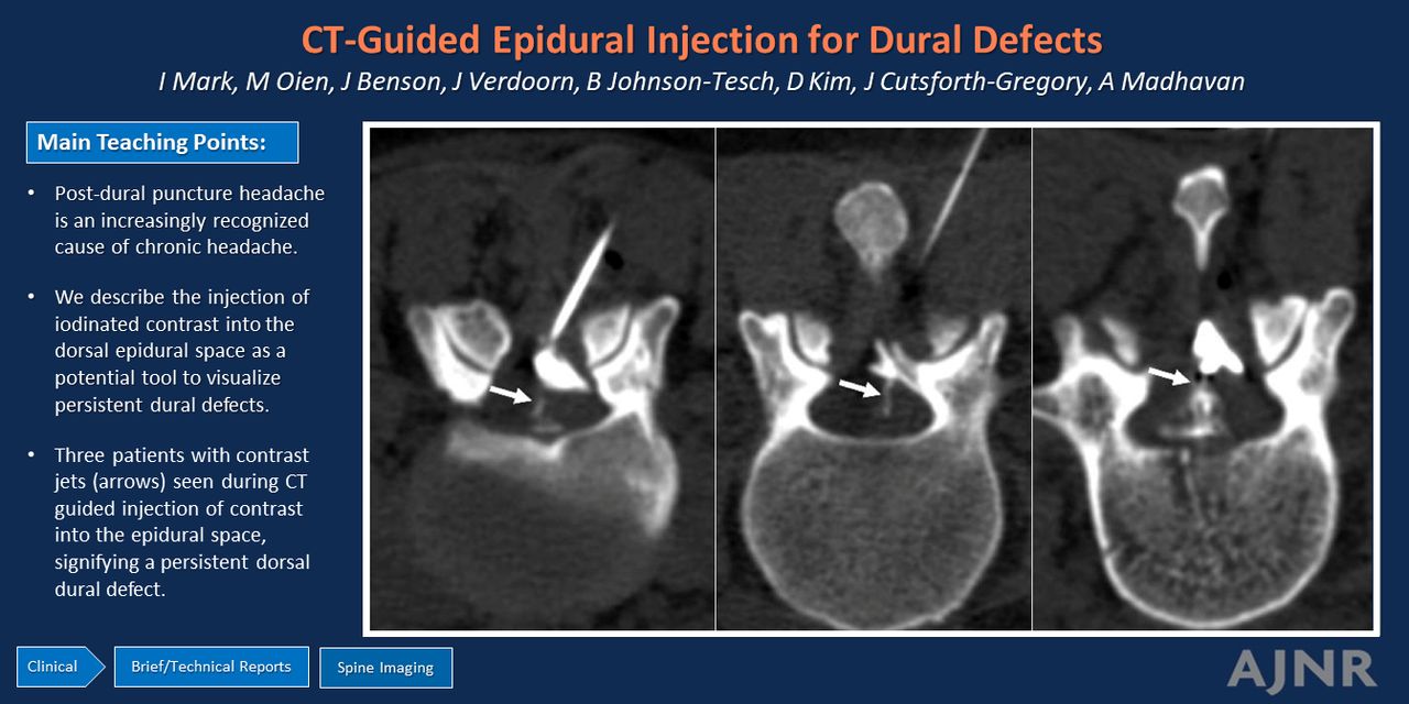
SUMMARY: Post–dural puncture headache is an increasingly recognized cause of chronic headache. Outside of clinical history and myelography that requires an additional dural puncture, there is no reliable diagnostic test to evaluate for persistent dural defects. We describe the injection of iodinated contrast into the dorsal epidural space under CT guidance in 5 patients as a potential tool to visualize persistent dural defects.

ABBREVIATIONS:

DSM
digital subtraction myelography
PDPH
Post–dural puncture headache
SIH
spontaneous intracranial hypotension

Spinal CSF leaks causing spontaneous intracranial hypotension (SIH) classically present with orthostatic headaches, but they can also present with nonspecific symptoms such as neck pain, tinnitus, dizziness, and behavioral changes mimicking dementia.1 Chronic leaks present more often with atypical symptoms, which can further complicate the diagnosis.2,3 Post–dural puncture headache (PDPH) is an increasingly recognized cause of chronic headaches.4 PDPH can occur in the setting of intentional dural puncture for a diagnostic lumbar puncture5 or unintended dural breach during epidural catheter placement. When the latter occurs, headache becomes chronic and debilitating in 30% of cases and may persist for longer than a year.6,7 One paper found that the 92.6% of PDPH cases have a reported normal brain MRI and 70.4% did not have orthostatic headaches.8 The atypical clinical presentation and insensitivity of brain MRI, and yet debilitating symptoms, underscore the need for better diagnostic tools to confirm PDPH.

Digital subtraction myelography (DSM) has previously been described as a technique to diagnose PDPH.9 This technique, as with CT myelography, relies on a new dural puncture for the administration of intrathecal contrast to identify the underlying dural defect. The purpose of this report is to describe CT-guided contrast injection into the epidural space as an alternative technique to definitively confirm a dural defect without additional dural puncture.

CASE SERIES

Our institution is a high-volume treatment center for spinal CSF leak, and our practice routinely performs targeted and nontargeted blood patches under CT guidance. For each blood patch, we inject iodinated contrast into the epidural space immediately before injecting the autologous blood in a sterile manner. This technique involves advancing a needle, most often a 20- or 22-gauge Tuohy, into the dorsal epidural space via an interlaminar approach. Iodinated contrast is injected to confirm correct needle tip position in the epidural space. Our practice most commonly uses diluted contrast (1:2–3 Omnipaque-300 to sterile saline) and injects approximately 1 to 2 mL per spinal level at which blood is to be placed. CT fluoroscopy is performed immediately after the injection of contrast to look for egress into the subarachnoid space, thus identifying a persistent dural defect.

Dural Defect Case 1

A 29-year-old man with Marfan syndrome had a fluoroscopic-guided lumbar drain placed by using a 14-gauge Touhy needle at L4–L5 in the setting of endovascular aortic aneurysm repair. One day after lumber drain removal, the patient reported new-onset headache (not further characterized) and emesis. The patient did not have a spine or brain MRI to evaluate for findings associated with spinal CSF leaks. An image-guided targeted epidural blood patch was ordered. As part of the procedure, a 20-gauge Tuohy needle was placed into the dorsal epidural space at L4–L5. Approximately 1 mL of dilute iodinated contrast was injected in the epidural space that demonstrated an immediate jet of contrast through the dural defect into the subarachnoid space (Fig 1). A targeted epidural blood patch was performed at this level, after which the patient had improvement of headaches and emesis.

FIG 1.
  • Download figure
  • Open in new tab
  • Download powerpoint
FIG 1.

Procedural images from case 1. The left image (A) demonstrates the needle tip in the dorsal aspect of the dorsal epidural space (dashed lines). Immediately after contrast injection to the epidural space, there is a jet of contrast (B, arrow) that goes into the subarachnoid space, identifying the dural defect.

Dural Defect Case 2

A 36-year-old woman underwent 2-day bilateral DSM.10,11 Each day included a dural puncture at L3–L4 with a 22-gauge spinal needle. The needle choice was based on provider preference. No definite evidence of a CSF-venous fistula or CSF leak was found on the DSM. Four days later, the patient underwent a multilevel blood patch as symptomatic relief for SIH (symptoms and diffuse dural enhancement on pre-DSM brain MRI) without a spinal leak identified. A 22-gauge Touhy needle was advanced into the dorsal epidural space at L3–L4 under CT-fluoroscopy guidance, a small amount of dilate Omnipaque-300 was injected, and there was an immediate contrast jet into the subarachnoid space (Fig 2).

FIG 2.
  • Download figure
  • Open in new tab
  • Download powerpoint
FIG 2.

CT fluoroscopy images from the injection of contrast into the epidural space at L3–L4 show the needle tip in the dorsal aspect of the dorsal epidural space (A, arrow), and the jet of contrast (B, arrow) from the epidural space through a dural defect into the subarachnoid space.

Additional Dural Defect Cases 3–5

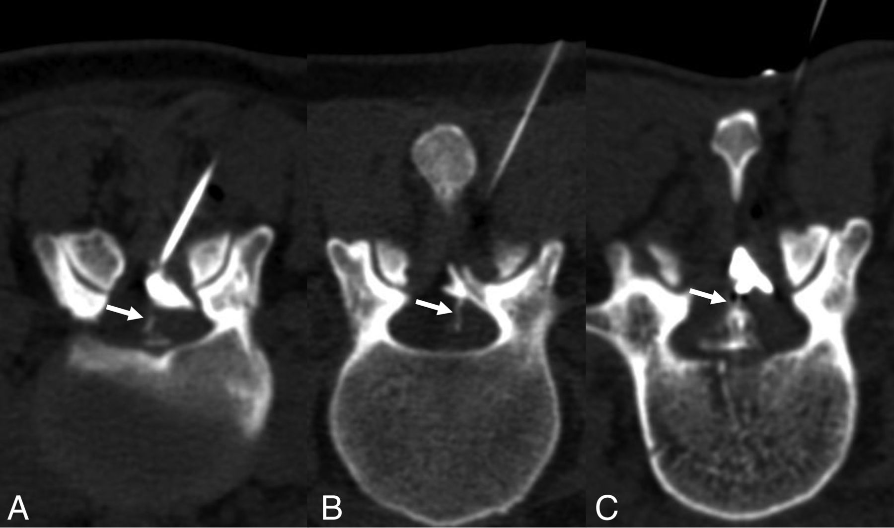
Three additional patients, 25 (Fig 3A), 36 (Fig 3B), and 47 (Fig 3C) years old underwent CT-guided epidural blood patches with a contrast jet after bilateral myelography that did not find a CVF or CSF leak. The blood patches occurred 1, 2, and 2 days after myelography, respectively. As part of our clinical workflow, patients with suspected SIH but no leak site identified undergo a multilevel blood patch after myelography. We include the lumbar puncture level as part of this procedure.

FIG 3.
  • Download figure
  • Open in new tab
  • Download powerpoint
FIG 3.

Three additional patients who had contrast jets (arrows) seen during CT-guided injection of contrast into the epidural space, signifying a persistent dorsal dural defect.

DISCUSSION

PDPH is underreported and can be debilitating. Prior work has used DSM to diagnose dural defects in PDPH, but this technique requires an extra dural puncture. Our technique of injecting contrast into the dorsal epidural space is different in that it does not require another puncture to be made in the dura. We present this technique for consideration in patients with suspected PDPH.

Performing myelography, be it DSM or CT myelography, for PDPH is problematic for several reasons. First, this requires a dural puncture that carries additional risk of PDPH. Second, contrast injection for a myelogram can cause leakage into the epidural space (Fig 4) that obscures the site of a pre-existing dural defect. Contrast injection into the epidural space does not have either of these limitations.

FIG 4.
  • Download figure
  • Open in new tab
  • Download powerpoint
FIG 4.

Two examples of extradural contrast after intrathecal injection that limit the ability of myelography to detect a dural defect. A, Image from a lateral decubitus photon-counting detector CT myelogram that shows contrast extending posteriorly along the needle tract (white arrow). B, CT imaging after DSM, after the needle was removed, shows extradural contrast at the site of prior needle placement (black arrow). Both findings would obscure the detection of contrast leakage through a dural defect present before myelography.

A case report from Callen et al12 described a similar contrast jet after epidural contrast in a postpartum patient with PDPH. Their patient had a focal dural outpouching on the lumbar spine MRI by using a 3D T2-weighted sequence. Although our patients had MR imaging only before the lumbar puncture, this MRI finding has not been described outside of this case report. Their patient did not have an adequate response to epidural fibrin and required surgical repair. An empiric lumbar epidural blood patch is standard practice for PDPH. In equivocal cases, treatment without diagnosis becomes complicated if the patient has atypical symptoms or has incomplete response to the blood patch that could potentially require surgery, as highlighted by the case from Callen et al.12

This technical report mimics the technique that we have recently described to diagnose spinal postoperative pseudomeningoceles by injecting extrathecal contrast to detect a dural defect.13 That report describes injecting contrast directly into a postoperative paraspinal fluid collection for the purposes of visualizing a contrast jet into the subarachnoid space to confirm and localize the leak site. Additionally, we separately described the injection of epidural contrast to propose an alternative mechanism for intrathecal hematoma after epidural blood patch, where epidural blood traverses from the epidural space to the subarachnoid space via a dural defect.14 If a contrast jet is seen, our practice tends to inject a smaller amount of blood, with the thought that high pressure of larger volumes could lead to more egress of blood into the subarachnoid space. The technique described in the present study is limited in that we only assess the dorsal dura, and a ventral dural defect caused by inadvertent deep needle position would not be evaluated.

Our technique, while it would remain the same for subacute versus chronic PDPH, was only demonstrated after recent lumbar puncture. This reflects our procedural practice, which performs multilevel blood patches after negative myelography work-up and does not see as many patients for chronic PDPH. Future work should be done to confirm this finding in subacute and chronic PDPH. Membrane formation has been described in the setting of spinal CSF leaks,15 and could occur at the site of dural defects in PDPH, particularly when long-standing. Our described technique could be specific but lacks sensitivity if obstructive membranes prevent the visualization of contrast jets. Even in the setting of potentially limited sensitivity, we favor epidural contrast injection rather than a new dural puncture and myelography to evaluate for persistent dural defects from a prior lumbar puncture. Our case examples had prior image-guided lumbar puncture with known puncture levels. If the prior puncture site is unknown secondary to absent records or nonimage guided technique, epidural contrast could be injected at multiple levels. Further study of this technique could also evaluate treatment outcomes related to the epidural blood patch. This CT fluoroscopy–guided procedure does require radiation exposure, but with only minimal increased dose when compared with an isolated CT fluoroscopy–guided epidural blood patch.

PDPH is increasingly recognized as an important cause of chronic debilitating headache. When the diagnosis is not recognized promptly after the spinal procedure, or when there are atypical symptoms, additional tests are often required to establish the diagnosis. Injecting contrast into the dorsal epidural space is a technique that can help to identify dural defects that cause PDPH.

Footnotes

  • Disclosure forms provided by the authors are available with the full text and PDF of this article at www.ajnr.org.

Indicates open access to non-subscribers at www.ajnr.org

References

  1. 1.↵
    1. D’Antona L,
    2. Jaime Merchan MA,
    3. Vassiliou A, et al
    . Clinical presentation, investigation findings, and treatment outcomes of spontaneous intracranial hypotension syndrome: a systematic review and meta-analysis. JAMA Neurol 2021;78:329–37 doi:10.1001/jamaneurol.2020.4799 pmid:33393980
    CrossRefPubMed
  2. 2.↵
    1. Williams J,
    2. Brinjikji W,
    3. Cutsforth-Gregory JK
    . Natural history of spontaneous intracranial hypotension: a clinical and imaging study. J Neurointerv Surg 2023;15:1124–28 doi:10.1136/jnis-2022-019300 pmid:36357170
    Abstract/FREE Full Text
  3. 3.↵
    1. Häni L,
    2. Fung C,
    3. Jesse CM, et al
    . Insights into the natural history of spontaneous intracranial hypotension from infusion testing. Neurology 2020;95:e247–55 doi:10.1212/WNL.0000000000009812 pmid:32522800
    Abstract/FREE Full Text
  4. 4.↵
    1. Carroll I,
    2. Callen AL
    . Beyond myths: transformative insights into spinal cerebrospinal fluid leaks and their role in persistent headache syndromes. Headache 2024;64:229–32 doi:10.1111/head.14677 pmid:38385703
    CrossRefPubMed
  5. 5.↵
    1. Philip JT,
    2. Flores MA,
    3. Beegle RD, et al
    . Rates of epidural blood patch following lumbar puncture comparing atraumatic versus bevel-tip needles stratified for body mass index. AJNR Am J Neuroradiol 2022;43:315–18 doi:10.3174/ajnr.A7397 pmid:35027350
    Abstract/FREE Full Text
  6. 6.↵
    1. Barad M,
    2. Carroll I,
    3. Reina MA, et al
    . Did she have an epidural? The long-term consequences of postdural puncture headache and the role of unintended dural puncture. Headache 2021;61:1314–23 doi:10.1111/head.14221 pmid:34570902
    CrossRefPubMed
  7. 7.↵
    1. Gauthama P,
    2. Kelkar A,
    3. Basar SMA, et al
    . Incidence of persistent headache at 18 months following accidental dural puncture in the obstetric population: a prospective service evaluation in 45 patients. Headache 2019;59:97–103 doi:10.1111/head.13442 pmid:30426481
    CrossRefPubMed
  8. 8.↵
    1. Niraj G,
    2. Critchley P
    . Management and outcomes of persistent headache after accidental dural puncture in the obstetric population: a 9-year prospective audit. Headache 2023;63:71–78 doi:10.1111/head.14447 pmid:36651506
    CrossRefPubMed
  9. 9.↵
    1. Schievink WI,
    2. Maya MM,
    3. Moser FG
    . Digital subtraction myelography in the investigation of post–dural puncture headache in 27 patients: technical note. SPI 2017;26:760–64 doi:10.3171/2016.11.SPINE16968
    CrossRefPubMed
  10. 10.↵
    1. Kim DK,
    2. Brinjikji W,
    3. Morris PP, et al
    . Lateral decubitus digital subtraction myelography: tips, tricks, and pitfalls. AJNR Am J Neuroradiol 2020;41:21–28 doi:10.3174/ajnr.A6368 pmid:31857327
    Abstract/FREE Full Text
  11. 11.↵
    1. Shlapak DP,
    2. Mark IT,
    3. Kim DK, et al
    . Incremental diagnostic yield and clinical outcomes of lateral decubitus CT myelogram immediately following negative lateral decubitus digital subtraction myelogram. Neuroradiol J 2023;36:593–600 doi:10.1177/19714009231173110 pmid:37128777
    CrossRefPubMed
  12. 12.↵
    1. Callen AL,
    2. Lennarson P,
    3. Carroll IR
    . A causative role for remote dural puncture and resultant arachnoid bleb in new daily persistent headache: a case report. Headache 2023;63:981–83 doi:10.1111/head.14584 pmid:37358488
    CrossRefPubMed
  13. 13.↵
    1. Mark IT,
    2. Madhavan AA,
    3. Delone DR, et al
    . Direct pseudomeningocele contrast injection for spinal CSF leak localization. Neuroradiol J 2023 Dec 26. Epub ahead of print. doi:10.1177/19714009231224413 pmid:38146822
    CrossRefPubMed
  14. 14.↵
    1. Mark I,
    2. Madhavan A,
    3. Carr C, et al
    . Intrathecal hematoma following epidural blood patch: an alternative mechanism to intrathecal puncture. Neuroradiol J 2024 Mar 23. Epub ahead of print. doi:10.1177/19714009241242587 pmid:38520266
    CrossRefPubMed
  15. 15.↵
    1. Häni L,
    2. Fung C,
    3. El Rahal A, et al
    . Distinct pattern of membrane formation with spinal cerebrospinal fluid leaks in spontaneous intracranial hypotension. Oper Neurosurg (Hagerstown) 2024;26:71–77 doi:10.1227/ons.0000000000000914 pmid:37747369
    CrossRefPubMed
  • Received June 11, 2024.
  • Accepted after revision July 26, 2024.
  • © 2025 by American Journal of Neuroradiology
PreviousNext
Back to top

In this issue

American Journal of Neuroradiology: 46 (1)
American Journal of Neuroradiology
Vol. 46, Issue 1
1 Jan 2025
  • Table of Contents
  • Index by author
  • Complete Issue (PDF)
Advertisement
Print
Download PDF
Email Article

Thank you for your interest in spreading the word on American Journal of Neuroradiology.

NOTE: We only request your email address so that the person you are recommending the page to knows that you wanted them to see it, and that it is not junk mail. We do not capture any email address.

Enter multiple addresses on separate lines or separate them with commas.
CT-Guided Epidural Contrast Injection for the Identification of Dural Defects
(Your Name) has sent you a message from American Journal of Neuroradiology
(Your Name) thought you would like to see the American Journal of Neuroradiology web site.
CAPTCHA
This question is for testing whether or not you are a human visitor and to prevent automated spam submissions.
Cite this article
Ian T. Mark, Michael Oien, John Benson, Jared Verdoorn, Ben Johnson-Tesch, D.K. Kim, Jeremy Cutsforth-Gregory, Ajay A. Madhavan
CT-Guided Epidural Contrast Injection for the Identification of Dural Defects
American Journal of Neuroradiology Jan 2025, 46 (1) 207-210; DOI: 10.3174/ajnr.A8437

Citation Manager Formats

  • BibTeX
  • Bookends
  • EasyBib
  • EndNote (tagged)
  • EndNote 8 (xml)
  • Medlars
  • Mendeley
  • Papers
  • RefWorks Tagged
  • Ref Manager
  • RIS
  • Zotero
0 Responses
Respond to this article
Share
Bookmark this article
CT-Guided Epidural Injection for Dural Defects
Ian T. Mark, Michael Oien, John Benson, Jared Verdoorn, Ben Johnson-Tesch, D.K. Kim, Jeremy Cutsforth-Gregory, Ajay A. Madhavan
American Journal of Neuroradiology Jan 2025, 46 (1) 207-210; DOI: 10.3174/ajnr.A8437
del.icio.us logo Twitter logo Facebook logo Mendeley logo
  • Tweet Widget
  • Facebook Like
  • Google Plus One
Purchase

Jump to section

  • Article
    • Graphical Abstract
    • Abstract
    • ABBREVIATIONS:
    • CASE SERIES
    • DISCUSSION
    • Footnotes
    • References
  • Figures & Data
  • Supplemental
  • Info & Metrics
  • Responses
  • References
  • PDF

Related Articles

  • PubMed
  • Google Scholar

Cited By...

  • No citing articles found.
  • Crossref
  • Google Scholar

This article has not yet been cited by articles in journals that are participating in Crossref Cited-by Linking.

More in this TOC Section

  • Imaging of Myxopapillary Ependymomas
  • Image-Guided Interventions for Low Back Pain
  • Technical Tips for CT-Guided Fibrin Glue Patching of CSF-Venous Fistulas
Show more Spine Imaging and Spine Image-Guided Interventions

Similar Articles

Advertisement

Indexed Content

  • Current Issue
  • Accepted Manuscripts
  • Article Preview
  • Past Issues
  • Editorials
  • Editors Choice
  • Fellow Journal Club
  • Letters to the Editor

Cases

  • Case Collection
  • Archive - Case of the Week
  • Archive - Case of the Month
  • Archive - Classic Case

Special Collections

  • Special Collections

Resources

  • News and Updates
  • Turn around Times
  • Submit a Manuscript
  • Author Policies
  • Manuscript Submission Guidelines
  • Evidence-Based Medicine Level Guide
  • Publishing Checklists
  • Graphical Abstract Preparation
  • Imaging Protocol Submission
  • Submit a Case
  • Become a Reviewer/Academy of Reviewers
  • Get Peer Review Credit from Publons

Multimedia

  • AJNR Podcast
  • AJNR SCANtastic
  • Video Articles

About Us

  • About AJNR
  • Editorial Board
  • Not an AJNR Subscriber? Join Now
  • Alerts
  • Feedback
  • Advertise with us
  • Librarian Resources
  • Permissions
  • Terms and Conditions

American Society of Neuroradiology

  • Not an ASNR Member? Join Now

© 2025 by the American Society of Neuroradiology All rights, including for text and data mining, AI training, and similar technologies, are reserved.
Print ISSN: 0195-6108 Online ISSN: 1936-959X

Powered by HighWire