Case of the Week
Section Editors: Matylda Machnowska1 and Anvita Pauranik2
1University of Toronto, Toronto, Ontario, Canada
2BC Children's Hospital, University of British Columbia, Vancouver, British Columbia, Canada
Sign up to receive an email alert when a new Case of the Week is posted.
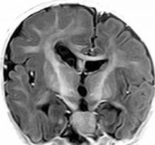
Figure Caption
Axial T2WI images (A, B) demonstrate enlarged right cerebral and right cerebellar hemispheres. Prominence of right lateral ventricle with straightening of right frontal horn is seen. Areas of T2 hyperintensity in right hemisphericwhite matter suggests poor myelination. Coronal T1 IR (C) shows diffuse gyral thickening with shallow sulci.


