Case of the Week
Section Editors: Matylda Machnowska1 and Anvita Pauranik2
1University of Toronto, Toronto, Ontario, Canada
2BC Children's Hospital, University of British Columbia, Vancouver, British Columbia, Canada
Sign up to receive an email alert when a new Case of the Week is posted.
Click images to view large versions
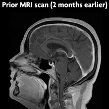
Pretreatment MRI scan: contrast-enhanced sagittal T1WI (A) shows a partially empty sella.
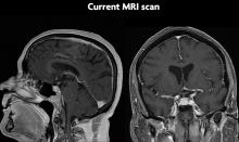
Post-treatment MRI scan: Contrast-enhanced sagittal (B, left) and coronal (B, right) T1WI show increased size of the pituitary gland (11.0 mm on the craniocaudal axis) and heterogeneous enhancement. The infundibulum is not involved.


