Advertisement

Main menu

  • Home
  • Content
    • Current Issue
    • Accepted Manuscripts
    • Article Preview
    • Past Issue Archive
    • AJNR Case Collection
    • Case of the Week Archive
    • Classic Case Archive
    • Case of the Month Archive
  • Special Collections
    • Spinal CSF Leak Articles (Jan 2020-June 2024)
    • 2024 AJNR Journal Awards
    • Most Impactful AJNR Articles
  • Multimedia
    • AJNR Podcast
    • AJNR Scantastics
    • Video Articles
  • For Authors
    • Submit a Manuscript
    • Author Policies
    • Fast publishing of Accepted Manuscripts
    • Graphical Abstract Preparation
    • Manuscript Submission Guidelines
    • Imaging Protocol Submission
    • Submit a Case for the Case Collection
  • About Us
    • About AJNR
    • Editorial Board
  • More
    • Become a Reviewer/Academy of Reviewers
    • Subscribers
    • Permissions
    • Alerts
    • Feedback
    • Advertisers
    • ASNR Home
  • Other Publications
    • ajnr

User menu

  • Alerts
  • Log in

Search

  • Advanced search
American Journal of Neuroradiology

American Journal of Neuroradiology

ASHNR American Society of Functional Neuroradiology ASHNR American Society of Pediatric Neuroradiology ASSR
  • Alerts
  • Log in

Advanced Search

  • Home
  • Content
    • Current Issue
    • Accepted Manuscripts
    • Article Preview
    • Past Issue Archive
    • AJNR Case Collection
    • Case of the Week Archive
    • Classic Case Archive
    • Case of the Month Archive
  • Special Collections
    • Spinal CSF Leak Articles (Jan 2020-June 2024)
    • 2024 AJNR Journal Awards
    • Most Impactful AJNR Articles
  • Multimedia
    • AJNR Podcast
    • AJNR Scantastics
    • Video Articles
  • For Authors
    • Submit a Manuscript
    • Author Policies
    • Fast publishing of Accepted Manuscripts
    • Graphical Abstract Preparation
    • Manuscript Submission Guidelines
    • Imaging Protocol Submission
    • Submit a Case for the Case Collection
  • About Us
    • About AJNR
    • Editorial Board
  • More
    • Become a Reviewer/Academy of Reviewers
    • Subscribers
    • Permissions
    • Alerts
    • Feedback
    • Advertisers
    • ASNR Home
  • Follow AJNR on Twitter
  • Visit AJNR on Facebook
  • Follow AJNR on Instagram
  • Join AJNR on LinkedIn
  • RSS Feeds

Welcome to the new AJNR, Updated Hall of Fame, and more. Read the full announcements.


AJNR is seeking candidates for the position of Associate Section Editor, AJNR Case Collection. Read the full announcement.


OtherHEAD AND NECK

CT and MRI Appearances of Primary Sphenoid Melanoma: A Rare Case

Kiran Batra, Avneesh Chhabra, Jaideep Rampure, Shangguio Tang, Robert Koenigsberg and Carlos Gonzales
American Journal of Neuroradiology November 2005, 26 (10) 2642-2644;
Kiran Batra
  • Find this author on Google Scholar
  • Find this author on PubMed
  • Search for this author on this site
Avneesh Chhabra
  • Find this author on Google Scholar
  • Find this author on PubMed
  • Search for this author on this site
Jaideep Rampure
  • Find this author on Google Scholar
  • Find this author on PubMed
  • Search for this author on this site
Shangguio Tang
  • Find this author on Google Scholar
  • Find this author on PubMed
  • Search for this author on this site
Robert Koenigsberg
  • Find this author on Google Scholar
  • Find this author on PubMed
  • Search for this author on this site
Carlos Gonzales
  • Find this author on Google Scholar
  • Find this author on PubMed
  • Search for this author on this site
  • Article
  • Figures & Data
  • Info & Metrics
  • Responses
  • References
  • PDF
Loading

Abstract

Summary: Primary sphenoid melanoma is a rare tumor. A 75-year-old man presented with complaints of headache and visual complaints for a few days. CT and MRI of the brain demonstrated a sphenoid sinus mass with appearances suggesting a diagnosis of melanoma. Fine-needle aspiration biopsy confirmed the diagnosis. Other differential diagnostic considerations on CT and MRI are discussed. We also discuss the further role of contrast MRI in determining the actual extent of the tumor in the critical skull base area.

Case Report

A 75-year-old black man with no significant past medical history presented with complaints of occipital headache and double vision of several days’ duration. Physical examination revealed ptosis and other signs of third nerve palsy in the left eye. He was initially referred by the clinician for a paranasal sinus (PNS) CT to rule out sinusitis.

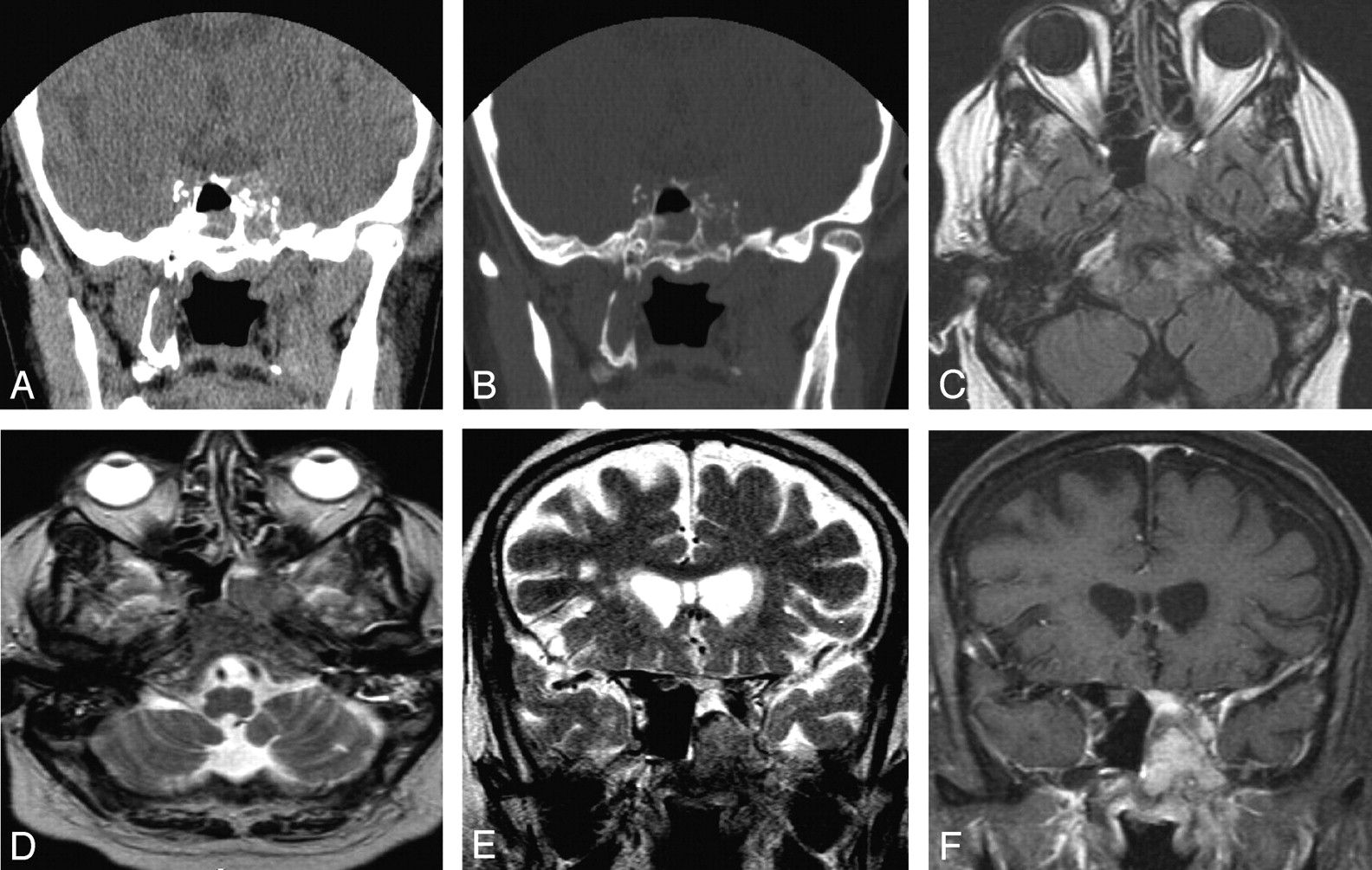
Noncontrast CT (NCCT) of the paranasal sinus (PNS) demonstrated a hyperattenuated mass in the left sphenoid sinus with destruction of the lateral sinus wall and extension into the left cavernous sinus (Fig 1A, -B). Follow-up MR imaging with contrast was performed to further characterize the lesion. The lesion appeared iso-hyperintense on T1-weighted image and hypointense to gray matter on T2-weighted image with diffuse homogenous enhancement on postgadolinium T1-weighted images. There was redemonstration of left parasellar extension into the cavernous sinus with encasement of the ipsilateral internal carotid artery. The lesion also involved the base of skull, in the region of foramen ovale, rotundum, and clivus. Associated diffuse dural thickening and enhancement was seen in the left temporal fossa (Fig 1C–F). The optic chiasma and pituitary gland appeared normal. The patient had no history of previously diagnosed cutaneous or mucosal melanomas at other sites.

Fig 1.
  • Download figure
  • Open in new tab
  • Download powerpoint
Fig 1.

Coronal NCCT of PNS at the level of sphenoid sinus (A and B), demonstrating a hyperattenuated mass in the left side of sphenoid sinus with destruction of the lateral sinus wall. Mucosal thickening from inflammatory disease was also seen in the right side of sphenoid sinus. Follow-up MR imaging with contrast redemonstrates the mass, appearing iso-hyperintense on axial T1-weighted image with extension in the left side of clivus (C). The mass appears hypointense on axial and coronal T2-weighted images (D and E) with diffuse homogenous enhancement in the mass and adjoining dura in left temporal fossa on postgadolinium T1-weighted images. Also seen is left parasellar extension into the cavernous sinus and the base of skull, in the region of foramen ovale.

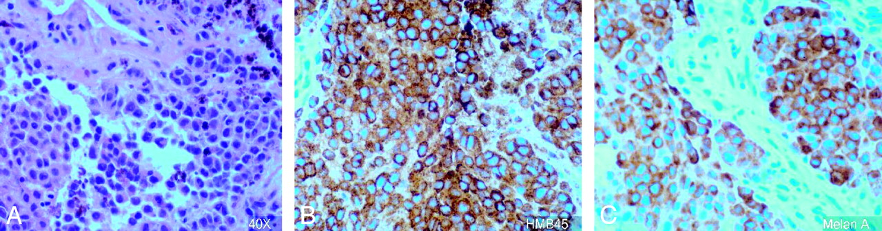
Surgical biopsy through trans-sphenoidal approach was performed, and anaplastic tumor cells that were positive for HMB45 and melan A and negative for S100, CK7, and CK 20 were identified (Fig 2).

Fig 2.
  • Download figure
  • Open in new tab
  • Download powerpoint
Fig 2.

Histopathology slide (magnification ×40) demonstrates melanoma cells (A), positive for HMB45 (B) and melan A (C).

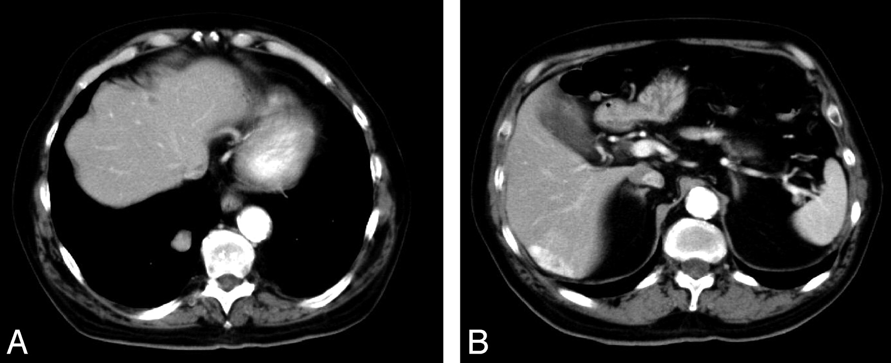
CT scans of chest, abdomen, and pelvis were performed to evaluate the extent of the disease. Multiple heterogeneously enhancing lesions were identified in the liver and in right lower lobe of lung, which suggested metastases (Fig 3A, -B). A bone scan was negative for bone metastasis. Stereotactic radiation therapy was initiated, and the patient is currently receiving palliative combination chemotherapy.

Fig 3.
  • Download figure
  • Open in new tab
  • Download powerpoint
Fig 3.

Hypervascular lesions in the liver and right lobe lung of lung suggest metastasis.

Discussion

Neoplasms arising in the PNS account for 0.2%–0.8% of all cancers and 2%–3% of head and neck cancers. Primary involvement of the sphenoidal sinus has been reported to occur in only 1%–2% of all PNS tumors (1). In one of the largest series of primary nasal and PNS tumors, Harbo et al (2) found the major histologic types to include squamous cell carcinoma (46%), lymphoma (14%), adenocarcinoma (13%), and malignant melanoma (9%). In a review of previously reported cases of sinonasal melanoma (SNM) by Batsakis et al (3), the most common site of origin in the sinonasal tract was the nasal cavity, followed by the maxillary sinus, ethmoid sinus, and frontal sinus. Primary melanoma of the sphenoid sinus is extremely rare, and, to the best of our knowledge, only 2 cases have been reported in the literature (4, 5).

In general, the mean age for SNM is later in life (5th–8th decades) than cutaneous malignant melanomas, and SNM is a more lethal disease in patients >60 years of age, a finding similar to cutaneous melanoma (6). Headache and visual symptoms—such as ptosis, blurred vision, and cranial nerve palsies as part of sphenocavernous syndrome—are common in sphenoid malignancies (7). Our patient also belonged to similar age category and presented with headache and left third nerve palsy.

The mass appeared iso-hyperattenuated on NCCT and caused destruction of the sphenoid sinus wall with left parasellar extension. CT is an excellent means of identifying subtle bone erosion in the sphenoid sinus, and this finding is important because bony destruction can expose critical neurovascular structures to the expanding mass lesion and there is an inherent risk of injury during surgery. The initial differential diagnostic consideration based on CT included a sphenoid meningioma, lymphoma, metastasis, fungal sinusitis, mucocele, and melanoma (8).

MR imaging is superior in defining the extent of the tumor, perineural spread, vascular encasement, and subtle intracranial spread. In our case, additional involvement of skull base foramina and clivus was identified on MR imaging. Variations in signal intensity also allow for the important differentiation between tumor involvement and obstructive or inflammatory sinus changes (9). The signal intensity changes on MR imaging appeared iso-hyperintense on T1-weighted images and hypointense on T2-weighted images with homogenous enhancement on postcontrast scans.

Meningioma usually causes hyperostosis of the adjacent bone and may result in pneumatosis dilatans and rarely causes frank bony destruction, and most meningiomas are iso-hyperintense on T2-weighted images. Dural enhancement was seen contiguous to the tumor in the left temporal fossa, giving the appearance of a dural tail. The presence of a dural tail can be seen in a meningioma (60%–72% cases). Scattered case reports have documented the presence of dural tail adjacent to chloroma, primary CNS lymphoma, sarcoidosis, vestibular schwannoma, metastatic tumor, syphilitic gumma, and an aggressive papillary middle ear tumor (10). To the best of our knowledge, dural tail in a case of melanoma has not been reported elsewhere. We could not prove, however, whether it was due to malignant infiltration of the dura or was a result of inflammatory hypervascular reaction, because the patient did not undergo surgery.

Fungal sinusitis and lymphoma may appear hypointense on T2-weighted images because of the presence of minerals in the former and high cellularity in the latter case. Fungal sinusitis occurs in a patient with underlying immunosuppression or diabetes mellitus. The lesions are usually infiltrative with heterogeneous enhancement and may be associated with frank bony destruction. Lymphoma lesions are usually multicentric and appear hypointense on T1-weighted images. Diffuse homogenous enhancement excluded the diagnosis of a mucocele, which usually shows a peripheral enhancement. Metastasis from hemorrhagic tumors and mucinous adenocarcinoma from a primary colon tumor can appear as hypointense lesions on T2-weighted image, though absence of a primary tumor and presence of homogenous enhancement suggested diagnosis of primary melanoma.

Surgical biopsy through the trans-sphenoidal approach revealed anaplastic tumor cells positive for HMB45 and melan A and negative for S100. Immunohistochemical studies confirm the diagnosis of sinonasal tract mucosal malignant melanomas with positive reactions for S-100 protein, tyrosinase, HMB-45, melan A, and microphthalmia transcription factor (8). Melanoma and neural tumors are usually positive for S-100 protein. A small number of melanomas may be negative for S-100, such as in our case. CK7 and CK20 are epithelial markers. Gastric cancer, breast cancer, and lung cancer may be positive for CK7. Colorectal cancer is positive for CK20. Our case demonstrated negative reactions to CK7 and CK20, thus excluding these possibilities.

The mainstay of treatment for mucosal melanoma is surgical resection. The surgical treatment of melanoma of the mucous membrane is more complex than the surgical treatment of cutaneous melanomas, and the prognoses of patients are in general poorer than for patients with cutaneous melanomas because of late presentation. In our case, because of the presence of distant metastasis, the patient began palliative radiation and chemotherapy.

In conclusion, primary melanoma can arise in the sphenoid sinus. CT scan reveals early bony destruction associated with the lesion. MR imaging signal intensity changes and enhancement patterns may be similar to melanoma elsewhere and may suggest the diagnosis. Contrast MR imaging also helps in determining further extent of the tumor in the critical skull base area.

References

  1. ↵
    Osguthorpe JD. Sinus neoplasia. Arch Otolaryngol Head Neck Surg 1994;120:19–25
    CrossRefPubMed
  2. ↵
    Harbo G, Grau C, Bundgaard T, et al. Cancer of the nasal cavity and paranasal sinuses: a clinico-pathological study of 277 patients. Acta Oncol 1997;36:45–50
    PubMed
  3. ↵
    Batsakis JG, Regezi JA, Solomon AR, et al. The pathology of head and neck tumors: mucosal melanomas, part 13. Head Neck Surg4 (1982 ):404–418
    PubMed
  4. ↵
    Tuttenberg J, Fink W, Back W, et al. A rare primary sellar melanoma: case report. J Neurosurg 2004;100:931–934
    PubMed
  5. ↵
    Pino Rivero V, Keituqwa Yanez T, Marcos Garcia M, et al. Melanoma of sphenoid sinus: case presentation and review of literature. Acta Otorrinolaringol Esp 2004;55:45–48
    PubMed
  6. ↵
    Chang AE, Karnell LH, Menck HR. The National Cancer Data Base report on cutaneous and noncutaneous melanoma: a summary of 84,836 cases from the past decade. The American College of Surgeons Commission on Cancer and the American Cancer Society. Cancer 1998;83:1664–1678
    CrossRefPubMed
  7. ↵
    DeMonte F, Ginsberg LE, Clayman GL. Primary malignant tumors of the sphenoidal sinus. Neurosurgery 200;46:1084–1091
  8. ↵
    Thompson LD, Wieneke JA, Miettinen M. Sinonasal tract and nasopharyngeal melanomas: a clinicopathologic study of 115 cases with a proposed staging system. Am J Surg Pathol 2003;27:594–611
    CrossRefPubMed
  9. ↵
    Ginsberg LE. Neoplastic diseases affecting the central skull base: CT and MR imaging. AJR Am J Roentgenol 1992;159:581–589
    CrossRefPubMed
  10. ↵
    Wallace EW. The dural tail sign. Radiology 2004;233:56–57
    CrossRefPubMed
  • Received January 14, 2005.
  • Accepted after revision January 20, 2005.
  • Copyright © American Society of Neuroradiology
View Abstract
PreviousNext
Back to top

In this issue

American Journal of Neuroradiology: 26 (10)
American Journal of Neuroradiology
Vol. 26, Issue 10
1 Nov 2005
  • Table of Contents
  • Index by author
Advertisement
Print
Download PDF
Email Article

Thank you for your interest in spreading the word on American Journal of Neuroradiology.

NOTE: We only request your email address so that the person you are recommending the page to knows that you wanted them to see it, and that it is not junk mail. We do not capture any email address.

Enter multiple addresses on separate lines or separate them with commas.
CT and MRI Appearances of Primary Sphenoid Melanoma: A Rare Case
(Your Name) has sent you a message from American Journal of Neuroradiology
(Your Name) thought you would like to see the American Journal of Neuroradiology web site.
CAPTCHA
This question is for testing whether or not you are a human visitor and to prevent automated spam submissions.
Cite this article
Kiran Batra, Avneesh Chhabra, Jaideep Rampure, Shangguio Tang, Robert Koenigsberg, Carlos Gonzales
CT and MRI Appearances of Primary Sphenoid Melanoma: A Rare Case
American Journal of Neuroradiology Nov 2005, 26 (10) 2642-2644;

Citation Manager Formats

  • BibTeX
  • Bookends
  • EasyBib
  • EndNote (tagged)
  • EndNote 8 (xml)
  • Medlars
  • Mendeley
  • Papers
  • RefWorks Tagged
  • Ref Manager
  • RIS
  • Zotero
0 Responses
Respond to this article
Share
Bookmark this article
CT and MRI Appearances of Primary Sphenoid Melanoma: A Rare Case
Kiran Batra, Avneesh Chhabra, Jaideep Rampure, Shangguio Tang, Robert Koenigsberg, Carlos Gonzales
American Journal of Neuroradiology Nov 2005, 26 (10) 2642-2644;
del.icio.us logo Twitter logo Facebook logo Mendeley logo
  • Tweet Widget
  • Facebook Like
  • Google Plus One

Jump to section

  • Article
    • Abstract
    • Case Report
    • Discussion
    • References
  • Figures & Data
  • Info & Metrics
  • Responses
  • References
  • PDF

Related Articles

  • No related articles found.
  • PubMed
  • Google Scholar

Cited By...

  • Melanoma of the Sinonasal Tract: Value of a Septate Pattern on Precontrast T1-Weighted MR Imaging
  • Crossref
  • Google Scholar

This article has not yet been cited by articles in journals that are participating in Crossref Cited-by Linking.

More in this TOC Section

  • Correlation of Apparent Diffusion Coefficient at 3T with Prognostic Parameters of Retinoblastoma
  • Parathyroid Lesions: Characterization with Dual-Phase Arterial and Venous Enhanced CT of the Neck
  • Efficacy of Diffusion-Weighted Imaging for the Differentiation between Lymphomas and Carcinomas of the Nasopharynx and Oropharynx: Correlations of Apparent Diffusion Coefficients and Histologic Features
Show more Head and Neck

Similar Articles

Advertisement

Indexed Content

  • Current Issue
  • Accepted Manuscripts
  • Article Preview
  • Past Issues
  • Editorials
  • Editors Choice
  • Fellow Journal Club
  • Letters to the Editor

Cases

  • Case Collection
  • Archive - Case of the Week
  • Archive - Case of the Month
  • Archive - Classic Case

Special Collections

  • Special Collections

Resources

  • News and Updates
  • Turn around Times
  • Submit a Manuscript
  • Author Policies
  • Manuscript Submission Guidelines
  • Evidence-Based Medicine Level Guide
  • Publishing Checklists
  • Graphical Abstract Preparation
  • Imaging Protocol Submission
  • Submit a Case
  • Become a Reviewer/Academy of Reviewers
  • Get Peer Review Credit from Publons

Multimedia

  • AJNR Podcast
  • AJNR SCANtastic
  • Video Articles

About Us

  • About AJNR
  • Editorial Board
  • Not an AJNR Subscriber? Join Now
  • Alerts
  • Feedback
  • Advertise with us
  • Librarian Resources
  • Permissions
  • Terms and Conditions

American Society of Neuroradiology

  • Not an ASNR Member? Join Now

© 2025 by the American Society of Neuroradiology All rights, including for text and data mining, AI training, and similar technologies, are reserved.
Print ISSN: 0195-6108 Online ISSN: 1936-959X

Powered by HighWire