Advertisement

Main menu

  • Home
  • Content
    • Current Issue
    • Accepted Manuscripts
    • Article Preview
    • Past Issue Archive
    • AJNR Case Collection
    • Case of the Week Archive
    • Classic Case Archive
    • Case of the Month Archive
  • Special Collections
    • Spinal CSF Leak Articles (Jan 2020-June 2024)
    • 2024 AJNR Journal Awards
    • Most Impactful AJNR Articles
  • Multimedia
    • AJNR Podcast
    • AJNR Scantastics
    • Video Articles
  • For Authors
    • Submit a Manuscript
    • Author Policies
    • Fast publishing of Accepted Manuscripts
    • Graphical Abstract Preparation
    • Manuscript Submission Guidelines
    • Imaging Protocol Submission
    • Submit a Case for the Case Collection
  • About Us
    • About AJNR
    • Editorial Board
  • More
    • Become a Reviewer/Academy of Reviewers
    • Subscribers
    • Permissions
    • Alerts
    • Feedback
    • Advertisers
    • ASNR Home
  • Other Publications
    • ajnr

User menu

  • Alerts
  • Log in

Search

  • Advanced search
American Journal of Neuroradiology

American Journal of Neuroradiology

ASHNR American Society of Functional Neuroradiology ASHNR American Society of Pediatric Neuroradiology ASSR
  • Alerts
  • Log in

Advanced Search

  • Home
  • Content
    • Current Issue
    • Accepted Manuscripts
    • Article Preview
    • Past Issue Archive
    • AJNR Case Collection
    • Case of the Week Archive
    • Classic Case Archive
    • Case of the Month Archive
  • Special Collections
    • Spinal CSF Leak Articles (Jan 2020-June 2024)
    • 2024 AJNR Journal Awards
    • Most Impactful AJNR Articles
  • Multimedia
    • AJNR Podcast
    • AJNR Scantastics
    • Video Articles
  • For Authors
    • Submit a Manuscript
    • Author Policies
    • Fast publishing of Accepted Manuscripts
    • Graphical Abstract Preparation
    • Manuscript Submission Guidelines
    • Imaging Protocol Submission
    • Submit a Case for the Case Collection
  • About Us
    • About AJNR
    • Editorial Board
  • More
    • Become a Reviewer/Academy of Reviewers
    • Subscribers
    • Permissions
    • Alerts
    • Feedback
    • Advertisers
    • ASNR Home
  • Follow AJNR on Twitter
  • Visit AJNR on Facebook
  • Follow AJNR on Instagram
  • Join AJNR on LinkedIn
  • RSS Feeds

Welcome to the new AJNR, Updated Hall of Fame, and more. Read the full announcements.


AJNR is seeking candidates for the position of Associate Section Editor, AJNR Case Collection. Read the full announcement.


Research ArticleBRAIN

MR Imaging of Human Herpesvirus-6 Encephalopathy after Hematopoietic Stem Cell Transplantation in Adults

T. Noguchi, F. Mihara, T. Yoshiura, O. Togao, K. Atsumi, T. Matsuura, T. Kuroiwa and H. Honda
American Journal of Neuroradiology November 2006, 27 (10) 2191-2195;
T. Noguchi
  • Find this author on Google Scholar
  • Find this author on PubMed
  • Search for this author on this site
F. Mihara
  • Find this author on Google Scholar
  • Find this author on PubMed
  • Search for this author on this site
T. Yoshiura
  • Find this author on Google Scholar
  • Find this author on PubMed
  • Search for this author on this site
O. Togao
  • Find this author on Google Scholar
  • Find this author on PubMed
  • Search for this author on this site
K. Atsumi
  • Find this author on Google Scholar
  • Find this author on PubMed
  • Search for this author on this site
T. Matsuura
  • Find this author on Google Scholar
  • Find this author on PubMed
  • Search for this author on this site
T. Kuroiwa
  • Find this author on Google Scholar
  • Find this author on PubMed
  • Search for this author on this site
H. Honda
  • Find this author on Google Scholar
  • Find this author on PubMed
  • Search for this author on this site
  • Article
  • Figures & Data
  • Info & Metrics
  • Responses
  • References
  • PDF
Loading

Article Figures & Data

Figures

  • Tables
  • Fig 1.
    • Download figure
    • Open in new tab
    • Download powerpoint
    Fig 1.

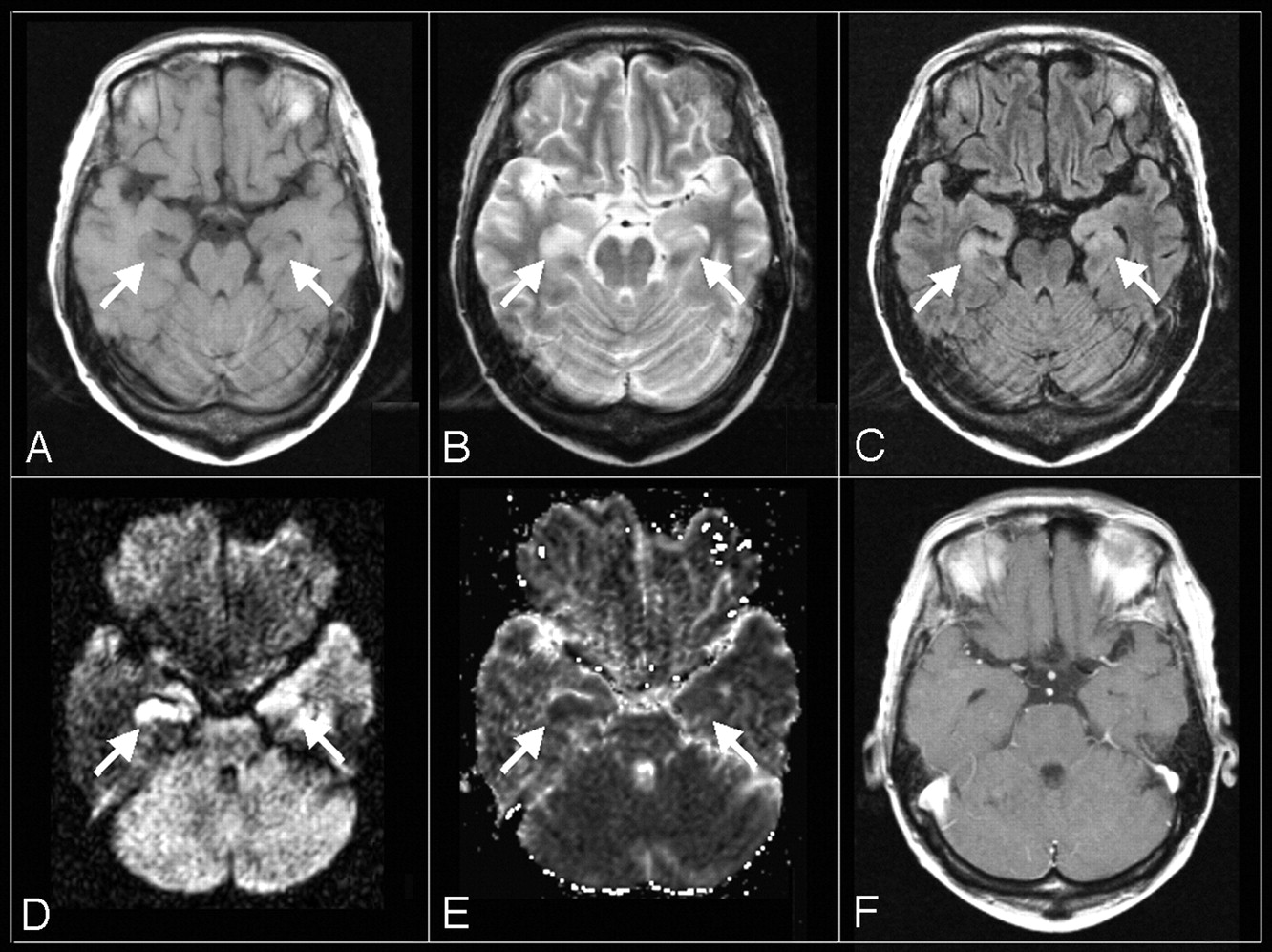
    Axial MR images of an HHV-6 encephalopathy patient (patient 2) obtained on the second day after the onset of neurologic symptoms.

    A, T1WI (TR/TE = 466/11 ms).

    B, T2WI (2650/93 ms).

    C, FLAIR (TR/TE/TI = 9000/97/2300 ms).

    D, DWI (TR/TE = 3100/119 ms, b = 1000).

    E, ADC map.

    F, postcontrast T1WI (TR/TE = 558/17 ms).

    An abnormal low signal intensity on T1WI (A) and high signal intensity on T2WI (B) and FLAIR (C) are shown in the bilateral amygdalae and hippocampi. High signal intensity on DWI (D) with ADC reduction (E) is also shown. However, no abnormal enhancement is seen on postcontrast T1WI (F).

  • Fig 2.
    • Download figure
    • Open in new tab
    • Download powerpoint
    Fig 2.

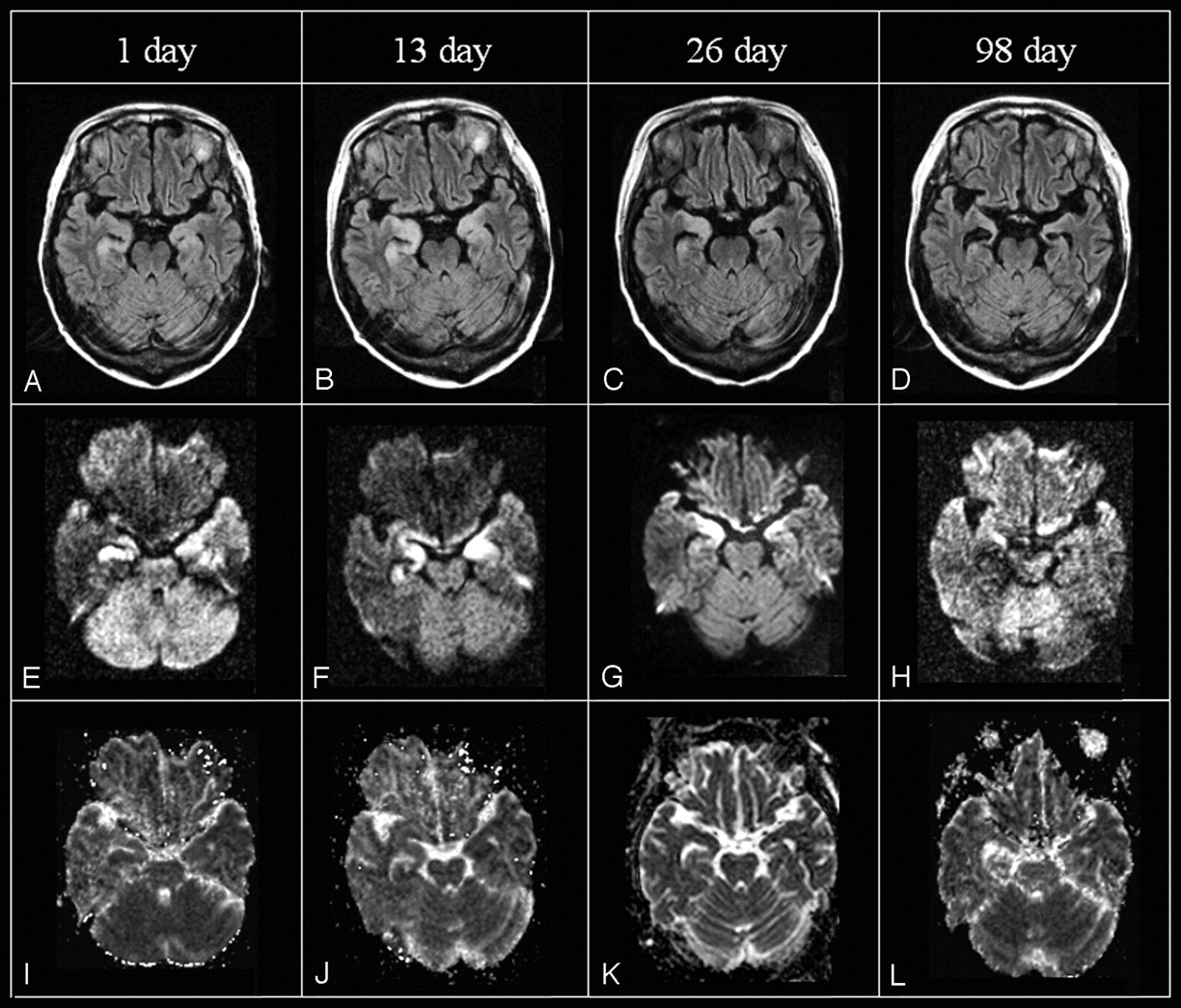
    Serial axial MR images of a 49-year-old woman (patient 1) including FLAIR (TR/TE/TI = 9000/110/2200 ms [A, B, D] or 9000/97/2300 ms [C]), DWI (TR/TE = 4000/137 ms [E, F, H] or 3100/119 ms [G]), and ADC maps on days 1, 13, 26, and 98 after the onset of neurologic symptoms (I-L). On FLAIR, high signal intensity in the bilateral amygdalae and hippocampi appears on day 1, peaks on day 13, and becomes less pronounced on day 26, disappearing on day 98 but leaving marked atrophy. High signal intensity on DWI is observed until day 26, whereas ADC value reduction is seen only on days 1 and 13.

Tables

  • Figures
    • View popup
    Table 1:

    Summary of patient histories

    Patient no: Age/SexPrimary DisorderType of TransplantInterval between Transplantation and Symptoms OnsetNeurological SymptomsSpecimen and Quantity of HHV-6 PCROutcome
    DisorientationShort-term Memory LossComaHypopneaConvulsion
    1:49/FAcute leukemiaCord blood stem cell27++9000 (CSF) and 3000 (blood*) copies/mLImproved (remaining disorientation and memory loss)
    2:41/FAcute leukemia(Peripheral blood stem cell)(979)++49,000 copies/mL (CSF)Improved (remaining disorientation and memory loss)
    3:55/MNon-Hodgkin lymphomaBone marrow22++400,000 copies/mL (blood)Dead (sepsis)
    4:52/MNon-Hodgkin lymphomaBone marrow14+++58,000 copies/mL (CSF)Improved
    5:36/MAcute leukemiaBone marrow19+++++2300 copies/mL (CSF)Dead (intestinal pneumonitis)
    6:37/FAcute leukemiaBone marrow17+++3000 copies/mL (CSF)Dead (liver failure)
    • Note:—HHV-6 indicates human herpesvirus 6; PCR; polymerase chain reaction.

    • * Peripheral blood.

    • View popup
    Table 2:

    MR findings in the amygdala/hippocampus and the time interval from the onset of neurological symptoms

    Time of Imaging with Reference to Symptom OnsetImaging Findings in the Amygdala/Hippocampus
    Plain CTT1WIT2WIFLAIRDWIADC ValueGd-T1WIOthers on MR
    Abnormal findingsLowHighHighHighReductionAbnormal enhancementAtophic change
    Early period, days 1–2 (n = 4 patients)0/3 (0%)2/4 (50%)2/4 (50%)2/3 (67%)2/2 (100%)2/2 (100%)0/2 (0%)0/4 (0%)
    Middle period, days 3–30 (n = 4 patients)5/6 (83%)4/6 (67%)5/6 (83%)3/5 (60%)1/3 (33%)0/2 (0%)2/6 (33%)
    Late period, days 31+ (n = 4 patients)0/4 (0%)0/4 (0%)0/4 (0%)0/4 (0%)0/2 (0%)0/2 (0%)4/4 (100%)
    • Note:—Gd-T1WI indicates gadolinium-diethylene triaminepentaacetic acid T1-weighted imaging; ADC, apparent diffusion coefficient; T1WI, T1-weighted imaging; T2WI, T2-weighted imaging; FLAIR, fluid-attenuated inversion recovery; DWI, diffusion-weighted imaging.

    • View popup
    Table 3:

    ADC ratio of abnormal to normal brain parenchyma

    Time of Imaging with Reference to Symptom OnsetPatients No.: Age/Sex
    1:49/F2:41/F3:55/M
    Early period, days 0–2 (n = 2 patients)59% (day 1)77% (day 2)
    Middle period, days 3–30 (n = 3 patients)83% (day 13)100% (day 22)114% (day 12)
    89% (day 26)
    Late period, days 31+ (n = 2 patients)137% (day 47)132% (day 45)
    115% (day 98)
    • Note:—ADC indicates apparent diffusion coefficient.

PreviousNext
Back to top

In this issue

American Journal of Neuroradiology: 27 (10)
American Journal of Neuroradiology
Vol. 27, Issue 10
November 2006
  • Table of Contents
  • Index by author
Advertisement
Print
Download PDF
Email Article

Thank you for your interest in spreading the word on American Journal of Neuroradiology.

NOTE: We only request your email address so that the person you are recommending the page to knows that you wanted them to see it, and that it is not junk mail. We do not capture any email address.

Enter multiple addresses on separate lines or separate them with commas.
MR Imaging of Human Herpesvirus-6 Encephalopathy after Hematopoietic Stem Cell Transplantation in Adults
(Your Name) has sent you a message from American Journal of Neuroradiology
(Your Name) thought you would like to see the American Journal of Neuroradiology web site.
CAPTCHA
This question is for testing whether or not you are a human visitor and to prevent automated spam submissions.
Cite this article
T. Noguchi, F. Mihara, T. Yoshiura, O. Togao, K. Atsumi, T. Matsuura, T. Kuroiwa, H. Honda
MR Imaging of Human Herpesvirus-6 Encephalopathy after Hematopoietic Stem Cell Transplantation in Adults
American Journal of Neuroradiology Nov 2006, 27 (10) 2191-2195;

Citation Manager Formats

  • BibTeX
  • Bookends
  • EasyBib
  • EndNote (tagged)
  • EndNote 8 (xml)
  • Medlars
  • Mendeley
  • Papers
  • RefWorks Tagged
  • Ref Manager
  • RIS
  • Zotero
0 Responses
Respond to this article
Share
Bookmark this article
MR Imaging of Human Herpesvirus-6 Encephalopathy after Hematopoietic Stem Cell Transplantation in Adults
T. Noguchi, F. Mihara, T. Yoshiura, O. Togao, K. Atsumi, T. Matsuura, T. Kuroiwa, H. Honda
American Journal of Neuroradiology Nov 2006, 27 (10) 2191-2195;
del.icio.us logo Twitter logo Facebook logo Mendeley logo
  • Tweet Widget
  • Facebook Like
  • Google Plus One

Jump to section

  • Article
    • Abstract
    • Materials and Methods
    • Results
    • Discussion
    • References
  • Figures & Data
  • Info & Metrics
  • Responses
  • References
  • PDF

Related Articles

  • No related articles found.
  • PubMed
  • Google Scholar

Cited By...

  • No citing articles found.
  • Crossref
  • Google Scholar

This article has not yet been cited by articles in journals that are participating in Crossref Cited-by Linking.

More in this TOC Section

  • Predictors of Reperfusion in Patients with Acute Ischemic Stroke
  • Qualitative and Quantitative Analysis of MR Imaging Findings in Patients with Middle Cerebral Artery Stroke Implanted with Mesenchymal Stem Cells
  • Multimodal CT Provides Improved Performance for Lacunar Infarct Detection
Show more Brain

Similar Articles

Advertisement

Indexed Content

  • Current Issue
  • Accepted Manuscripts
  • Article Preview
  • Past Issues
  • Editorials
  • Editors Choice
  • Fellow Journal Club
  • Letters to the Editor

Cases

  • Case Collection
  • Archive - Case of the Week
  • Archive - Case of the Month
  • Archive - Classic Case

Special Collections

  • Special Collections

Resources

  • News and Updates
  • Turn around Times
  • Submit a Manuscript
  • Author Policies
  • Manuscript Submission Guidelines
  • Evidence-Based Medicine Level Guide
  • Publishing Checklists
  • Graphical Abstract Preparation
  • Imaging Protocol Submission
  • Submit a Case
  • Become a Reviewer/Academy of Reviewers
  • Get Peer Review Credit from Publons

Multimedia

  • AJNR Podcast
  • AJNR SCANtastic
  • Video Articles

About Us

  • About AJNR
  • Editorial Board
  • Not an AJNR Subscriber? Join Now
  • Alerts
  • Feedback
  • Advertise with us
  • Librarian Resources
  • Permissions
  • Terms and Conditions

American Society of Neuroradiology

  • Not an ASNR Member? Join Now

© 2025 by the American Society of Neuroradiology All rights, including for text and data mining, AI training, and similar technologies, are reserved.
Print ISSN: 0195-6108 Online ISSN: 1936-959X

Powered by HighWire