Advertisement

Main menu

  • Home
  • Content
    • Current Issue
    • Accepted Manuscripts
    • Article Preview
    • Past Issue Archive
    • AJNR Case Collection
    • Case of the Week Archive
    • Classic Case Archive
    • Case of the Month Archive
  • Special Collections
    • Spinal CSF Leak Articles (Jan 2020-June 2024)
    • 2024 AJNR Journal Awards
    • Most Impactful AJNR Articles
  • Multimedia
    • AJNR Podcast
    • AJNR Scantastics
    • Video Articles
  • For Authors
    • Submit a Manuscript
    • Author Policies
    • Fast publishing of Accepted Manuscripts
    • Graphical Abstract Preparation
    • Manuscript Submission Guidelines
    • Imaging Protocol Submission
    • Submit a Case for the Case Collection
  • About Us
    • About AJNR
    • Editorial Board
  • More
    • Become a Reviewer/Academy of Reviewers
    • Subscribers
    • Permissions
    • Alerts
    • Feedback
    • Advertisers
    • ASNR Home
  • Other Publications
    • ajnr

User menu

  • Alerts
  • Log in

Search

  • Advanced search
American Journal of Neuroradiology

American Journal of Neuroradiology

ASHNR American Society of Functional Neuroradiology ASHNR American Society of Pediatric Neuroradiology ASSR
  • Alerts
  • Log in

Advanced Search

  • Home
  • Content
    • Current Issue
    • Accepted Manuscripts
    • Article Preview
    • Past Issue Archive
    • AJNR Case Collection
    • Case of the Week Archive
    • Classic Case Archive
    • Case of the Month Archive
  • Special Collections
    • Spinal CSF Leak Articles (Jan 2020-June 2024)
    • 2024 AJNR Journal Awards
    • Most Impactful AJNR Articles
  • Multimedia
    • AJNR Podcast
    • AJNR Scantastics
    • Video Articles
  • For Authors
    • Submit a Manuscript
    • Author Policies
    • Fast publishing of Accepted Manuscripts
    • Graphical Abstract Preparation
    • Manuscript Submission Guidelines
    • Imaging Protocol Submission
    • Submit a Case for the Case Collection
  • About Us
    • About AJNR
    • Editorial Board
  • More
    • Become a Reviewer/Academy of Reviewers
    • Subscribers
    • Permissions
    • Alerts
    • Feedback
    • Advertisers
    • ASNR Home
  • Follow AJNR on Twitter
  • Visit AJNR on Facebook
  • Follow AJNR on Instagram
  • Join AJNR on LinkedIn
  • RSS Feeds

Welcome to the new AJNR, Updated Hall of Fame, and more. Read the full announcements.


AJNR is seeking candidates for the position of Associate Section Editor, AJNR Case Collection. Read the full announcement.


OtherHEAD & NECK

MR Spectroscopy in Sinus Mucocele: N-Acetyl Mimics of Brain N-Acetylaspartate

E. Andre, M. Xu, D. Yang, J.K. Siow, T.T. Yeo, Y. Xu and C.C.T. Lim
American Journal of Neuroradiology November 2006, 27 (10) 2210-2213;
E. Andre
  • Find this author on Google Scholar
  • Find this author on PubMed
  • Search for this author on this site
M. Xu
  • Find this author on Google Scholar
  • Find this author on PubMed
  • Search for this author on this site
D. Yang
  • Find this author on Google Scholar
  • Find this author on PubMed
  • Search for this author on this site
J.K. Siow
  • Find this author on Google Scholar
  • Find this author on PubMed
  • Search for this author on this site
T.T. Yeo
  • Find this author on Google Scholar
  • Find this author on PubMed
  • Search for this author on this site
Y. Xu
  • Find this author on Google Scholar
  • Find this author on PubMed
  • Search for this author on this site
C.C.T. Lim
  • Find this author on Google Scholar
  • Find this author on PubMed
  • Search for this author on this site
  • Article
  • Figures & Data
  • Info & Metrics
  • Responses
  • References
  • PDF
Loading

Article Figures & Data

Figures

  • Fig 1.
    • Download figure
    • Open in new tab
    • Download powerpoint
    Fig 1.

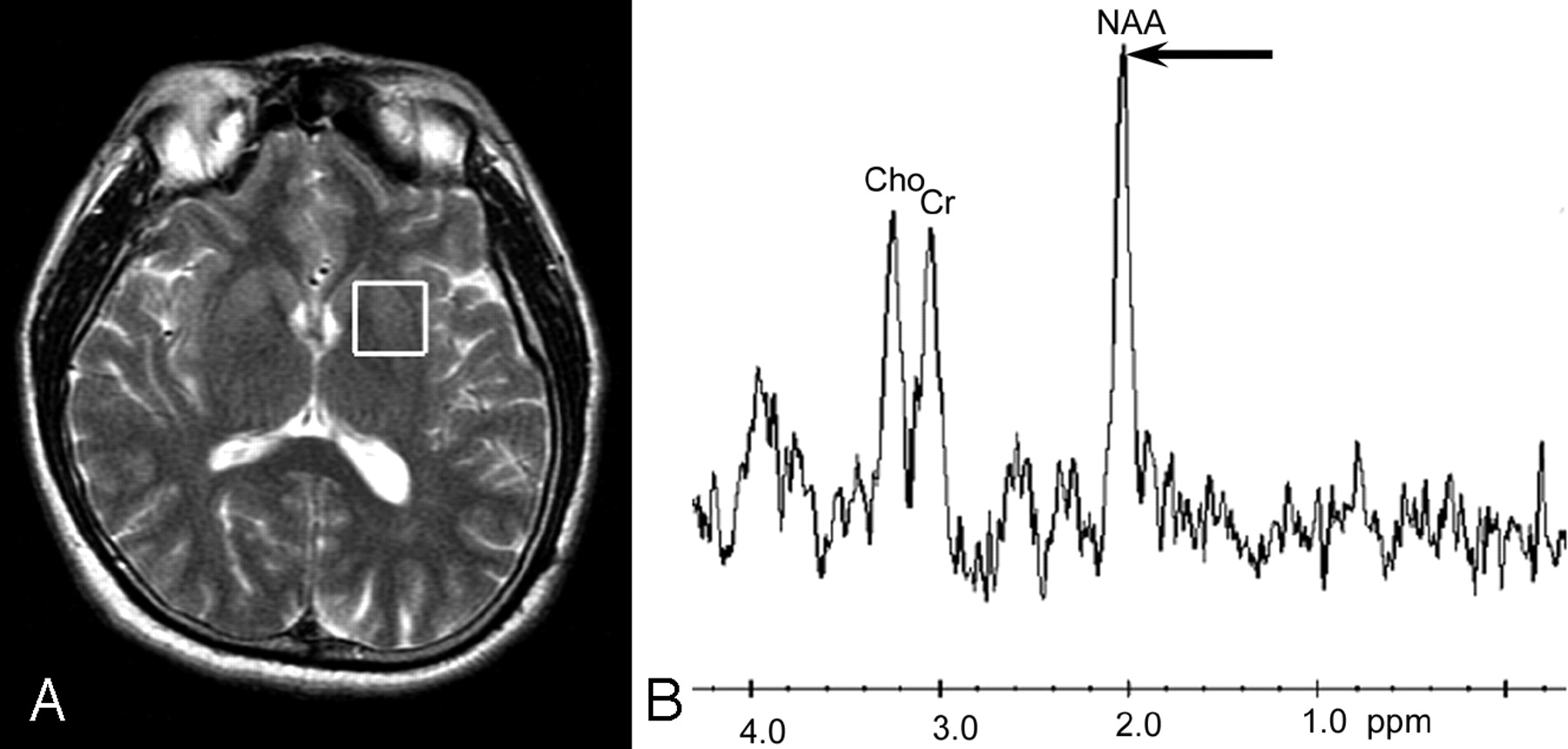
    In vivo MR spectroscopy of normal brain. Axial T2-weighted MR image shows a single voxel of interest for MR spectroscopy (white box) placed within the brain parenchyma in a healthy volunteer (A). The corresponding in vivo MR spectrum shows the normal dominant peaks of NAA at 2.0 ppm (arrow), Cr at 3.0 ppm, and Cho at 3.2 ppm (B).

  • Fig 2.
    • Download figure
    • Open in new tab
    • Download powerpoint
    Fig 2.

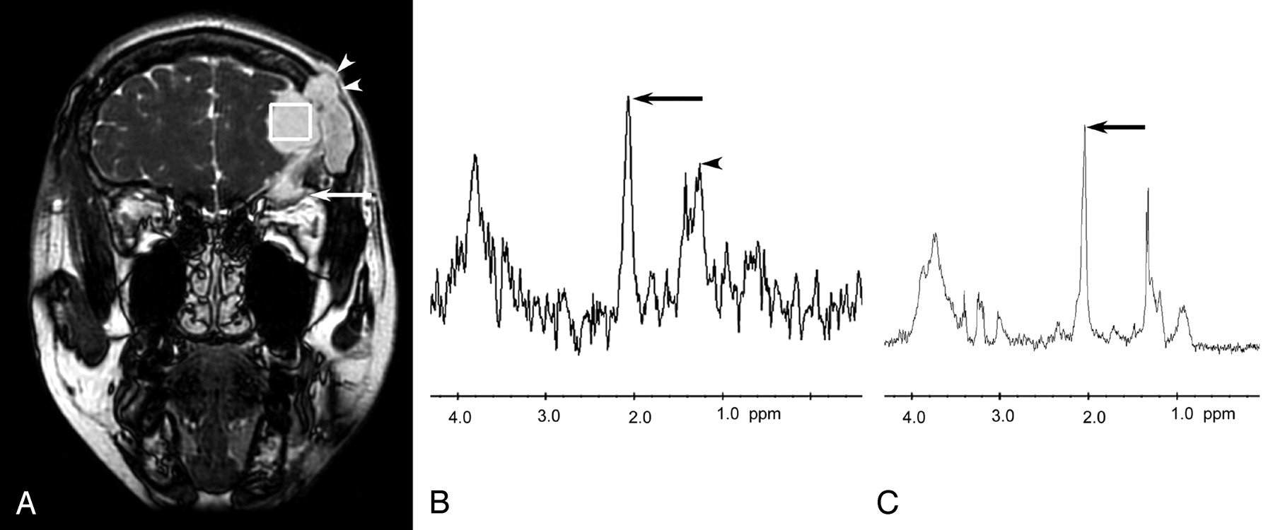
    33-year-old woman with frontal sinus mucocele. Coronal T2-weighted localizer image shows the voxel of interest for MR spectroscopy (white box) within a mucocele of high signal intensity (A). The mass is clearly extra-axial and compresses the frontal lobe brain parenchyma, which is excluded from the voxel. Note the involvement of the superior aspect of the right orbit (arrow) and destruction of the outer table of the skull (arrowheads). In vivo MR spectrum (B) shows a dominant peak at 2.0 ppm (arrow) and another at 3.9 ppm. There is also a complex multiplet between 1.2 and 1.4 ppm (arrowhead), probably from a combination of lipid and lactate. In vitro 1D proton NMR spectroscopy (C) of the mucocele contents removed at surgery confirms the dominant peak at 2.0 ppm (arrow) and other peaks at various positions of chemical shift.

  • Fig 3.
    • Download figure
    • Open in new tab
    • Download powerpoint
    Fig 3.

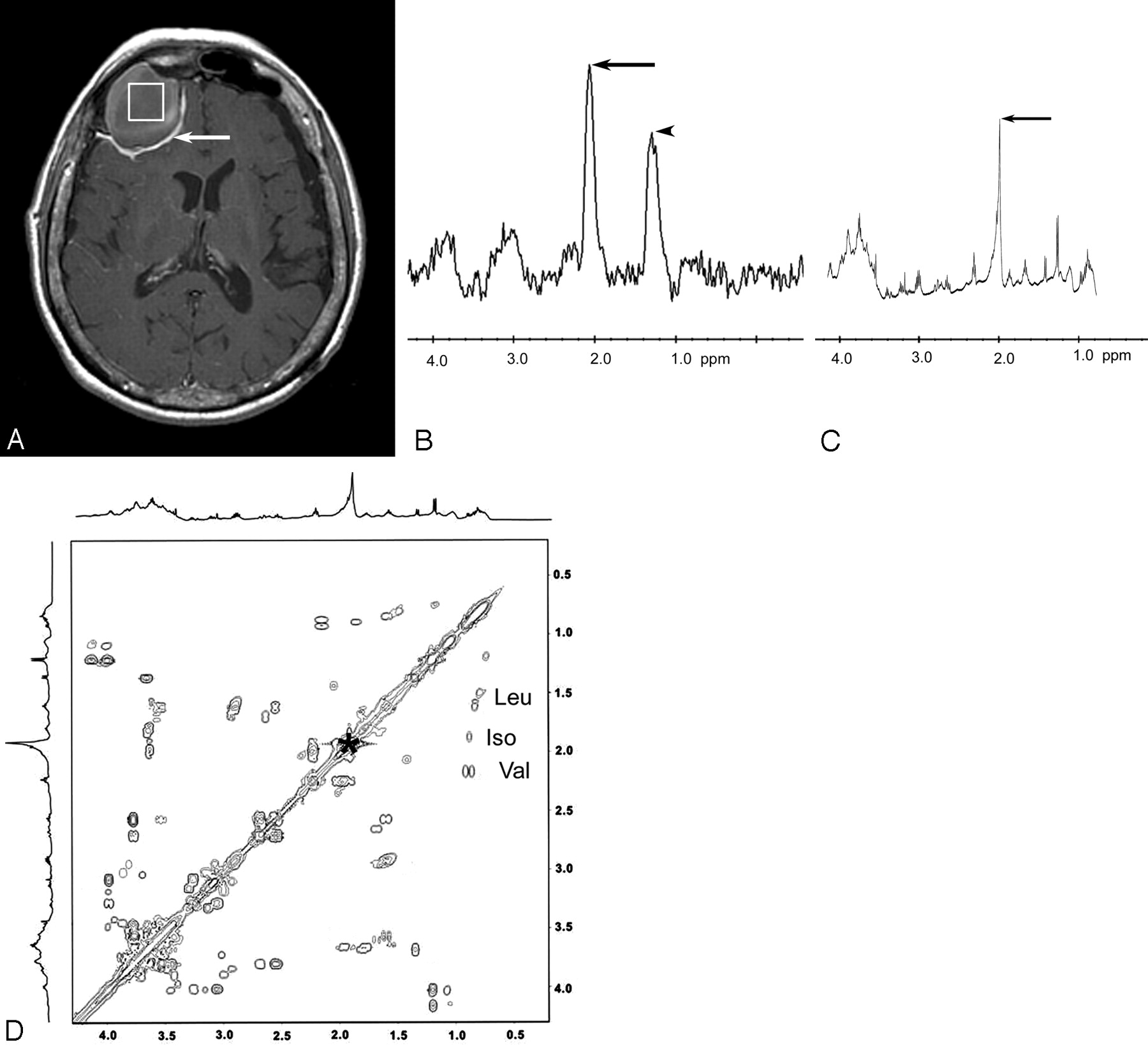
    74-year-old woman with frontal sinus mucocele. Axial postcontrast MR image (A) shows the voxel of interest placed completely within the right frontal mucocele. Dural contrast enhancement (arrow) confirms the extra-axial location of the mucocele. There is no visible contamination of the voxel by normal brain tissue or scalp fat. In vivo proton MR spectrum (B) shows dominant peaks at 2.0 ppm (arrow) and 1.2 ppm (arrowhead), the latter assigned as lipid breakdown products. In vitro 1D NMR spectrum (C) of the mucocele sample after surgery confirms the main metabolite peak at 2.0 ppm (arrow). The 2D COSY spectra (D) show an N-acetyl compound at 2.0 ppm (asterisk), as well as cross-peaks for leucine (Leu), isoleucine (Iso) and valine (Val).

  • Fig 4.
    • Download figure
    • Open in new tab
    • Download powerpoint
    Fig 4.

    Chemical structure of NAA and N-acetylated glycoproteins. The common CH3 moiety of N-acetyl compounds (box) in NAA (A) and N-acetyl glycoproteins (B and C) is highlighted. MR spectroscopy will detect the electrons in the CH3 chemical bond, and the metabolite peak at 2.0 ppm will be positive for NAA as well as N-acetyl glycoproteins.

PreviousNext
Back to top

In this issue

American Journal of Neuroradiology: 27 (10)
American Journal of Neuroradiology
Vol. 27, Issue 10
November 2006
  • Table of Contents
  • Index by author
Advertisement
Print
Download PDF
Email Article

Thank you for your interest in spreading the word on American Journal of Neuroradiology.

NOTE: We only request your email address so that the person you are recommending the page to knows that you wanted them to see it, and that it is not junk mail. We do not capture any email address.

Enter multiple addresses on separate lines or separate them with commas.
MR Spectroscopy in Sinus Mucocele: N-Acetyl Mimics of Brain N-Acetylaspartate
(Your Name) has sent you a message from American Journal of Neuroradiology
(Your Name) thought you would like to see the American Journal of Neuroradiology web site.
CAPTCHA
This question is for testing whether or not you are a human visitor and to prevent automated spam submissions.
Cite this article
E. Andre, M. Xu, D. Yang, J.K. Siow, T.T. Yeo, Y. Xu, C.C.T. Lim
MR Spectroscopy in Sinus Mucocele: N-Acetyl Mimics of Brain N-Acetylaspartate
American Journal of Neuroradiology Nov 2006, 27 (10) 2210-2213;

Citation Manager Formats

  • BibTeX
  • Bookends
  • EasyBib
  • EndNote (tagged)
  • EndNote 8 (xml)
  • Medlars
  • Mendeley
  • Papers
  • RefWorks Tagged
  • Ref Manager
  • RIS
  • Zotero
0 Responses
Respond to this article
Share
Bookmark this article
MR Spectroscopy in Sinus Mucocele: N-Acetyl Mimics of Brain N-Acetylaspartate
E. Andre, M. Xu, D. Yang, J.K. Siow, T.T. Yeo, Y. Xu, C.C.T. Lim
American Journal of Neuroradiology Nov 2006, 27 (10) 2210-2213;
del.icio.us logo Twitter logo Facebook logo Mendeley logo
  • Tweet Widget
  • Facebook Like
  • Google Plus One

Jump to section

  • Article
    • Abstract
    • Materials and Methods
    • Case Reports
    • Discussion
    • Footnotes
    • References
  • Figures & Data
  • Info & Metrics
  • Responses
  • References
  • PDF

Related Articles

  • No related articles found.
  • PubMed
  • Google Scholar

Cited By...

  • No citing articles found.
  • Crossref
  • Google Scholar

This article has not yet been cited by articles in journals that are participating in Crossref Cited-by Linking.

More in this TOC Section

  • Chondrosarcoma vs Synovial Chondromatosis: Imaging
  • NI-RADS for HEAD&NECK Cancer Recurrence
  • WHO Classification Update: Nasal&Skull Base Tumors
Show more HEAD & NECK

Similar Articles

Advertisement

Indexed Content

  • Current Issue
  • Accepted Manuscripts
  • Article Preview
  • Past Issues
  • Editorials
  • Editors Choice
  • Fellow Journal Club
  • Letters to the Editor

Cases

  • Case Collection
  • Archive - Case of the Week
  • Archive - Case of the Month
  • Archive - Classic Case

Special Collections

  • Special Collections

Resources

  • News and Updates
  • Turn around Times
  • Submit a Manuscript
  • Author Policies
  • Manuscript Submission Guidelines
  • Evidence-Based Medicine Level Guide
  • Publishing Checklists
  • Graphical Abstract Preparation
  • Imaging Protocol Submission
  • Submit a Case
  • Become a Reviewer/Academy of Reviewers
  • Get Peer Review Credit from Publons

Multimedia

  • AJNR Podcast
  • AJNR SCANtastic
  • Video Articles

About Us

  • About AJNR
  • Editorial Board
  • Not an AJNR Subscriber? Join Now
  • Alerts
  • Feedback
  • Advertise with us
  • Librarian Resources
  • Permissions
  • Terms and Conditions

American Society of Neuroradiology

  • Not an ASNR Member? Join Now

© 2025 by the American Society of Neuroradiology All rights, including for text and data mining, AI training, and similar technologies, are reserved.
Print ISSN: 0195-6108 Online ISSN: 1936-959X

Powered by HighWire