
Article Figures & Data
Figures
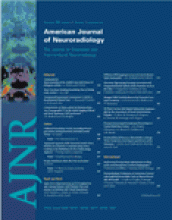
- Fig 1.
Circumferential compression of the cauda equina. The cauda equina was constricted outside the dura mater (D) by using a silicone tube (S), which caused 30% constriction of the diameter of the dura mater by using a silicone tube at L6/7 disk level.
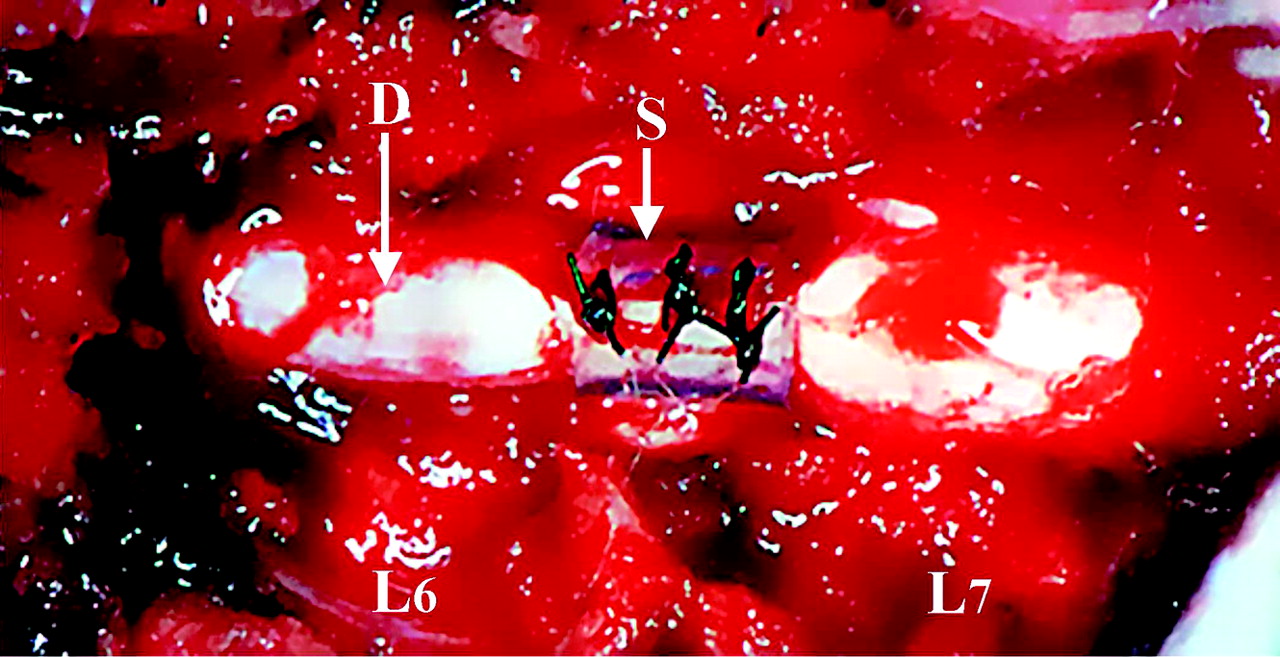
- Fig 2.
Dural sac measurements on MR imaging. A, Control group. B, 30% constriction group. C, Cross-sectional area (mm2) of the dural sac in control and 30% constriction model.
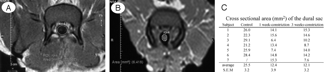
- Fig 3.
Comparison between enhanced MR imaging and fluorescent micrograph of the cauda equina.
A and B, Control group. No enhancement of a healthy cauda equina (CE) was found on a gadolinium-enhanced MR image (T1-weighted spin-echo [SE] image, 600/25 [TR/TE]). The cauda equina and epidural root sleeves (ERS) showed moderate signal intensities and the signal intensity was similar to that of muscle in normal conditions (A), EBA emits a bright red fluorescence in clear contrast to the green fluorescence of the nerve tissue. EBA was limited inside the blood vessels, and the blood-nerve barrier was maintained as seen under fluorescent microscopy (B).
C and D, After 1 week constriction.
E and F, After 3 weeks constriction. Clear enhancement was seen inside the cauda equina constricted by a silicon tube (S) as seen on gadolinium-enhanced MR image. No enhancement of epidural root sleeves (ERS) was found on gadolinium-enhanced MR image (T1-weighted spin-echo [SE] image, 600/25 [TR/TE]) (C and E). In the cauda equina, where enhancement was found on MR imaging, EBA emits a bright red fluorescence, which leaked outside the blood vessels, and intraradicular edema was seen under a fluorescent microscope (D and F). BV, blood vessel; CE, cauda equine; ENS, epidural root sleeves; NR, nerve root; RS, root sheath; SS, subarachnoid space.
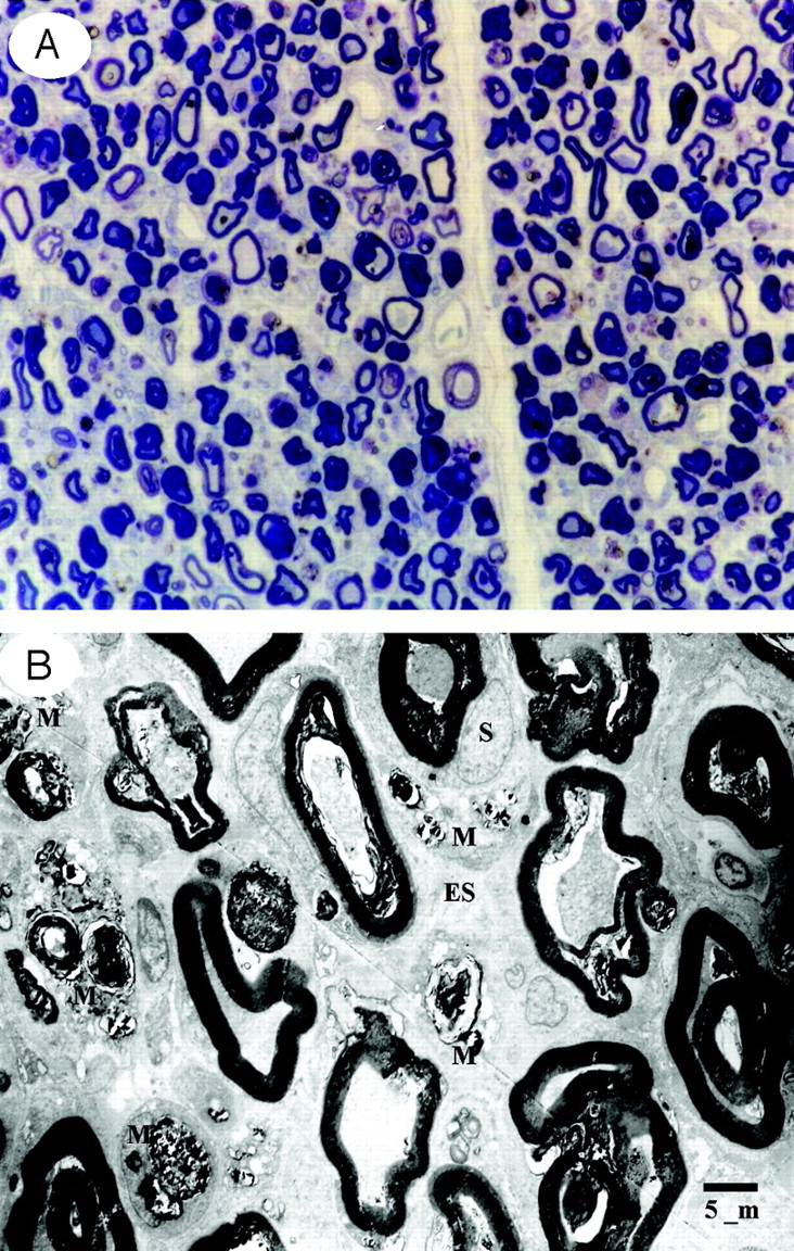
- Fig 4.
Light (A) and electron micrographs (B) in the cauda equina at the site of constriction after 1 week. One week after cauda equina constriction, Wallerian degeneration was apparent in the constriction area. Histologic and electron microscopic examination of these regions showed deformation of the myelin sheath and the nerve fibers were separated. In addition, macrophages phagocytizing myelin debris were seen between the separated nerve fibers. A, Toluidin blue stain: ×50. B, ×1500 (ES, endoneurial space; M, macrophage; S, Schwann cell).
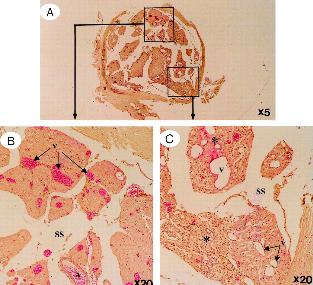
- Fig 5.
Light micrographs in the cauda equina at the site of constriction after 3 weeks (H&E stain).
A, A whole view of cauda equina showing as high intensity on gadolinium-enhanced MR imaging. B, Ventral roots. C, Dorsal roots. Light microscopy revealed congestion and dilation of the radicular veins (B and C, arrows) inside the cauda equina, inflammatory cell infiltration, and Wallerian degeneration (C, asterisks) observed in the entrapped region. This situation reflected breakdown of blood–nerve barrier on fluorescent microscopy and high intensity on gadolinium-enhanced MR imaging.
- Fig 6.
Light micrographs in the cauda equina central (A and B) and peripheral (C and D). to the site of compression after 3 weeks. Proximal to the constriction region, venous dilation and Wallerian degeneration were apparent in the dorsal root, but there was no nerve fiber degeneration in the ventral root (A and B). In contrast, degeneration of nerve fibers was observed in the ventral root distal to the constriction region, a change attributed to arachnoiditis resulting from Wallerian degeneration (C and D). A, artery; DR, dorsal root; SS, subarachnoid space; V, vein; VR, ventral root.
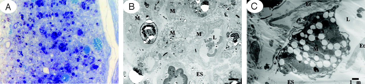
- Fig 7.
Light and electron micrographs in the cauda equina at the site of constriction after 3 weeks.
A, Light microscopy revealed nerve fiber degeneration. (Toluidin blue stain, ×50).
B, Electron microscopy revealed abundance of macrophages phagocytosing the degenerated myelin sheath (×1500).
C, High magnification of a capillary revealed separation of the tight junction to allow communication between the vascular lumen and endoneurial space, indicating breakdown of the blood-nerve barrier and a macrophage capable of passing through the vessel walls (×5000).
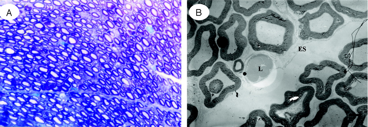
- Fig 8.
Light (A) and electron (B) micrographs in the cauda equina of control group. After 3 weeks, no Wallerian degeneration was evident in the nerve root.
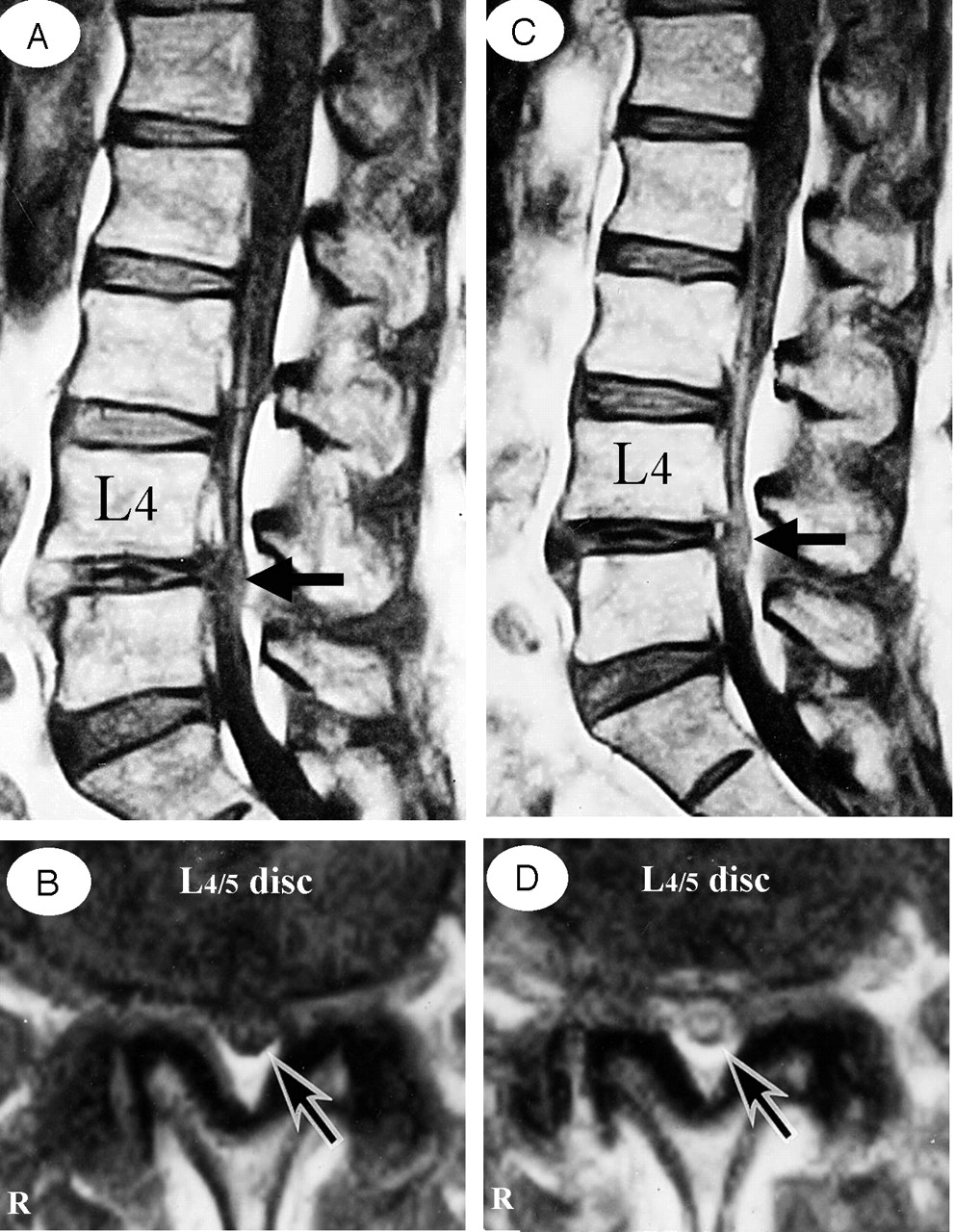
- Fig 9.
A 73-year-old man complained of weakness and numbness of the lower extremities after walking about 300 meters, but no obvious sensory loss and muscle weakness were noted. A and B, Precontrast T1-weighted (500/35) sagittal (A) and axial (B) conventional spin-echo MR images indicated a diagnosis of LSCS at L4/5 disk level (arrows). C and D, T1-weighted (500/35) sagittal (C) and axial (D) MR images acquired at L4/5 disk level obtained after 0.1 mmol/kg intravenous Gd-DTPA administration showing the generalized central canal stenosis as well as punctuate areas of intrathecal enhancement (arrows), which indicates a breakdown in the blood-nerve barrier.


In this issue


Jump to section
Related Articles
Cited By...
- No citing articles found.