
Article Figures & Data
Figures
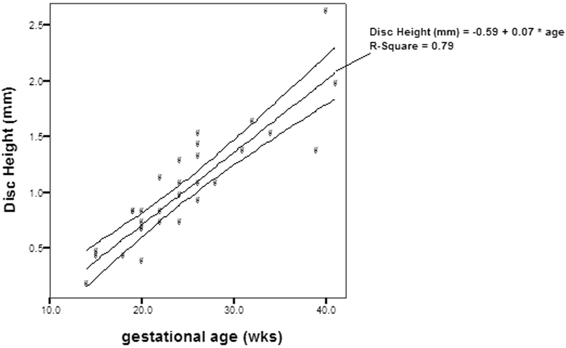
- Fig 1.
Disk height and gestational age, demonstrating significant correlation (P <.01) between the 2 parameters.
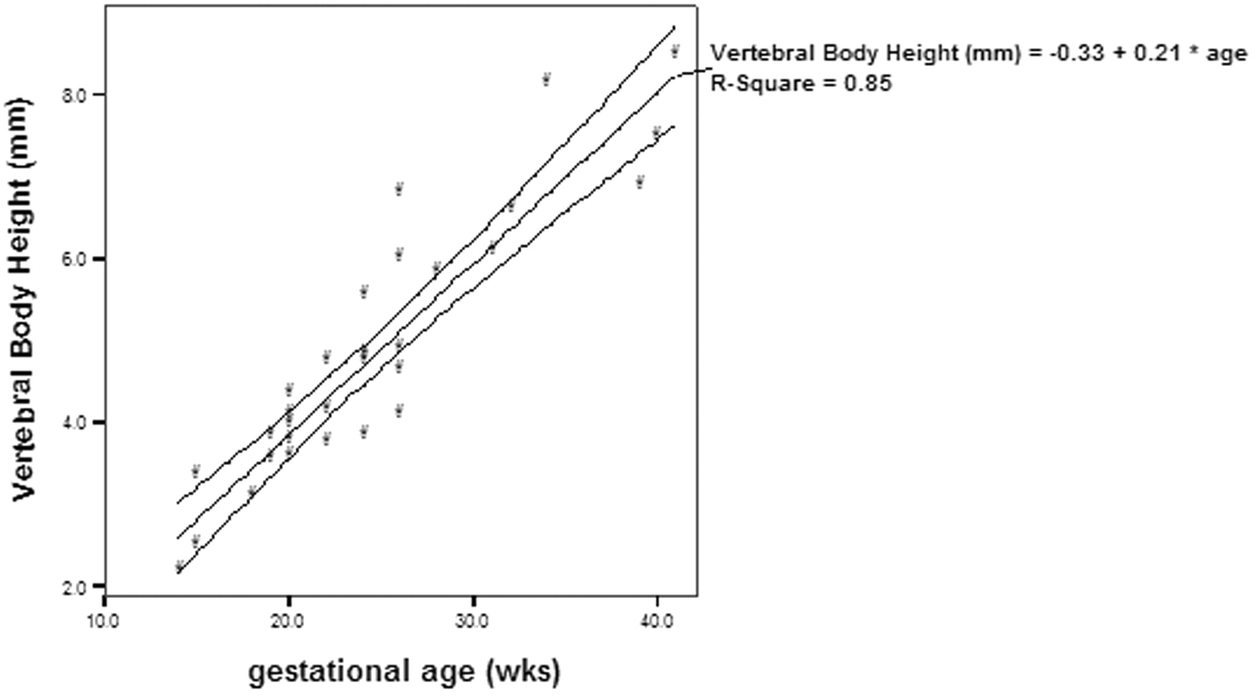
- Fig 2.
Vertebral body height and gestational age, demonstrating significant correlation (P <.01) between the 2 parameters.
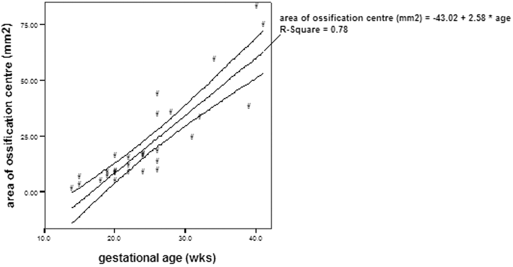
- Fig 3.
Area of ossification center of the vertebral body and gestational age, demonstrating significant correlation (P <.01) between the 2 parameters.
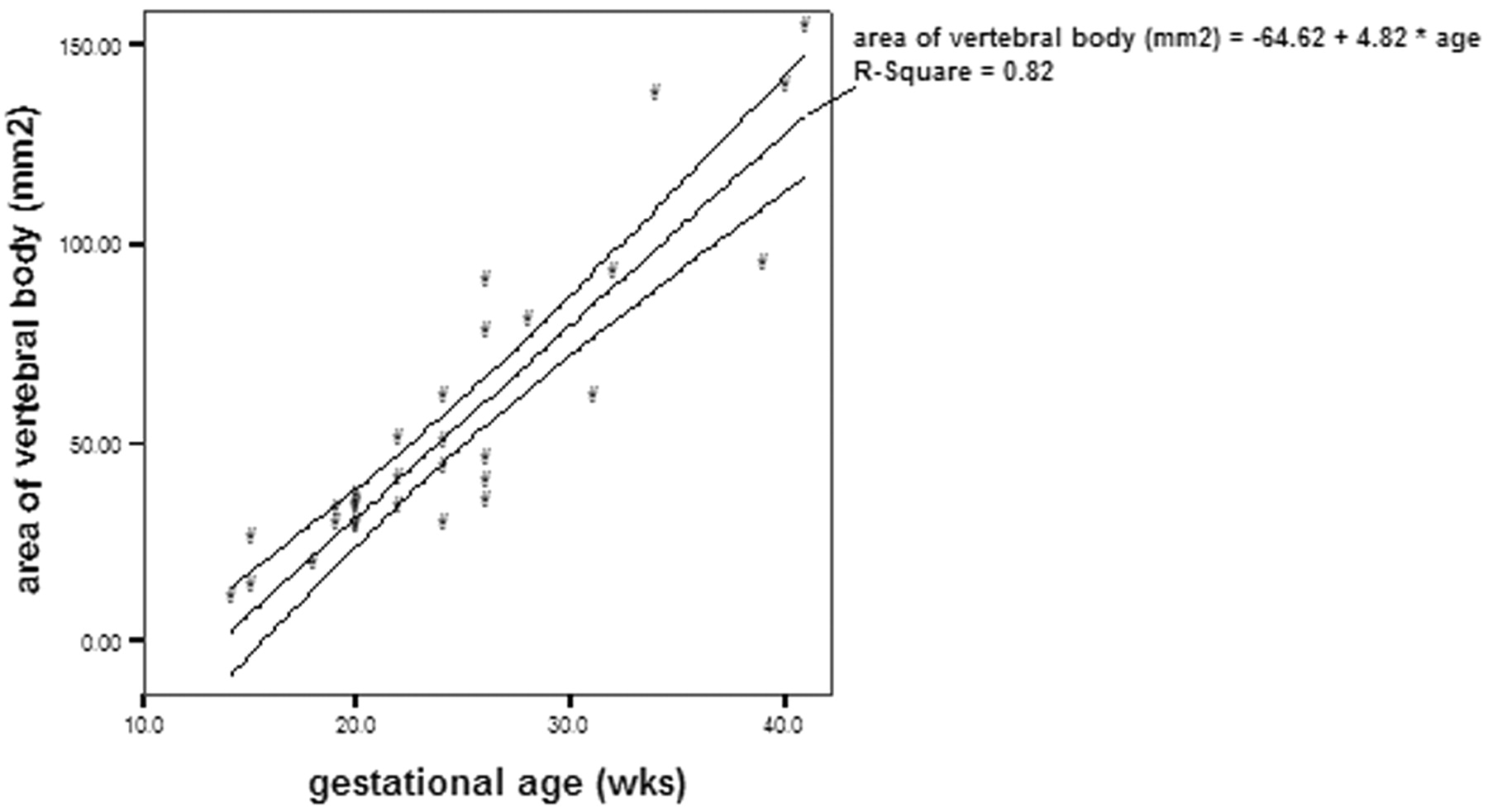
- Fig 4.
Area of the vertebral body taken at midsection of the vertebra and the gestational age, demonstrating significant correlation (P <.01) between the 2 parameters.
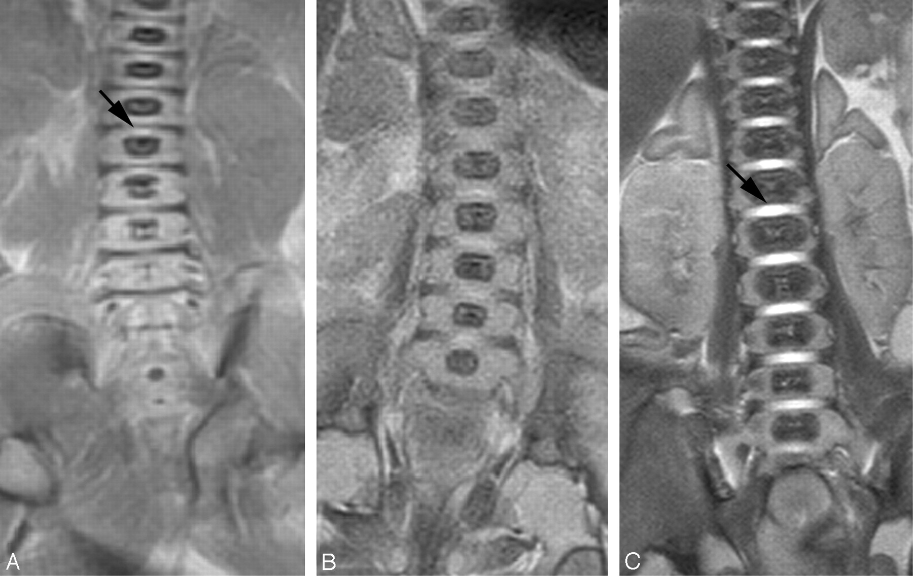
- Fig 5.
Coronal T2 sequence from (A) 18-week, (B) 22-week, and (C) 40-week fetuses. The disk space appears as a linear low-signal-intensity area in the 18-week fetus (small arrow). High signal intensity is seen in the disk space of the 22-week and 40-week fetuses (large arrow). The disk height and vertebral body increase in size with increasing gestational age. The proportion of the vertebral body that is ossified increases with increasing gestational age.
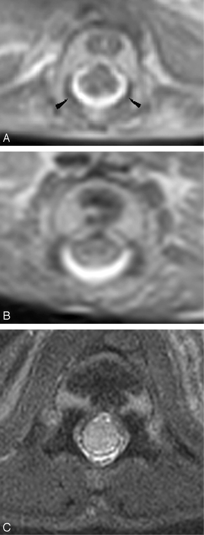
- Fig 6.
Axial T2 of the spine from (A) 18-week, (B) 22-week, and (C) 41-week fetuses. The ossification centers of the posterior elements are first seen at the base of the lamina (arrowhead). With increasing gestation, ossification is seen to proceed posteriorly to the remaining lamina, anteriorly to the pedicle and posterior aspect of the vertebral body, and laterally to the transverse process.
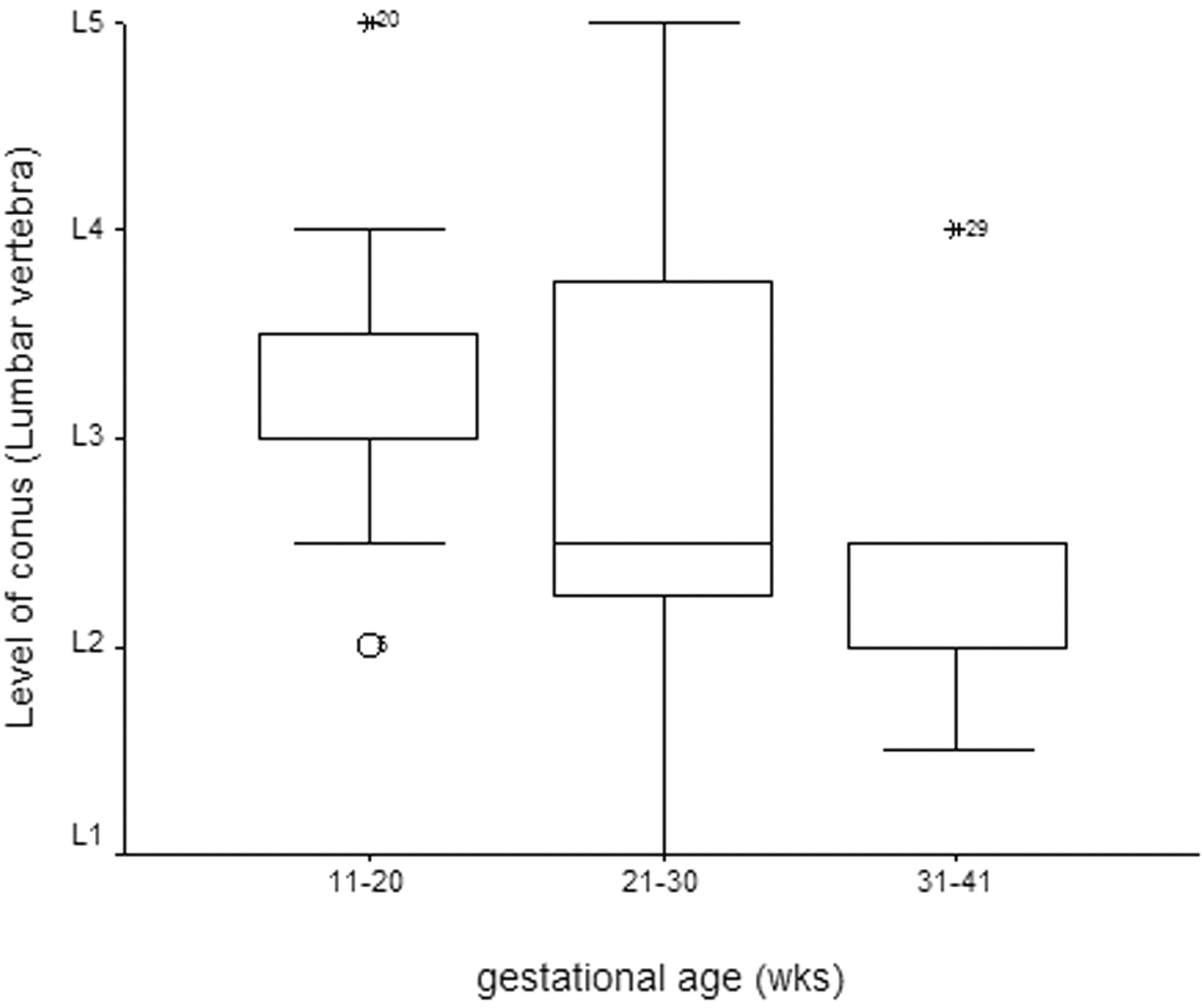
- Fig 7.
The level of the conus at different gestational age.
- Fig 8.
Coronal T2 images from (A) 20-week and (B) 40-week fetuses. The conus lies at L4 level in the 20-week fetus and at L2 level in the 40-week fetus.


In this issue


Jump to section
Related Articles
Cited By...
- No citing articles found.