
Article Figures & Data
Figures
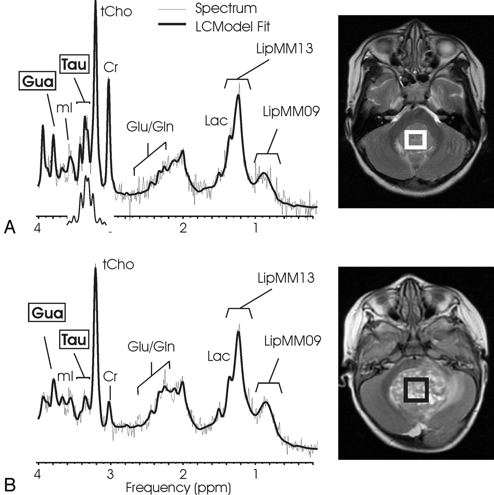
- Fig 1.
1H-MR spectroscopy of medulloblastoma. 1H spectra of a solid-appearing medulloblastoma (A) and of a medulloblastoma with necrotic/cystic areas (B) and corresponding T2-weighted transverse fast spin-echo MR image [repetition time (TR)/echo time (TE), 3500/85 ms; 256 × 192 matrix; 2 signals acquired; echo-train length of 16; acquisition time of 2 minutes 48 seconds] indicating the region of interest. Absolute quantitation included a correction for the different fraction of cystic tissue (6% versus 48%), and spectra are scaled to allow direct comparison of peak areas. A singlet at 3.78 ppm consistent with guanidinoacetate (Gau) is observed, and taurine (Tau) is detected as a complex signal intensity at ≈3.4 ppm. Note the different levels of Tau, creatine (Cr), and glutamate/glutamine (Glu/Gln) signal intensity in the spectra. Spectra also exhibit broad lipid and macromolecule resonances at 0.9 and 1.3 ppm. One peak of the lactate (Lac) doublet at 1.33 ppm is detected as a shoulder of the broad LipMM13 resonance. N-acetylaspartate (NAA) is depleted, and total choline (tCho) is prominent in all spectra of medulloblastoma. The insert in spectrum A shows the spectrum of Tau scaled to the size of the fitted Tau components. Shown are the unfiltered raw data (thin line) and the LCModel fit to the data (thick line).
- Fig 2.
1H MR spectroscopy of pilocytic astrocytoma. Short echo time (TE) spectra of a supratentorial (A) and infratentorial (B) pilocytic astrocytoma and corresponding MR images indicating the regions of interest are shown. Pilocytic astrocytoma spectra are noisy because of the low cellularity of the lesions. Absolute quantitation revealed low creatine (Cr) concentrations. High lactate (Lac) and prominent total choline (tCho) and N-acetylaspartate (NAA) relative to creatine were noted.
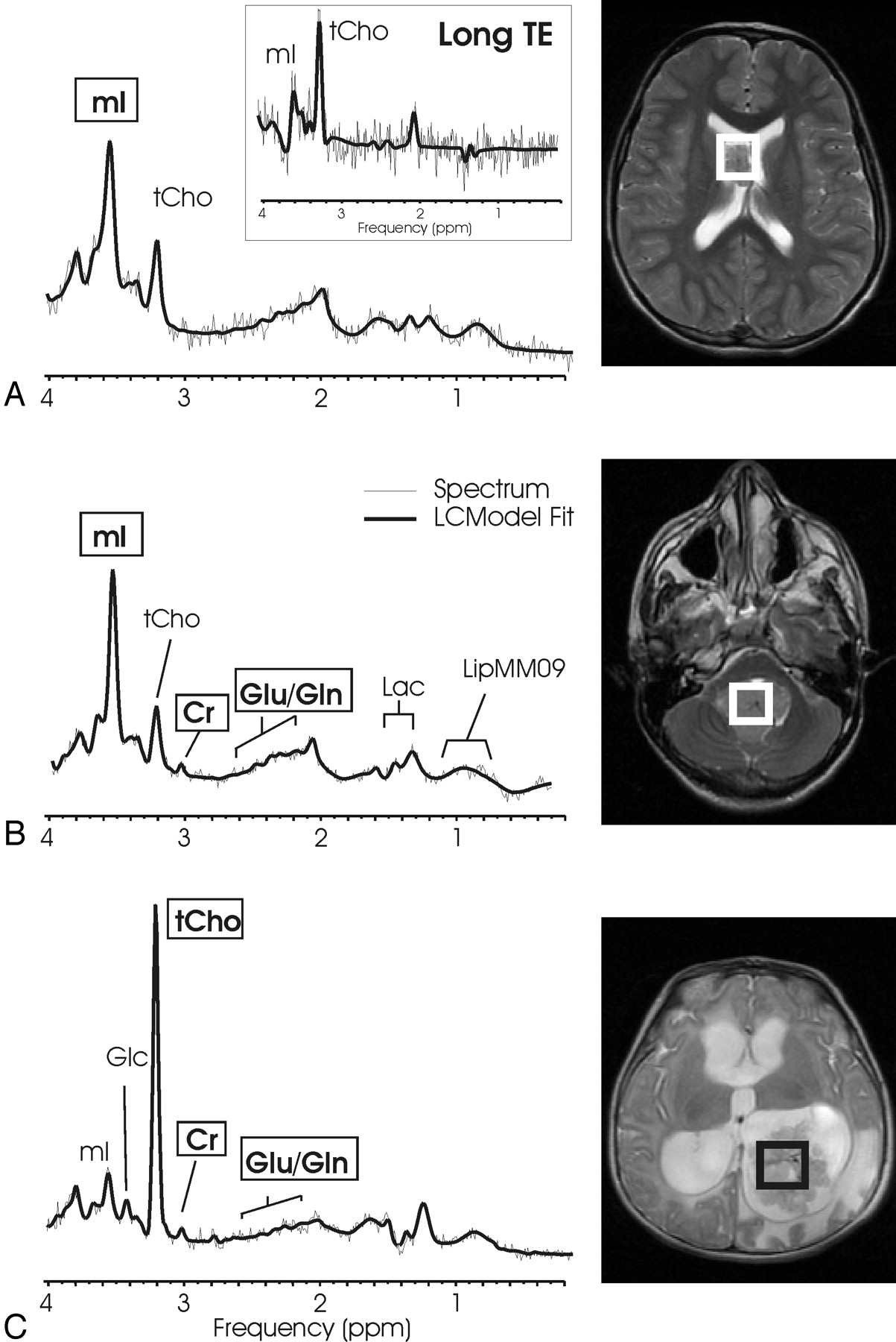
- Fig 3.
Choroid plexus papilloma and choroid plexus carcinoma. Short echo time (TE) MR spectra of a supratentorial (A) and a infratentorial (B) choroid plexus papilloma and of a choroid plexus carcinoma (C) with corresponding MR images indicating the regions of interest. Choroid plexus papilloma shows a prominent myo-inositol peak, whereas creatine (Cr) is hardly detectable. In contrast, more malignant choroid plexus carcinoma shows a prominent choline peak, whereas myo-inositol is not elevated. The insert in spectrum A displays a long TE spectrum obtained from the same region of interest. The long TE spectrum is used to rule out glycine contributing to the mI peak.
- Fig 4.
Low-grade astrocytoma and anaplastic astrocytoma. Short echo time (TE) MR spectra of 2 anaplastic astrocytomas (A, -B) and of low-grade astrocytoma (C) with corresponding MR images indicating the regions of interest are shown. The 3 spectra are scaled according to measured concentrations to allow direct comparison. Astrocytomas appear to be quite heterogeneous. Note the striking difference in total choline (tCho) in spectra A and B, tumors of the same diagnostic name. The prominent scyllo-inositol (sI) observed in spectrum C is not representative for all astrocytoma.
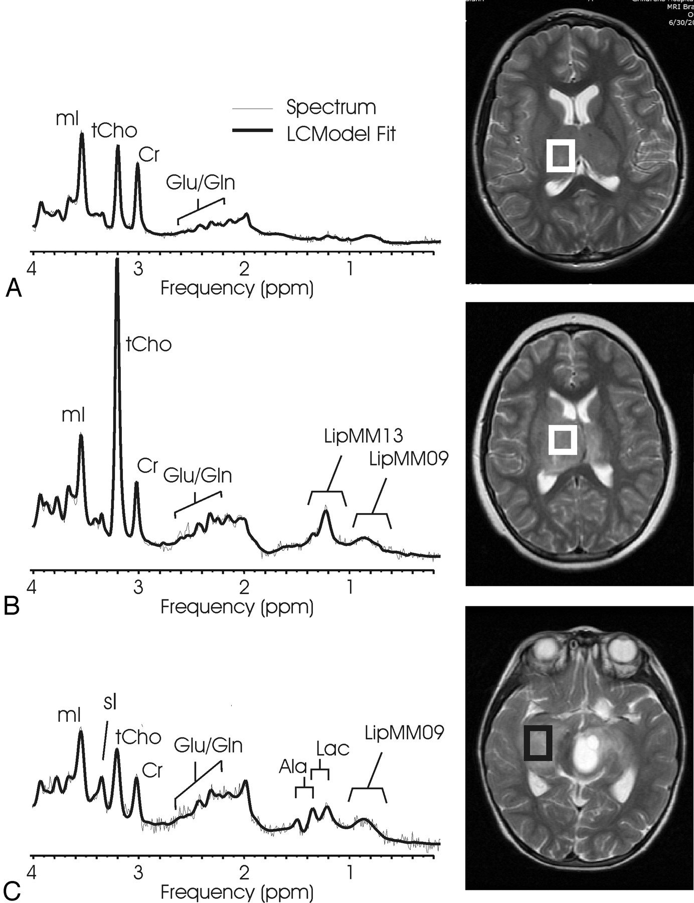
- Fig 5.
Low-grade ependymoma and anaplastic ependymoma. Short echo time (TE) MR spectra of a low-grade ependymoma (A) and of an anaplastic ependymoma (B) with corresponding MR images indicating the regions of interest. The 2 spectra are scaled according to measured concentration to allow direct comparison. A feature of ependymoma is very low N-acetylaspartate (NAA) signal intensity. In particular, no trace of NAA was found in any of the anaplastic ependymoma. Note that glutamate + glutamine (Glx) relative to creatine (Cr) is more prominent in anaplastic ependymoma.
- Fig 6.
Pineal germinoma. Short echo time (TE) MR spectra of a pineal germinoma and MR imaging indicating the region of interest. Taurine (Tau) and guanidinoacetate (Gau) are, as in medulloblastoma, consistently observed in all pineal germinomas.
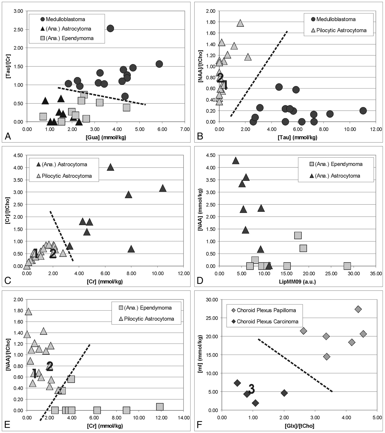
- Fig 7.
Differentiation of common pediatric brain tumors by quantitative 1H-MR spectroscopy. Shown are measured concentrations and concentration ratios of tumors obtained from individual spectra. Labels 1, 2, and 3 in B, C, E, and F indicate data points obtained from individual spectra of the 3 cases presented in more detail in the Results section.


Tables
WHO Grade Subjects S/I No. of Spectra Sex (F/M) Age (mean ± SD) Study subjects 60 27/33 78 25/35 7.22 ± 5.18 PNET (medulloblastoma) IV 14 0/14 16 6/8 6.2 ± 3.6 Anaplastic astrocytoma III 5 5/0 5 2/3 12.4 ± 6.5 Low-grade astrocytoma II 3 2/1 4 2/1 5.2 ± 1.6 Pilocytic astrocytoma I 17 6/11 21 8/9 6.2 ± 4.9 Anaplastic ependymoma III 4 3/1 5 2/2 5.6 ± 3.3 Ependymoma II 5 0/5 8 3/2 4.7 ± 3.4 Choroid plexus papilloma I 3 2/1 6 0/3 9.2 ± 6.5 Choroid plexus carcinoma IV 3 3/0 4 1/2 1.1 ± 1.4 Germinoma (pineal) —* 6 6/0 9 1/5 14.4 ± 2.4 Note:—S indicates supratentorial; I, infratentorial; PNET, primitive neuroectodermal tumor.
* There is no WHO grade for germinoma. Germinomas sometimes come mixed with other primitive germ cell components such as endodermal sinus tumor, choriocarcinoma, immature teratoma, and mature teratoma. All germinoma cases included in this study were pure germinomas and responded well to radiation therapy.
- Table 2:
Absolute concentrations and concentration ratios of untreated pediatric brain tumors
Medulloblastoma Anaplastic Astrocytoma/Astrocytoma Pilocytic Astrocytoma Anaplastic Ependymoma/Ependymoma Choroid Plexus Papilloma Choroid Plexus Carcinoma Pineal Germinoma All Tumors Subjects 14 8 17 9 3 3 6 60 Absolute concentrations (mmol/kg tissue) [NAA] 0.9 ± 0.8 2.3 ± 1.5 1.9 ± 0.8 0.2 ± 0.4**** 2.4 ± 1.3 0.2 ± 0.1***** 1.3 ± 1.1 1.3 ± 1.1 [Cr] 4.7 ± 1.7 6.2 ± 2.4 1.0 ± 0.8***** 5.3 ± 3.1 1.3 ± 0.7* 1.2 ± 0.7* 3.1 ± 0.9 3.5 ± 2.6 [tCho] 5.1 ± 1.5*** 3.9 ± 3.1 2.2 ± 1.1* 3.2 ± 1.5 1.8 ± 0.3*** 5.1 ± 2.1 2.2 ± 0.5* 3.4 ± 2.0 [mI] 7.4 ± 4.0 12.3 ± 5.3 3.8 ± 2.1***** 13.1 ± 5.7 20.4 ± 3.7* 4.1 ± 2.0 5.2 ± 1.1* 8.2 ± 5.8 [Gln] 6.3 ± 3.5 9.51 ± 4.1 6.4 ± 3.2 8.4 ± 3.9 2.6 ± 0.6**** 2.6 ± 1.5 4.4 ± 2.6 6.5 ± 3.7 [Glu] 7.5 ± 4.0 4.8 ± 2.9 3.3 ± 1.8* 2.9 ± 3.2 4.2 ± 2.4 2.7 ± 1.6 8.7 ± 2.7 5.0 ± 3.5 [Glx]† 13.8 ± 5.2 14.4 ± 4.5 9.7 ± 3.5 11.3 ± 4.4 6.8 ± 2.0 5.4 ± 0.9**** 13.1 ± 3.1 11.5 ± 4.7 [Tau] 5.8 ± 2.3**** 1.7 ± 1.1 0.4 ± 0.6***** 1.5 ± 1.1 0.8 ± 0.9 2.5 ± 0.6 3.0 ± 1.3 2.3 ± 2.5 [Glc] 1.0 ± 0.8 2.2 ± 1.1 2.5 ± 1.8 0.8 ± 0.7 1.6 ± 0.8 3.4 ± 2.6 0.9 ± 1.0 1.7 ± 1.5 [Lac] 3.6 ± 1.6 2.8 ± 3.9 4.3 ± 2.2 2.1 ± 2.1 0.6 ± 0.5** 1.5 ± 1.4 0.9 ± 1.6 3.0 ± 2.5 [sI] 0.2 ± 0.2 0.5 ± 0.5 0.2 ± 0.3 0.3 ± 0.3 0.9 ± 0.2 0.0 ± 0.1*** 0.3 ± 0.3 0.3 ± 0.3 [Ala] 2.7 ± 1.2* 0.9 ± 0.9 1.4 ± 0.7 1.7 ± 1.0 1.0 ± 0.7 0.9 ± 1.0 2.0 ± 0.9 1.7 ± 1.1 [Gua] 3.7 ± 1.1* 1.5 ± 0.5**** 2.6 ± 1.0 2.4 ± 1.0 3.0 ± 1.0 3.1 ± 0.4 3.7 ± 1.1 2.8 ± 1.2 LipMM09‡ 16.7 ± 9.7 7.0 ± 2.6** 11.4 ± 5.2 14.5 ± 6.8 4.4 ± 1.6** 12.5 ± 7.5 12.0 ± 5.4 12.3 ± 7.2 LipMM13‡ 64.3 ± 73.2 15.9 ± 13.4* 21.8 ± 16.1 49.6 ± 31.4 8.3 ± 2.4**** 61.8 ± 73.9 63.4 ± 53.9 40.6 ± 47.9 [NAA]/[tCho] 0.2 ± 0.2*** 0.9 ± 0.9 0.9 ± 0.4** 0.1 ± 0.2*** 1.3 ± 0.5 0.1 ± 0.0***** 0.6 ± 0.5 0.6 ± 0.6 [Crl]/[tCho] 1.0 ± 0.4 2.1 ± 1.2 0.5 ± 0.3***** 1.8 ± 0.8 0.7 ± 0.3 0.3 ± 0.2*** 1.5 ± 0.5 1.1 ± 0.8 [mI]/[tCho] 1.5 ± 0.9** 4.0 ± 2.0 1.8 ± 0.7* 4.8 ± 2.4 11.9 ± 3.7 0.9 ± 0.6* 2.5 ± 1.1 3.0 ± 2.8 [Gln]/[tCho] 1.2 ± 0.6**** 2.8 ± 1.0 3.2 ± 1.7 2.9 ± 1.6 1.5 ± 0.5 0.7 ± 0.6 2.0 ± 1.2 2.3 ± 1.5 [Glu]/[tcho] 1.5 ± 0.8 1.8 ± 1.7 1.7 ± 0.9 1.3 ± 1.5 2.3 ± 1.0 0.6 ± 0.3* 4.0 ± 1.1* 1.8 ± 1.4 [Glx]/[tCho] 2.8 ± 0.9*** 4.7 ± 2.2 4.8 ± 1.7 4.2 ± 2.6 3.8 ± 0.5 1.2 ± 0.7* 6.0 ± 0.8** 4.1 ± 2.0 [Tau]/[tCho] 1.2 ± 0.5** 0.3 ± 0.3 0.2 ± 0.4* 0.5 ± 0.3 0.5 ± 0.6 0.6 ± 0.3 1.4 ± 0.6 0.6 ± 0.6 [NAA]/[Cr] 0.2 ± 0.2** 0.4 ± 0.3 1.8 ± 1.2** 0.1 ± 0.1**** 1.8 ± 0.4 0.1 ± 0.1*** 0.4 ± 0.3* 0.7 ± 1.0 [mI]/[Cr] 1.7 ± 0.9* 2.0 ± 0.5* 3.6 ± 1.9 2.8 ± 1.4 16.6 ± 6.6 7.0 ± 7.3 1.8 ± 0.5* 3.5 ± 4.0 [Gln]/[Cr] 1.3 ± 0.7*** 1.8 ± 1.0 5.5 ± 2.5*** 1.7 ±0.7 2.0 ± 1.2 4.1 ± 4.0 1.3 ± 1.0 2.7 ± 2.4 [Glu]/[Cr] 1.9 ± 1.5 0.8 ± 0.5*** 3.5 ± 2.5 0.8 ± 1.0* 3.1 ± 0.4 2.7 ± 1.0 2.9 ± 0.9 2.2 ± 1.8 [Glx]/[Cr] 3.3 ± 1.6 2.6 ± 1.1* 9.1 ± 4.6** 2.5 ± 1.4* 5.1 ± 1.1 7.0 ± 4.8 4.2 ± 1.1 4.9 ± 3.8 [Tau]/[Cr] 1.3 ± 0.4* 0.2 ± 0.3* 0.3 ± 0.4* 0.3 ± 0.3 0.5 ± 0.9 3.6 ± 2.8 1.2 ± 0.8 0.8 ± 1.1 [Gau]/[Cr] 0.9 ± 0.4 0.3 ± 0.1**** 2.7 ± 2.3 0.6 ± 0.3* 2.4 ± 0.9 5.1 ± 4.3 1.4 ± 0.5 1.6 ± 1.9 [Glc]/[Cr] 0.2 ± 0.2* 0.4 ± 0.2 2.4 ± 1.5 0.2 ± 0.2* 1.5 ± 1.3 6.5 ± 7.9 0.3 ± 0.3 1.2 ± 2.3 Note:—
* P < .01,
** P < .001,
*** P < .0001,
**** P < .00001,
***** P< .000001 versus All Other tumors
† [Glx] = [Glu] + [Gln].
‡ Absolute intensity (arbitrary units).
Ana. Astrocytoma/Astrocytoma Pilocytic Astrocytoma Ana. Ependymoma/Ependymoma Choroid PlexusPapilloma Choroid PlexusCarcioma Pineal Germinoma Medulloblastoma ↑[Tau]/[Cr]***** ↑[Tau]**** ↑[Tau]/[Cr]**** ↑[tCho]**** ↑[Glx]*** ↑[tCho]**** ↑[Tau]**** ↓[NAA]/[tCho]**** ↑[Tau]**** [↑[Lac]** ↑[Cr]** ↓[Glx]/[tCho]**** ↑[Gua]**** ↑[Tau]/[Cr]**** ↑[Tau]/[tCho]** ↑[Tau]** ↑[Tau]** ↑[Tau]* Ana. Astro./Astrocytoma ↑[Gln]/[Cr]*** ↑[NAA]* ↑[Cr]** ↑[Cr]** ↓[Glu]/[Cr]* ↑[Cr]/[tCho]*** ↓[Gua]/[Cr]* ↓[Glu]/[Cr]** ↑[Glx]** ↓[Gua]/[Cr]* ↑[Cr]** ↓LipMM09* ↓[Gln]* ↓[Gua]* ↓[Gua]* Pilocytic Astrocytoma [NAA]/[tCho]***** ↑[Lac]*** ↑[NAA]/[tCho]***** ↑[Gln]/[Cr]*** ↑[NAA]***** ↑[Gln]** ↑[NAA]***** ↑[Glc]/[Cr]*** ↑[Gln]/[Cr]*** ↑LipMM09** ↑[Glx]** ↑[tCho]/[Cr]** Ana. Epend./Ependymoma ↓[Glu]/[Cr]** ↑[Cr]/[tCho]** ↑[Glu//[tCho]* ↑[Gln]* ↑[mI]* ↓[Glu]/[Cr]* ↑LipMM09* ↑[Glx]* ↑[mI]* Choroid ↑[mI]* ↓[Glx]/[tCho]* Plexus ↑[tCho]* ↓[Glx]* Papilloma ↑[NAA]/[Cr]* ↑[NAA]/[Cr]* Choroid Plexus Carcinoma ↓[Glu]/[tCho]** ↓[Glx]** ↓[Cr]/[tCho]* * P < .01,
** P < .001,
*** P < .0001,
**** P < .00001,
***** P < .000001.
In this issue


Jump to section
Related Articles
Cited By...
- Pediatric Atypical Teratoid/Rhabdoid Tumors of the Brain: Identification of Metabolic Subgroups Using In Vivo 1H-MR Spectroscopy
- What is MR spectroscopy?
- Multimodality Brain Tumor Imaging: MR Imaging, PET, and PET/MR Imaging
- Comparison of Perfusion, Diffusion, and MR Spectroscopy between Low-Grade Enhancing Pilocytic Astrocytomas and High-Grade Astrocytomas