
Article Figures & Data
Figures
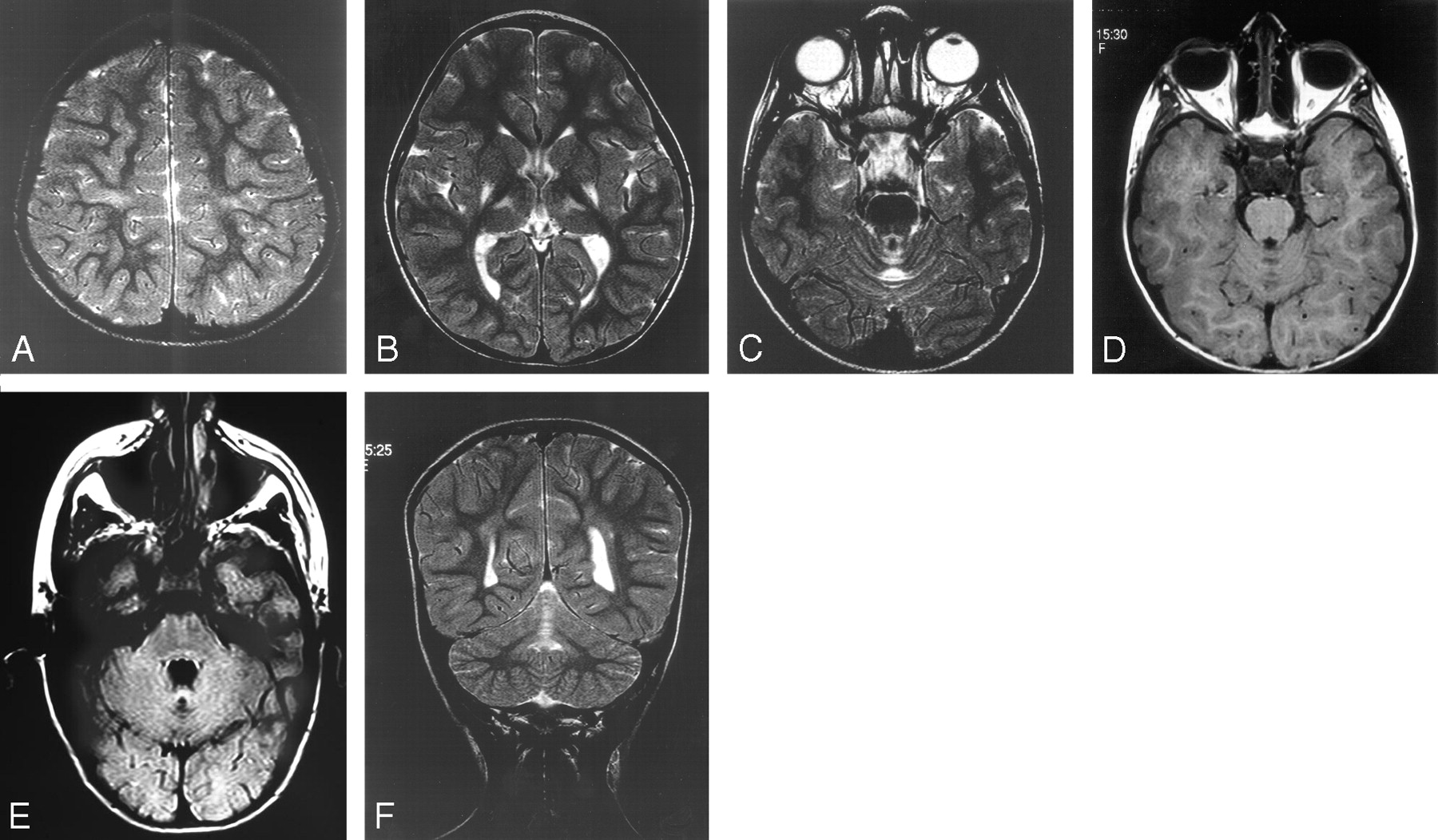
- Fig 1.
Five-year-old girl with athetoid spastic quadriplegia.
A, Axial T2. Mild signal intensity change in the perirolandic white matter.
B, Axial T2. Typical signal intensity change in the posterior putamen and the ventrolateral thalamus bilaterally.
C, Axial T2. Typical signal intensity change in the anterior lobe of the vermis.
D, Axial T1. Low T1 signal intensity change in the vermis.
E, Axial FLAIR. High T2 signal intensity and cystic change.
F, Coronal T2. Anterior lobe of vermis high T2 signal intensity change.
- Fig 2.
Nine-month-old girl with dyskinetic cerebral palsy.
A, Coronal T2. Central lobule of anterior lobe of the vermis high T2 signal intensity.
B, Axial FLAIR. High T2 signal intensity and cystic change of anterior lobe of vermis.
C, Sagittal T2. Central lobule of anterior lobe of vermis high T2 signal intensity.
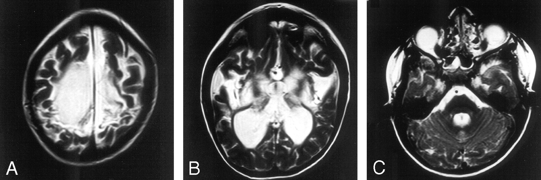
- Fig 3.
Seven-year-old boy with severe spastic quadriplegia.
A, Axial T2. Severe signal intensity change and volume loss in the perirolandic white matter.
B, Axial T2. Severe putaminal and thalamic volume loss and high T2 signal intensity bilaterally.
C, Axial T2. Mild vermian high T2 signal intensity change.


Tables
Patient clinical information and scores for brain damage secondary to hypoxic ischemic encephalopathy
Case Age (years) Apgar Heart Rate (bpm) Putamen Thalamus PCWM Total Score* Vermis Others 1 minute 5 minutes 1 4 6 6 >100 2 2 1 5 0 0 2 2 1 3 80 3 3 2 8 1 Hippocampus, caudate 3 3 0 2 70 2 3 2 7 1 0 4 15 0 1 UR 1 3 2 6 1 0 5 4 1 7 60 3 3 3 9 1 Hippocampus 6 1 1 2 60 2 1 1 4 1 Hippocampus 7 1 1 5 <20 3 2 2 7 1 Hippocampus 8 14 1 2 UR 2 2 2 6 1 Hippocampus 9 6 0 3 UR 2 1 1 4 0 0 10 7 6 9 2 1 2 5 0 0 11 5 2 1 3 2 6 1 Caudate 12 5 3 5 1 2 1 4 1 Hippocampus 13 5 0 4 UR 3 3 2 8 1 0 14 12 1 6 1 0 3 4 0 0 15 3 1 5 40–60 2 2 1 5 1 Hippocampus 16 24 1 5 2 2 0 4 1 0 17 1 3 5 3 0 2 5 0 Caudate 18 5 1 0 3 3 3 9 1 0 19 1 0 0 UR 2 2 1 5 0 0 20 2 UR 2 2 0 4 1 Hippocampus 21 2 2 3 70 2 3 2 7 0 0 22 2 5 6 80 3 3 3 9 1 0 23 1 1 1 60–80 2 2 2 6 1 Hippocampus 24 4 4 8 70 0 0 2 2 0 Caudate 25 2 4 6 60 2 1 3 6 0 Caudate 26 22 0 2 UR 1 2 0 3 1 0 27 15 6 7 UR 2 2 1 5 0 0 28 4 6 8 2 2 2 6 1 Hippocampus 29 7 1 4 60 2 2 0 4 0 0 30 5 2 7 <80 3 2 3 8 0 0 Note:—bpm indicates beats per minute; PCWM, paracentral white matter; UR, unrecordable.
* Total score of the abnormalities seen in putamen, thalamus, and paracentral white matter.
In this issue


Jump to section
Related Articles
Cited By...
- Injury to the Cerebellum in Term Asphyxiated Newborns Treated with Hypothermia
- Low Cerebellar Vermis Volumes and Impaired Neuropsychologic Performance in Children Treated for Brain Tumors and Leukemia
- Anatomic Localization of Dyskinesia in Children with "Profound" Perinatal Hypoxic-Ischemic Injury
- Injury to the Developing Cerebellum: Mechanisms and Consequences