
Article Figures & Data
Figures
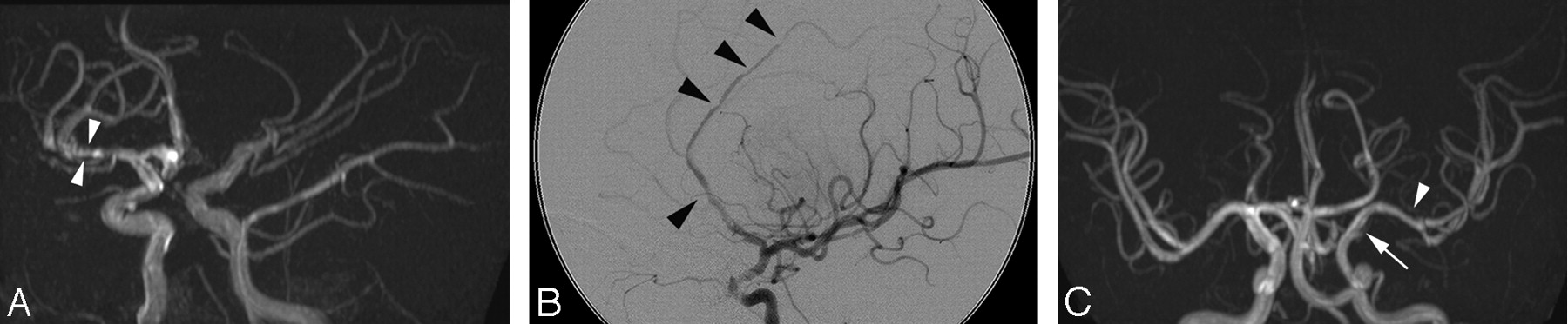
- Fig 1.
Representative MRA and angiographic images. Aggressive appearing lesions were those that demonstrated multiple short segment irregularity with alternating narrowing and dilation (beading) (white arrow) (A) or multiple longer segmental narrowing with normal intervening vessel (black arrowheads) (B). Aneurysms (not shown) were included in this definition. Benign appearing lesions (C) had smooth (white arrows), often solitary, tapered (white arrowhead) narrowing that could be concentric or eccentric. Incidental hypoplasia of the ipsilateral A1 is noted.
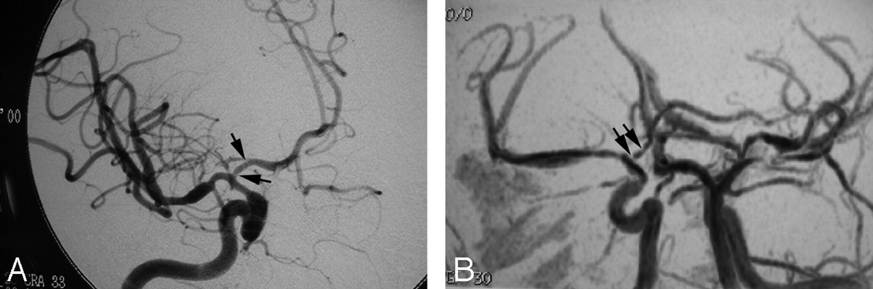
- Fig 2.
Good agreement of CA (A) and (B) MRA for right ICA. Both modalities demonstrate tapered narrowing of the terminal carotid and proximal M1 and A1 (carotid terminus) with focal midM1 dilation and distal narrowing. Both modalities identify beading of the proximal A1 (black arrows).
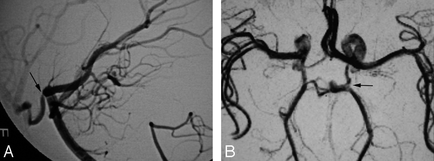
- Fig 3.
Abnormal CA (A) in the context of normal MRA (B) in a patient with a lone focal stenosis of the left PcomA. No other CA abnormality was present. MR imaging was abnormal maintaining suspicion for vasculitis despite a negative MRA.
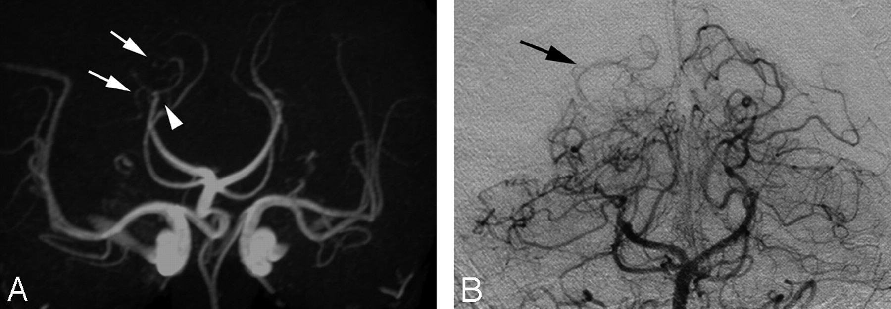
- Fig 4.
A, Occlusion of the P3 segment of the right PCA on TOF MRA (white arrowhead). Inferior temporal branches (white arrows) are slightly more prominent than on the contralateral side but B, extent of collaterals and reconstitution of the distal PCA (black arrow) best seen on CA.


Tables
MRA Patients (%) CA Patients (%) P Value Unilateral lesions* 19 (79.2) 19 (90.5) 1.0 Bilateral lesions 5 (20.8) 2 (9.5) .42 Any proximal lesion† regardless of whether distal lesion 22 (87.5) 18 (85.7) .28 Distal lesion only 2 (8.3) 3 (14.3) .18 Anterior circulation only‡ 19 (79.2) 15 (71.4) .36 Posterior circulation only 2 (8.3) 3 (14.3) 1.00 Anterior and posterior 3 (12.5) 3 (14.3) 1.00 Ipsilateral ≥ 2 lesions§ 16 (66.7) 16 (76.2) 1.00 Total lesions ≥ 5 3 (12.5) 4 (19.0) .29 Benign appearance 19 (79.2) 16 (76.2) .50 Aggressive appearance 5 (20.8) 5 (23.8) 1.00 Note:— MRA indicates MR angiography; CA, conventional angiography. Twenty-one CA and 24 MRA studies were abnormal and compared in 25 patients.
* Unilateral vs bilateral, P <0.05.
† Proximal vs distal, P = .08.
‡ Anterior vs posterior, P = .06.
§ Multifocal vs unifocal, P = .09.
Lesions MRA (n = 62) CA (n = 64) P Value ICA 11 (17.7%) 11 (17.5%) .94 ACA 11 (17.7%) 17 (27.0%) .23 MCA 34 (54.8%) 26 (41.3%) .11 M1 24 16 .10 M2 10 9 .75 M4 0 1 .32 PCA 5 (8.1%) 9 (14.3%) .28 P1 2 2 .97 P2 3 7 .21 Vertebrobasilar 0 0 Anterior choroidal 0 0 PcomA 1 (1.6%) 1 (1.6%) .98 Note:— MRA indicates MR angiography; CA, conventional angiography; ICA, internal carotid artery; ACA, anterior cerebral artery; MCA, middle cerebral artery; PCA, posterior cerebral artery; PcomA, posterior communicating artery.
Morphology MRA (n = 56) CA (n = 54) P Value Smooth 45 (80.4%) 45 (83.3%) .80 Irregular 11 (19.6%) 9 (16.7%) .80 Concentric 54 (96.4%) 50 (92.6%) .57 Eccentric 2 (3.6%) 4 (7.4%) .57 Graduated 11 (19.6%) 11 (20.4%) .95 Single 52 (92.9%) 48 (88.9%) .64 Beading 6 (10.7%) 6 (11.1%) .97 Multiple 2 (3.6%) 6 (11.1%) .33 Note:— CA indicates conventional angiography; MRA, MR angiography. Data exclude occlusions and aneurysms.
Location Number (n = 19) A1 4 (21.1%) Cavernous carotid 3 (15.8%) M1 2 (10.5%) M2 4 (21.1%) M4 1 (5.3%) P1 1 (5.3%) P2 1 (5.3%) PcomA 1 (5.3%) Pericallosal 2 (10.5%) Note:— MRA indicates MR angiography; PcomA, posterior communicating artery.
CA Stenosis Number of Lesions on CA MRA Agreement Lesions MRA Falsely Upgraded Lesions MRA Falsely Downgraded Lesions MRA False-Negatives <50% 20 4 (20%) 7 (35%) 9 (45%) 50–<75% 25 12 (48%) 6 (24%) 1 (4%) 6 (24%) 75–99% 9 7 (78%) 1 (11%) 1 (11%) 100% 8 1 (13%) 4 (50%) 3 (38%) Note:— MRA indicates MR angiography; CA, conventional angiography. Total MRA agreement is 43 lesions, excluding 2 aneurysms. Number of lesions correctly graded or up- and downgraded by MRA when compared with CA (n = 62 CA lesions; excludes 2 aneurysms)


