Advertisement

Main menu

  • Home
  • Content
    • Current Issue
    • Accepted Manuscripts
    • Article Preview
    • Past Issue Archive
    • AJNR Case Collection
    • Case of the Week Archive
    • Classic Case Archive
    • Case of the Month Archive
  • Special Collections
    • Spinal CSF Leak Articles (Jan 2020-June 2024)
    • 2024 AJNR Journal Awards
    • Most Impactful AJNR Articles
  • Multimedia
    • AJNR Podcast
    • AJNR Scantastics
    • Video Articles
  • For Authors
    • Submit a Manuscript
    • Author Policies
    • Fast publishing of Accepted Manuscripts
    • Graphical Abstract Preparation
    • Manuscript Submission Guidelines
    • Imaging Protocol Submission
    • Submit a Case for the Case Collection
  • About Us
    • About AJNR
    • Editorial Board
  • More
    • Become a Reviewer/Academy of Reviewers
    • Subscribers
    • Permissions
    • Alerts
    • Feedback
    • Advertisers
    • ASNR Home
  • Other Publications
    • ajnr

User menu

  • Alerts
  • Log in

Search

  • Advanced search
American Journal of Neuroradiology

American Journal of Neuroradiology

ASHNR American Society of Functional Neuroradiology ASHNR American Society of Pediatric Neuroradiology ASSR
  • Alerts
  • Log in

Advanced Search

  • Home
  • Content
    • Current Issue
    • Accepted Manuscripts
    • Article Preview
    • Past Issue Archive
    • AJNR Case Collection
    • Case of the Week Archive
    • Classic Case Archive
    • Case of the Month Archive
  • Special Collections
    • Spinal CSF Leak Articles (Jan 2020-June 2024)
    • 2024 AJNR Journal Awards
    • Most Impactful AJNR Articles
  • Multimedia
    • AJNR Podcast
    • AJNR Scantastics
    • Video Articles
  • For Authors
    • Submit a Manuscript
    • Author Policies
    • Fast publishing of Accepted Manuscripts
    • Graphical Abstract Preparation
    • Manuscript Submission Guidelines
    • Imaging Protocol Submission
    • Submit a Case for the Case Collection
  • About Us
    • About AJNR
    • Editorial Board
  • More
    • Become a Reviewer/Academy of Reviewers
    • Subscribers
    • Permissions
    • Alerts
    • Feedback
    • Advertisers
    • ASNR Home
  • Follow AJNR on Twitter
  • Visit AJNR on Facebook
  • Follow AJNR on Instagram
  • Join AJNR on LinkedIn
  • RSS Feeds

Welcome to the new AJNR, Updated Hall of Fame, and more. Read the full announcements.


AJNR is seeking candidates for the position of Associate Section Editor, AJNR Case Collection. Read the full announcement.


Getting new auth cookie, if you see this message a lot, tell someone!
OtherHEAD & NECK

Cervical Vertebral Artery Variations: An Anatomic Study

S.R. Satti, C.A. Cerniglia and R.A. Koenigsberg
American Journal of Neuroradiology May 2007, 28 (5) 976-980;
S.R. Satti
  • Find this author on Google Scholar
  • Find this author on PubMed
  • Search for this author on this site
C.A. Cerniglia
  • Find this author on Google Scholar
  • Find this author on PubMed
  • Search for this author on this site
R.A. Koenigsberg
  • Find this author on Google Scholar
  • Find this author on PubMed
  • Search for this author on this site
  • Article
  • Figures & Data
  • Info & Metrics
  • Responses
  • References
  • PDF
Loading

Article Figures & Data

Figures

  • Fig 1.
    • Download figure
    • Open in new tab
    • Download powerpoint
    Fig 1.

    Digital subtraction angiography. There is opacification of the left vertebral artery (LV) as the 2nd branch off the left subclavian artery (LS, arrow). The 1st branch of the left subclavian artery is the thyroid, and the 3rd branch is the cervical artery (CA).
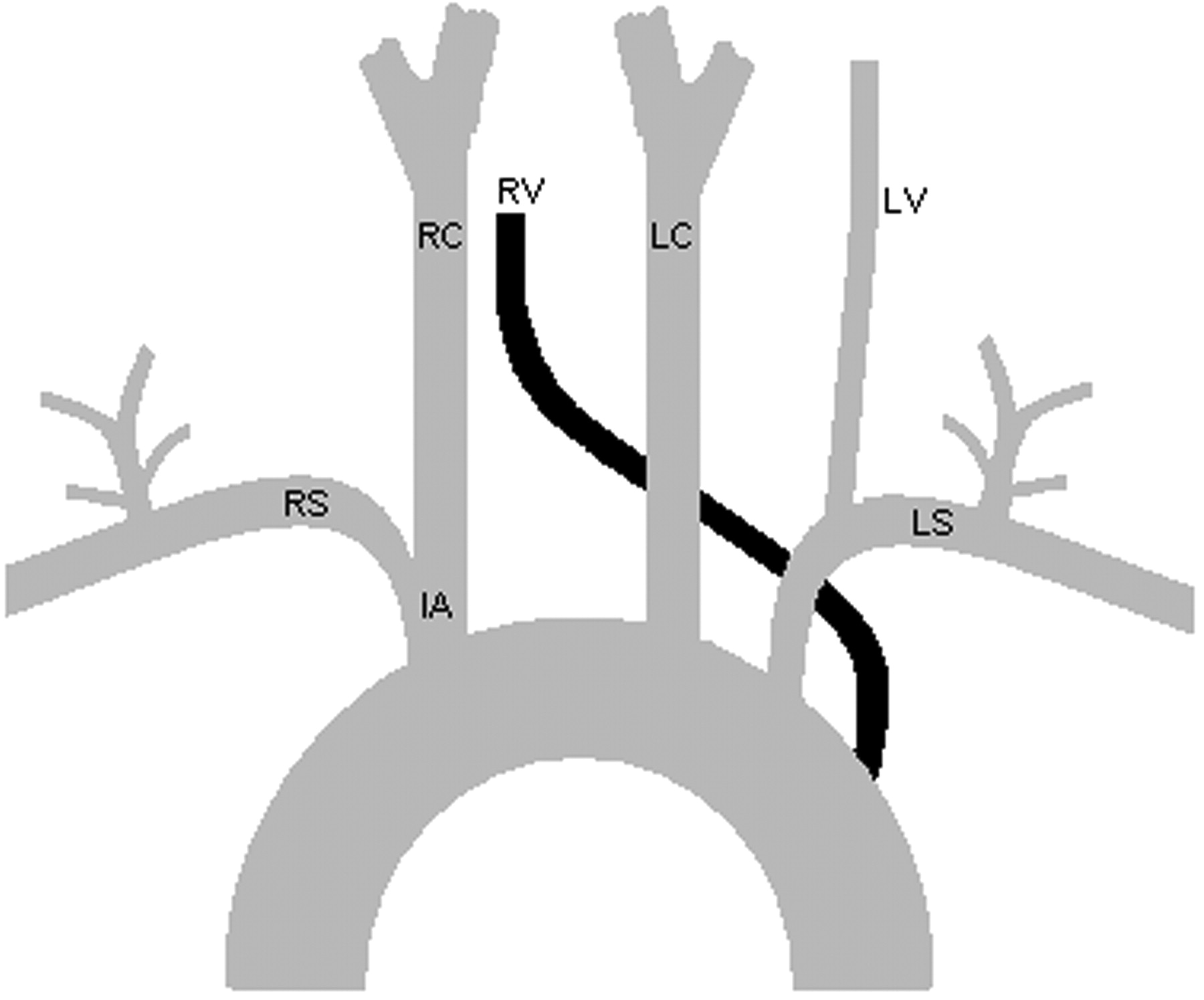
  • Fig 2.
    • Download figure
    • Open in new tab
    • Download powerpoint
    Fig 2.

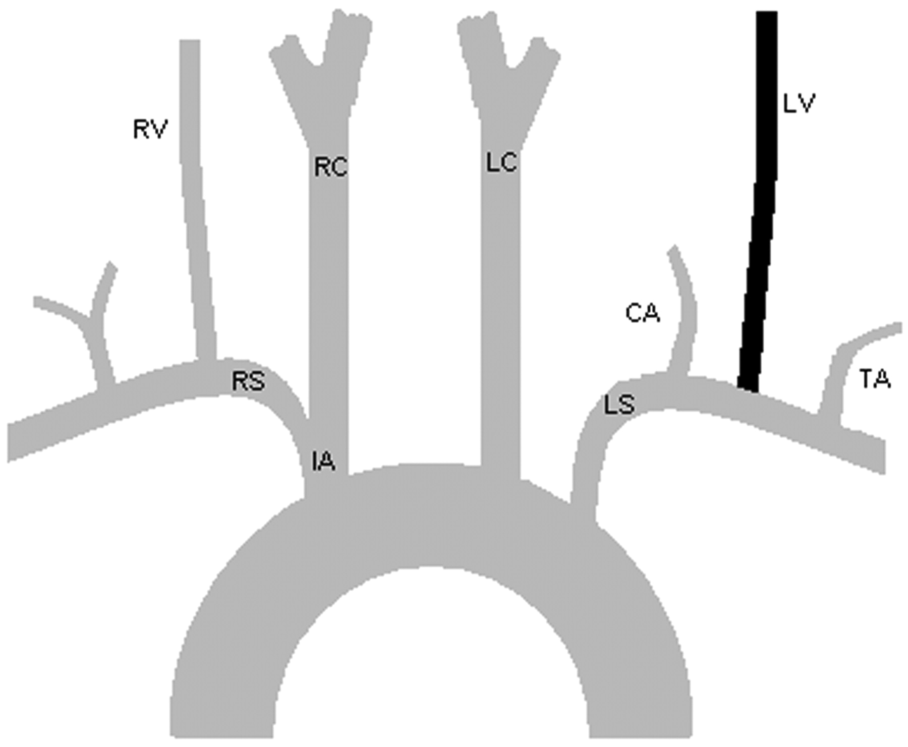
    LV as the 2nd branch off the LS between the TA (thyroid artery) and the CA. RV indicates the right vertebral artery; RS, right subclavian artery; IA, innominate artery; RC, right common carotid artery; LC, left common carotid artery; TA, inferior thyroid artery.

  • Fig 3.
    • Download figure
    • Open in new tab
    • Download powerpoint
    Fig 3.

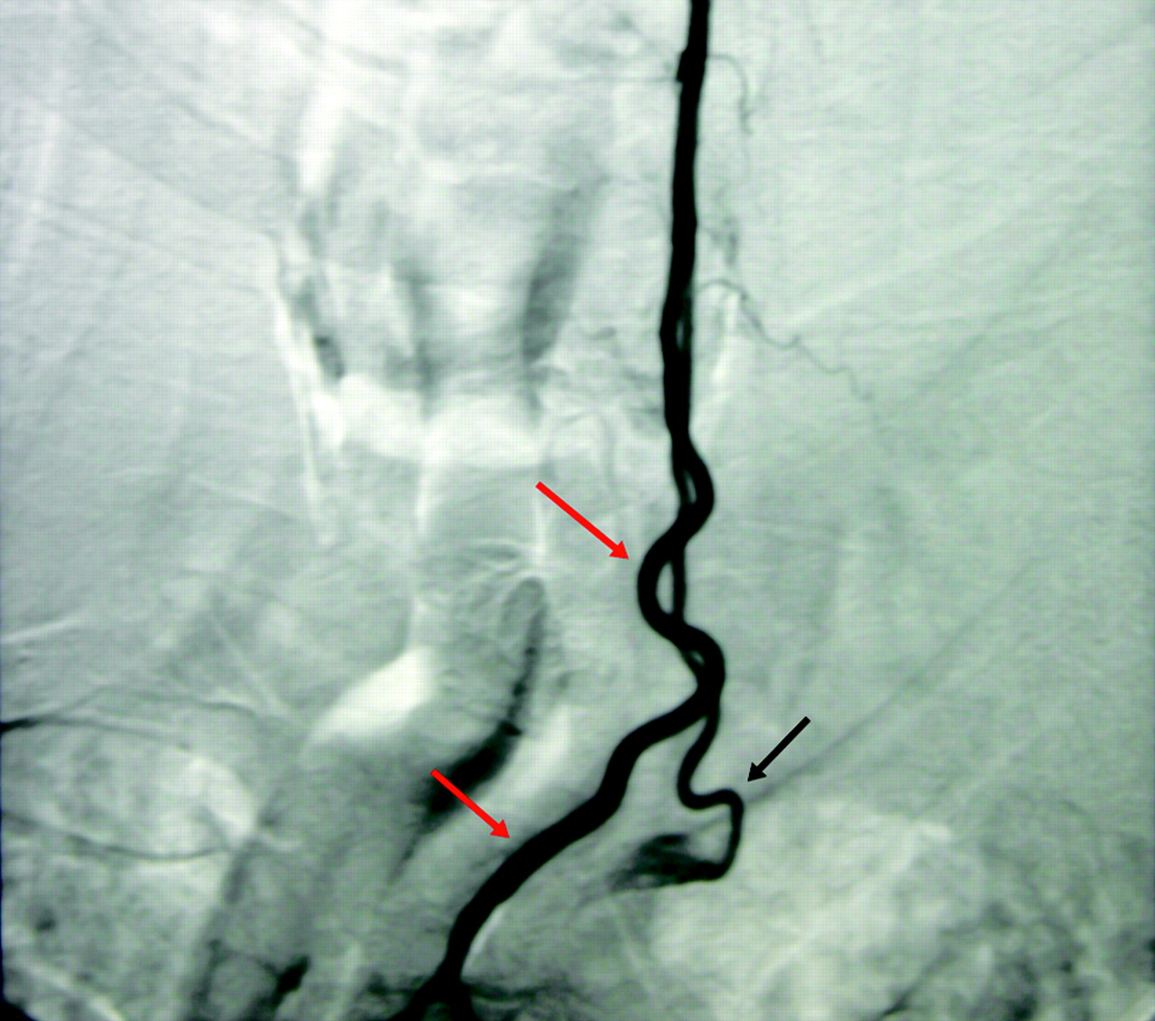
    Digital subtraction angiography. There is opacification of the LV as the 3rd branch off the aortic arch (red arrows) with retrograde flow into a hypoplastic duplicated limb (black arrow), which arose as the 2nd branch of the LS.
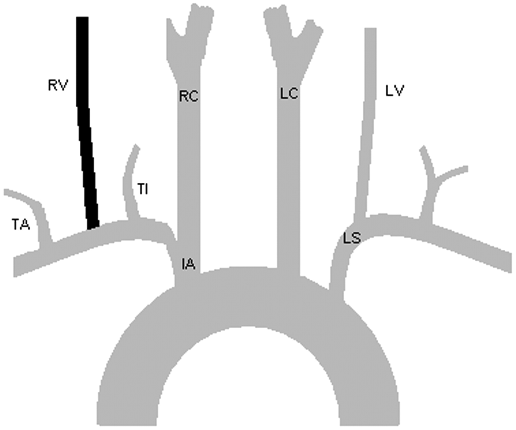
  • Fig 4.
    • Download figure
    • Open in new tab
    • Download powerpoint
    Fig 4.

    LV has 2 origins. The 1st originates as the 3rd branch of the aortic arch, and the 2nd, as the 1st branch of the LS. There is an incidentally noted bovine arch. RTC indicates right thyrocervical trunk; LTC, left thyrocervical trunk.
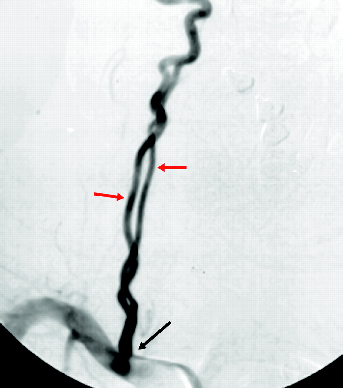
  • Fig 5.
    • Download figure
    • Open in new tab
    • Download powerpoint
    Fig 5.

    Digital subtraction angiography. Aortogram with the pigtail catheter placed in the proximal descending aorta demonstrates filling of right vertebral artery (red arrows) as the last great vessel off of the arch. The right vertebral artery crosses the midline and extends cranially along the right lateral aspect of the neck.
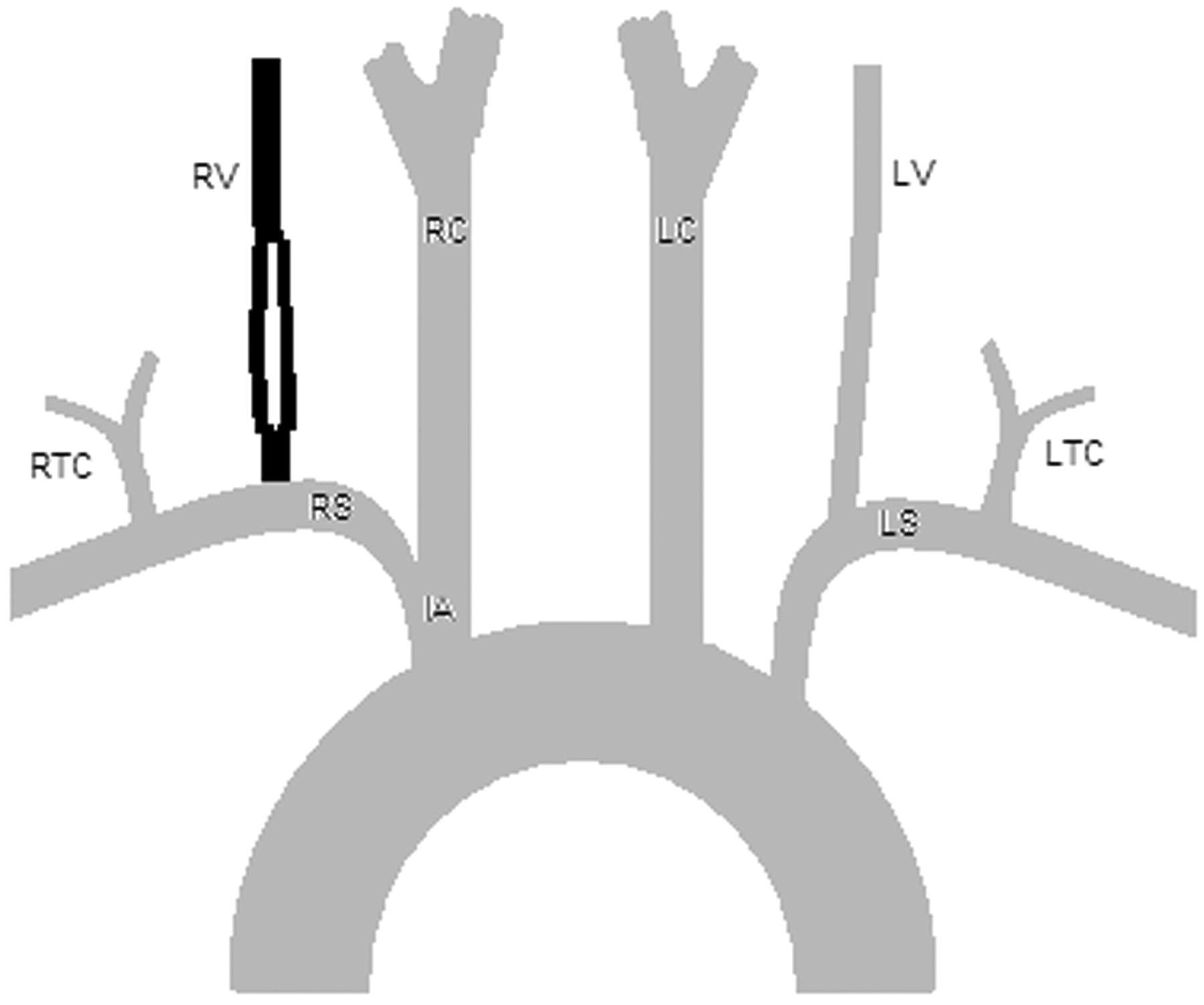
  • Fig 6.
    • Download figure
    • Open in new tab
    • Download powerpoint
    Fig 6.

    RV arises as the last branch off of the aorta. RS indicates right subclavian artery; RC, right common carotid artery.

  • Fig 7.
    • Download figure
    • Open in new tab
    • Download powerpoint
    Fig 7.

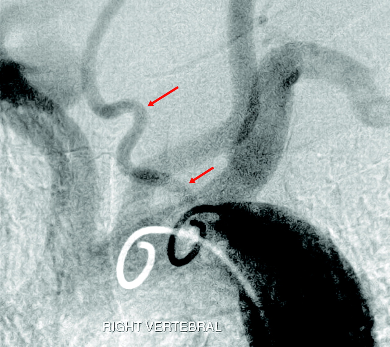
    Digital subtraction angiography. There is opacification of the RV (red arrows) as the 2nd branch off the RS. The 1st branch of the RS is the thyroidea ima artery (TI) (black arrowheads), and the 3rd branch is the TA.

  • Fig 8.
    • Download figure
    • Open in new tab
    • Download powerpoint
    Fig 8.

    RV is the 2nd branch off the RS between the TI and the TA.

  • Fig 9.
    • Download figure
    • Open in new tab
    • Download powerpoint
    Fig 9.

    Digital subtraction angiography. There is opacification of a duplicated RV (red arrows). The origin (black arrow) is at the normal position as the 2nd branch of the RS.

  • Fig 10.
    • Download figure
    • Open in new tab
    • Download powerpoint
    Fig 10.

    The RV is partially duplicated with a common origin as the 2nd branch off the RS. LTC indicates left thyrocervical trunk.
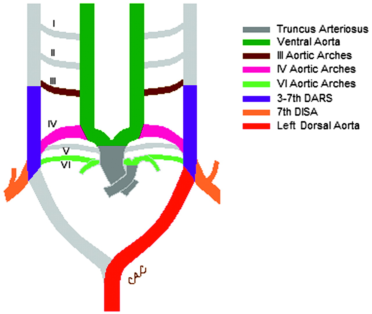
  • Fig 11.
    • Download figure
    • Open in new tab
    • Download powerpoint
    Fig 11.

    Normal schematic diagram of the primitive ventral and dorsal aorta, 6 aortic arches, dorsal aortic root segments (DARS), and 7th dorsal intersegmental artery (DISA).

  • Fig 12.
    • Download figure
    • Open in new tab
    • Download powerpoint
    Fig 12.

    Normal schematic diagram of the aortic arch and the great vessels demonstrates the embryologic origins of the arch and its major branches. RIC indicates right internal carotid artery; REC, right external carotid artery, LIC, left internal carotid; LEC, left external carotid artery.

PreviousNext
Back to top

In this issue

American Journal of Neuroradiology: 28 (5)
American Journal of Neuroradiology
Vol. 28, Issue 5
May 2007
  • Table of Contents
  • Index by author
Advertisement
Print
Download PDF
Email Article

Thank you for your interest in spreading the word on American Journal of Neuroradiology.

NOTE: We only request your email address so that the person you are recommending the page to knows that you wanted them to see it, and that it is not junk mail. We do not capture any email address.

Enter multiple addresses on separate lines or separate them with commas.
Cervical Vertebral Artery Variations: An Anatomic Study
(Your Name) has sent you a message from American Journal of Neuroradiology
(Your Name) thought you would like to see the American Journal of Neuroradiology web site.
CAPTCHA
This question is for testing whether or not you are a human visitor and to prevent automated spam submissions.
Cite this article
S.R. Satti, C.A. Cerniglia, R.A. Koenigsberg
Cervical Vertebral Artery Variations: An Anatomic Study
American Journal of Neuroradiology May 2007, 28 (5) 976-980;

Citation Manager Formats

  • BibTeX
  • Bookends
  • EasyBib
  • EndNote (tagged)
  • EndNote 8 (xml)
  • Medlars
  • Mendeley
  • Papers
  • RefWorks Tagged
  • Ref Manager
  • RIS
  • Zotero
0 Responses
Respond to this article
Share
Bookmark this article
Cervical Vertebral Artery Variations: An Anatomic Study
S.R. Satti, C.A. Cerniglia, R.A. Koenigsberg
American Journal of Neuroradiology May 2007, 28 (5) 976-980;
del.icio.us logo Twitter logo Facebook logo Mendeley logo
  • Tweet Widget
  • Facebook Like
  • Google Plus One

Jump to section

  • Article
    • Abstract
    • Case Reports
    • Discussion
    • Conclusion
    • References
  • Figures & Data
  • Info & Metrics
  • Responses
  • References
  • PDF

Related Articles

  • No related articles found.
  • PubMed
  • Google Scholar

Cited By...

  • Differences and Similarities Between Spontaneous Dissections of the Internal Carotid Artery and the Vertebral Artery
  • Crossref
  • Google Scholar

This article has not yet been cited by articles in journals that are participating in Crossref Cited-by Linking.

More in this TOC Section

  • WHO Classification Update: Nasal&Skull Base Tumors
  • Peritumoral Signal in Vestibular Schwannomas
  • Chondrosarcoma vs Synovial Chondromatosis: Imaging
Show more HEAD & NECK

Similar Articles

Advertisement

Indexed Content

  • Current Issue
  • Accepted Manuscripts
  • Article Preview
  • Past Issues
  • Editorials
  • Editors Choice
  • Fellow Journal Club
  • Letters to the Editor

Cases

  • Case Collection
  • Archive - Case of the Week
  • Archive - Case of the Month
  • Archive - Classic Case

Special Collections

  • Special Collections

Resources

  • News and Updates
  • Turn around Times
  • Submit a Manuscript
  • Author Policies
  • Manuscript Submission Guidelines
  • Evidence-Based Medicine Level Guide
  • Publishing Checklists
  • Graphical Abstract Preparation
  • Imaging Protocol Submission
  • Submit a Case
  • Become a Reviewer/Academy of Reviewers
  • Get Peer Review Credit from Publons

Multimedia

  • AJNR Podcast
  • AJNR SCANtastic
  • Video Articles

About Us

  • About AJNR
  • Editorial Board
  • Not an AJNR Subscriber? Join Now
  • Alerts
  • Feedback
  • Advertise with us
  • Librarian Resources
  • Permissions
  • Terms and Conditions

American Society of Neuroradiology

  • Not an ASNR Member? Join Now

© 2025 by the American Society of Neuroradiology All rights, including for text and data mining, AI training, and similar technologies, are reserved.
Print ISSN: 0195-6108 Online ISSN: 1936-959X

Powered by HighWire