Advertisement

Main menu

  • Home
  • Content
    • Current Issue
    • Accepted Manuscripts
    • Article Preview
    • Past Issue Archive
    • AJNR Case Collection
    • Case of the Week Archive
    • Classic Case Archive
    • Case of the Month Archive
  • Special Collections
    • Spinal CSF Leak Articles (Jan 2020-June 2024)
    • 2024 AJNR Journal Awards
    • Most Impactful AJNR Articles
  • Multimedia
    • AJNR Podcast
    • AJNR Scantastics
    • Video Articles
  • For Authors
    • Submit a Manuscript
    • Author Policies
    • Fast publishing of Accepted Manuscripts
    • Graphical Abstract Preparation
    • Manuscript Submission Guidelines
    • Imaging Protocol Submission
    • Submit a Case for the Case Collection
  • About Us
    • About AJNR
    • Editorial Board
  • More
    • Become a Reviewer/Academy of Reviewers
    • Subscribers
    • Permissions
    • Alerts
    • Feedback
    • Advertisers
    • ASNR Home
  • Other Publications
    • ajnr

User menu

  • Alerts
  • Log in

Search

  • Advanced search
American Journal of Neuroradiology

American Journal of Neuroradiology

ASHNR American Society of Functional Neuroradiology ASHNR American Society of Pediatric Neuroradiology ASSR
  • Alerts
  • Log in

Advanced Search

  • Home
  • Content
    • Current Issue
    • Accepted Manuscripts
    • Article Preview
    • Past Issue Archive
    • AJNR Case Collection
    • Case of the Week Archive
    • Classic Case Archive
    • Case of the Month Archive
  • Special Collections
    • Spinal CSF Leak Articles (Jan 2020-June 2024)
    • 2024 AJNR Journal Awards
    • Most Impactful AJNR Articles
  • Multimedia
    • AJNR Podcast
    • AJNR Scantastics
    • Video Articles
  • For Authors
    • Submit a Manuscript
    • Author Policies
    • Fast publishing of Accepted Manuscripts
    • Graphical Abstract Preparation
    • Manuscript Submission Guidelines
    • Imaging Protocol Submission
    • Submit a Case for the Case Collection
  • About Us
    • About AJNR
    • Editorial Board
  • More
    • Become a Reviewer/Academy of Reviewers
    • Subscribers
    • Permissions
    • Alerts
    • Feedback
    • Advertisers
    • ASNR Home
  • Follow AJNR on Twitter
  • Visit AJNR on Facebook
  • Follow AJNR on Instagram
  • Join AJNR on LinkedIn
  • RSS Feeds

Welcome to the new AJNR, Updated Hall of Fame, and more. Read the full announcements.


AJNR is seeking candidates for the position of Associate Section Editor, AJNR Case Collection. Read the full announcement.


Getting new auth cookie, if you see this message a lot, tell someone!
Research ArticlePediatrics
Open Access

White Matter Impairment in Rett Syndrome: Diffusion Tensor Imaging Study with Clinical Correlations

A. Mahmood, G. Bibat, A.-L. Zhan, I. Izbudak, L. Farage, A. Horska, S. Mori and S. Naidu
American Journal of Neuroradiology February 2010, 31 (2) 295-299; DOI: https://doi.org/10.3174/ajnr.A1792
A. Mahmood
  • Find this author on Google Scholar
  • Find this author on PubMed
  • Search for this author on this site
G. Bibat
  • Find this author on Google Scholar
  • Find this author on PubMed
  • Search for this author on this site
A.-L. Zhan
  • Find this author on Google Scholar
  • Find this author on PubMed
  • Search for this author on this site
I. Izbudak
  • Find this author on Google Scholar
  • Find this author on PubMed
  • Search for this author on this site
L. Farage
  • Find this author on Google Scholar
  • Find this author on PubMed
  • Search for this author on this site
A. Horska
  • Find this author on Google Scholar
  • Find this author on PubMed
  • Search for this author on this site
S. Mori
  • Find this author on Google Scholar
  • Find this author on PubMed
  • Search for this author on this site
S. Naidu
  • Find this author on Google Scholar
  • Find this author on PubMed
  • Search for this author on this site
  • Article
  • Figures & Data
  • Info & Metrics
  • Responses
  • References
  • PDF
Loading

Abstract

BACKGROUND AND PURPOSE: RTT, caused by mutations in the methyl CPG binding protein 2 (MeCP2) gene, is a disorder of neuronal maturation and connections. Our aim was to prospectively examine FA by DTI and correlate this with certain clinical features in patients with RTT.

MATERIALS AND METHODS: Thirty-two patients with RTT underwent neurologic assessments and DTI. Thirty-seven age-matched healthy female control subjects were studied for comparison. With use of a 1.5T MR imaging unit, DTI data were acquired, and FA was evaluated to investigate multiple regional tract–specific abnormalities in patients with RTT.

RESULTS: In RTT, significant reductions in FA were noted in the genu and splenium of the corpus callosum and external capsule, with regions of significant reductions in the cingulate, internal capsule, posterior thalamic radiation, and frontal white matter. In contrast, FA of visual pathways was similar to control subjects. FA in the superior longitudinal fasciculus, which is associated with speech, was equal to control subjects in patients with preserved speech (phrases and sentences) (P = .542), whereas FA was reduced in those patients who were nonverbal or speaking only single words (P < .001). No correlations between FA values for tracts and clinical features such as seizures, gross or fine motor skills, and head circumference were identified.

CONCLUSIONS: DTI, a noninvasive technique to assess white matter tract pathologic features, may add specificity to the assessment of RTT clinical severity that is presently based on the classification of MeCP2 gene mutation and X-inactivation.

Abbreviations

AIR
automated image registration
ALIC
anterior limb of internal capsule
CC
corpus callosum
CG1, CG3, CG4
cingulate gyrus
CG2
cingulum
CP
cerebral peduncle
CPG
cytosine guanosine dinucleotide
DTI
diffusion tensor imaging
EC
external capsule
FA
fractional anisotropy
FW
frontal white matter
FX/ST
fornix/stria terminalis
GC
genu of corpus callosum
ICBM
International Consortium of Brain Mapping
IFO
inferior fronto-occipital fasciculus
LI
laterality index
MCP
middle cerebellar peduncle
PCR
posterior corona radiata
PLIC
posterior limb of internal capsule
PTR
posterior thalamic radiation
ROI
region of interest
RTT
Rett syndrome
SCC
splenium of corpus callosum
SCP
superior cerebellar peduncle
SLF
superior longitudinal fasciculus
SS
sagittal stratum.

RTT, a neurodevelopmental disorder that predominantly affects female patients, is caused by mutations in the methyl CPG binding protein 2 (MeCP2) gene located at Xq28.1,2 RTT occurs in 1 in 22,000 girls, and patients with RTT seem to develop normally up to 6 months of life.3,4 Nonetheless, deceleration in the velocity of head growth occurs as early as 4 months of life up to 2 years, leading to microcephaly in most patients.5 Patients lose the ability to use words and experience poor hand use with cognitive and motor deficits.

Pathologic studies show that brain weight in RTT is reduced compared with control subjects.6 This decrease in the weight of the brain is not generalized. It is more significant in the cerebral hemispheres and less prominent in the cerebellum. Brain volume measurements in vivo show regional alterations, with relative preservation in the posterior occipital and posterior temporal regions.7 The regional reduction in volumes is not attributed to atrophy8 but to poor neuronal maturation9,10 and connections.11 Increased cell-packing attenuation because of poor dendritic arborization has been attributed to poor neuronal interconnectivity resulting in microcephaly.6,11,12

Structure-specific pathologic features of white matter can be assessed noninvasively by DTI.13,14 Different quantitative measures can be derived from a DTI study. Our prospective study used the directionality of water diffusion, quantified by FA,15 to investigate the tract-specific abnormalities and their correlations with some clinical features in patients with RTT.

Materials and Methods

Subjects

Thirty-two girls with RTT syndrome were included in the study based on MeCP2 gene mutation analysis and clinical parameters. They underwent neuroimaging and neurologic evaluation as part of the Natural History study in RTT being conducted at the Kennedy Krieger Institute. In-depth clinical assessments included neurologic status, head circumference, history of seizures, respiratory irregularities, gait, and speech. Imaging and clinical tests were performed during the same admission. Thirty-seven age-matched healthy female control subjects were included for comparison. There was no significant age difference between patients with RTT vs control subjects (5.5 ± 0.7 vs 6.1 ± 0.7 years, P = .58). All patients were sedated for MR imaging scan, and control subjects were not.

The Institutional Review Board at Johns Hopkins University approved the study, and informed consent was obtained from legal guardians.

Data Acquisition

A 1.5T MR unit (Philips Medical Systems, Best, the Netherlands) was used for data acquisition. Conventional MR imaging consisted of sagittal T1-weighted, axial T2 fast spin-echo, and axial fluid-attenuated inversion recovery images. Conventional brain MR images were obtained in the patients and control subjects. We acquired DTI data using a single-shot echo-planar imaging sequence with the sensitivity encoding, parallel-imaging scheme (reduction factor, 2.5).16 The FOV was 240 × 240 mm; 96 × 96 imaging matrix was zero-filled to 256 × 256 pixels. Transverse sections of 2.5 mm in thickness were acquired parallel to the anterior/posterior commissure line. DTI was encoded along 30 independent orientations,17 and the b-value was 700 s/mm2. Three DTI datasets were acquired for each participant, and the acquisition time per dataset was approximately 6 minutes.

Data Processing

For DTI processing, software (DTIStudio) built in-house was used (H. Jiang, S. Mori; Johns Hopkins University and Kennedy Krieger Institute, Baltimore, Maryland; http://lbam.med.jhmi.edu).18 We first realigned the images using 12-mode affine transformation of the AIR program for coregistration and eddy-current distortion correction.19 We then calculated the 6 elements of the diffusion tensor for each pixel by using multivariate linear fitting.13 After the diagonalization, 3 eigenvalues and eigenvectors were obtained. For the anisotropy map, FA was used.15

Strategy for Delineation of Region of Interest

The image analysis was based on manually delineated ROIs. To enhance reproducibility of the structure identification, we adopted a method aided by a brain normalization scheme (a “hybrid approach” method) in our previous publication.20 First, all brains were normalized to ICBM-DTI-81 atlas (http://www.loni.ucla.edu/Atlases/Atlas_Detail.jsp?atlas_id = 15) by use of 12-mode affine transformation. To drive the registration, we used non-diffusion-weighted images. This adjusted the overall brain sizes and orientations across the subjects so that 2D observation sections with consistent orientations and locations could be extracted for the ROI drawing. We performed this initial brain registration using Landmarker (www.mristudio.org).20 After the registration, predefined 2D observation places (2 axial section at z = 82 and 65 and 2 coronal sections at y = 89 and 125 in the atlas space) were extracted. In this study, we placed multiple ROIs for large-fiber bundles. These multiple ROIs on the same white matter structures do not always lead to the same observations. The FA values of the same white matter tracts vary along their path.21 Individual axons may merge and exit a tract at many points. It is, therefore, anatomically possible that abnormal FAs are found only at a portion of white matter tracts.21

Fig 1 and the accompanying Table show all 45 ROIs placed at the specific anatomic locations. The shape of these ROIs followed the white matter parcellation map defined in the ICBM-DTI-81 atlas.

Fig 1.
  • Download figure
  • Open in new tab
  • Download powerpoint
Fig 1.

Total of 42 ROIs (21 in each hemisphere) and 3 in the corpus callosum are shown.

View this table:
  • View inline
  • View popup

Comparisons of FA between patients with RTT and control subjects

Statistical Method

The t test and Mann-Whitney test were used for comparison of diffusion parameters between the patients and age-matched female control subjects. A P value of .05 or less was considered statistically significant. To investigate the asymmetry between FA of the left and right hemispheres, we used 2 methods: 1) comparison of left and right FA values, and 2) LI for FA in all examined fiber tracts by using the formula LI = (left − right)/[(left + right)]. Significance of the hemispheric differences was assessed with the paired t test for left-to-right comparison and the unpaired t test for LI comparison. Bonferroni correction was applied. We performed all statistical analyses using STATA version 8.0 (StataCorp, College Station, Texas).

Results

A total of 32 patients with RTT and 37 control subjects were age matched when compared as 1 cohort (P = .58) or in 3 subgroups: 1) younger than 5 years (2.8 [SD, 0.86] vs 2.7 [SD, 1.45]; P = .9), 2) ages 5 to 10 years (7 [SD, 1.09] vs 7.6 [SD, 1.34]; P = .414), 3) older than 10 years (13.5 [SD, 2.72] vs 12.8 [SD, 1.2]; P = .472). Conventional MR imaging of patients and control subjects was obtained, and results were found to be unremarkable in control subjects. However, patients were noted to have microcephaly without other abnormalities. The Table shows the comparisons of FA between patients and control subjects. FA was not different in regions comprising the limbic system, namely the fornix and posterior cingulate gyrus. However, in the patients, FA for the anterior cingulum bundles (CG1) was significantly reduced. Of the cerebellar tracts, only the right middle cerebellar tract showed a trend of reduced FA. Significant reductions in FA were noted in the genu and splenium of the corpus callosum, and external capsule, with regional reductions in the anterior and posterior limbs of the internal capsule, posterior thalamic radiation, and frontal white matter in RTT. In addition, there was no difference in FA between patients with RTT and control subjects in 3 of the 4 ROIs in the posterior thalamic radiation with significantly higher FA in the posterior corona radiata of the patients (Table).

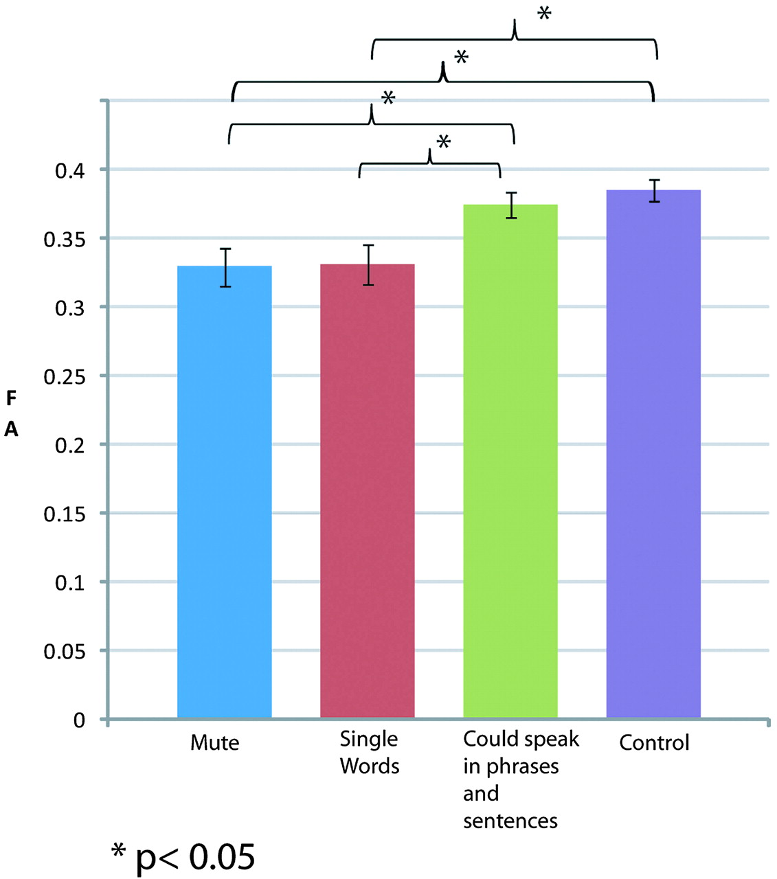
Patients were categorized by their capacity to speak: 1) Mute (n = 13), 2) Single words (n = 13), and 3) Could speak in phrases or sentences (n = 6). As shown in Fig 2, mean FA of the superior longitudinal fasciculus in control subjects and group 3 patients was not different (0.38 ± 0.04 vs 0.375 ± 0.02; t = 0.615; P = .542). In a similar setting, there was no difference in the FA of the patients with RTT in group 1 and group 2 (0.32 ± 0.05 vs 0.33 ± 0.05; t = −0.089; P = .929). But groups 1 and 2 with minimal speech were significantly different from group 3 and the control subjects (group 1 vs group 3: t = −2.12; P = .046; group 1 vs control: t = 3.77; P < .001; group 2 vs group 3: t = −2.03; P = .055; group 2 vs control: t = 3.58; P < .001). This trend was not seen in any other region.

Fig 2.
  • Download figure
  • Open in new tab
  • Download powerpoint
Fig 2.

Comparison of FA among patients categorized by speech capacity in the superior longitudinal fasciculus and control subjects.

Our study did not find any correlation between FA values in the examined tracts and seizure status, head circumference, or walking ability. Hemispheric asymmetry analysis evaluating left/right differences showed that, in contrast to control subjects, asymmetry was not present in the inferior fronto-occipital fasciculus (P > .99), external capsule (P > .99), superior longitudinal fasciculus (P > .99), and posterior thalamic radiation (P > .99) in patients with RTT. Additional LI analysis only showed a significant trend in the inferior fronto-occipital fasciculus tracts (P = .082) compared with control subjects. We noted hemispheric asymmetry in CG3 (posterior cingulate gyrus) of the patients (P = .044) compared with control subjects, who showed no asymmetry (P = .505).

Discussion

In the studied group of patients with RTT, the FA was reduced in the corpus callosum, internal capsule, and frontal white matter compared with control subjects. In contrast, mean FA in most of the regions comprising the limbic system and cerebellar tracts was similar in both groups. The posterior corona radiata was the only region in the patients with RTT where FA was greater than that of control subjects and this finding is in keeping with the observation in volumetric studies by Carter et al.7 Without histologic results, it is difficult to conclude about the exact mechanism of the FA decrease observed in this patient population. Past studies have suggested axonal involvement to explain FA reduction,20,22 but loss of myelin has not been shown to be a contributing factor.23 In patients with RTT, axonal vulnerability must be an important contributor to the reduced FA, as shown by reduced levels of N-acetylaspartate in MR spectroscopic imaging.24

We noted 3 major correlations with clinical findings in RTT, the most significant of which was a correlation of FA in the superior longitudinal fasciculus with the ability to speak. The superior longitudinal fasciculus is one of the main association bundles that connect the external surface of temporoparieto-occipital regions with the convexity of the frontal lobe and, in part, is associated with phonologic speech.25,26 The left-more-than-right asymmetry in the superior longitudinal fasciculus, as seen in the control subjects, was not found in the patients.

Second, the presence of intact visual capabilities, which are used by some girls with RTT to communicate, may be in accordance with normal to increased FA in the posterior corona radiata. This observation was in contrast to the FA that was significantly reduced by greater margins in all regions of the corpus callosum affecting interhemispheric connectivity, probably because of immature neurons and their poor dendritic arborization.27 However, other white matter structures that are known to be associated with visual functions such as the sagittal stratum did not show increased FA. The increased FA of the posterior thalamic radiation, therefore, needs careful interpretation with increased numbers of patients. Study data also argue that, with few exceptions, white matter tracts in RTT are diffusely affected compared with control subjects. Except for the right middle cerebellar tract, other cerebellar tracts were generally spared. Right-to-left differences in some brain regions of patients with RTT suggest that neuronal connectivity may have become restricted before development of specific skills contributing to interhemispheric asymmetry in these patients.

The third significant observation is that of reduced FA in the anterior cingulate gyrus, which is particularly revealing in view of the characteristic mood and behavioral changes seen in patients with RTT. In a similar setting, changes in the anterior cingulate gyrus and other components of the limbic system are also reported in schizophrenic28 and autistic29 patients. In addition, the posterior cingulate gyrus showed hemispheric asymmetry in the patients, which was not present in the control subjects. Larger studies are required, however, to investigate the correlation between severe behavioral and emotional problems in patients with RTT and limbic system components.

Volumetric MR imaging studies in RTT have shown a significantly reduced cerebral volume affecting both gray and white matter.7,30,31 In patients with RTT, MR imaging spectroscopy has shown significant decreases in average concentrations of N-acetylaspartate in both gray and white matter of the frontal and parietal lobes and insula compared with control subjects, demonstrating axonal involvement in RTT.24,23 Our in vivo observation confirms diffuse involvement of white matter tracts with areas of selective vulnerability in the limbic system components.

Longitudinal evaluations and studies in older subjects are needed for better understanding of changes in FA and its relationship to motor regression, and loss of speech noted in the older girls with RTT. Increased sample size will also allow better correlation of function to FA measurements. Improved technology with use of 3T or higher MR imaging modalities may provide better definition of tracts.

Clinical variability is currently assessed by neurologic examinations and mutation type. DTI studies would add specificity to these observations and help to understand the effects of individual mutations whose clinical effects are modified by age and X-inactivation in female patients. Therapeutic benefits may also be ascertained by their effects on neuronal tracts, either as improvement or lack of progressive reduction in FA.

Acknowledgments

We thank Terri Brawner and Kathleen Kahl for their technical support in acquiring the MR imaging scans as well as Carolyn Gillen for nursing assistance during the MR imaging examinations.

Footnotes

  • Supported by National Institute for Child Health and Human Development grant P01-HD-24448; National Institute on Aging grant AG-20012; National Center for Research Resources grant RR-15241; and Johns Hopkins Institute for Clinical and Translational Research grant UL1RR025005.

Indicates open access to non-subscribers at www.ajnr.org

References

  1. 1.↵
    1. Amir RE,
    2. Van den Veyver IB,
    3. Wan M,
    4. et al
    . Rett syndrome is caused by mutations in X-linked MECP2, encoding methyl-CpG-binding protein 2. Nature Genet 1999;23:185–88
    CrossRefPubMed
  2. 2.↵
    1. Sirianni N,
    2. Naidu S,
    3. Pereira J,
    4. et al
    . Rett syndrome: confirmation of X-linked dominant inheritance, and localization of the gene to Xq28. Am J Human Genet 1998;63:1552–58
    CrossRefPubMed
  3. 3.↵
    1. Kozinetz CA,
    2. Skender ML,
    3. MacNaughton N,
    4. et al
    . Epidemiology of Rett syndrome: a population-based registry. Pediatrics 1993;91:445–50
    Abstract/FREE Full Text
  4. 4.↵
    1. Hagberg B,
    2. Aicardi J,
    3. Dias K,
    4. et al
    . A progressive syndrome of autism, dementia, ataxia, and loss of purposeful hand use in girls: Rett's syndrome: report of 35 cases. Ann Neurol 1983;14:471–79
    CrossRefPubMed
  5. 5.↵
    1. Naidu S
    . Rett syndrome: a disorder affecting early brain growth. Ann Neurol 1997;42:3–10
    CrossRefPubMed
  6. 6.↵
    1. Armstrong D,
    2. Dunn JK,
    3. Antalffy B,
    4. et al
    . Selective dendritic alterations in the cortex of Rett syndrome. J Neuropathol Exp Neurol 1995;54:195–201
    CrossRefPubMed
  7. 7.↵
    1. Carter JC,
    2. Lanham DC,
    3. Pham D,
    4. et al
    . Selective cerebral volume reduction in Rett syndrome: a multiple-approach MR imaging study. AJNR Am J Neuroradiol 2008;29:436–41
    Abstract/FREE Full Text
  8. 8.↵
    1. Kerr A,
    2. Engerstrom I
    1. Kaufmann WE
    . Cortical development in Rett syndrome: molecular, neurochemical, and anatomic aspects. In: Kerr A, Engerstrom I eds. Rett Disorder and the Developing Brain. Oxford, UK: Oxford University Press; 2001
  9. 9.↵
    1. Ronnett GV,
    2. Leopold D,
    3. Cai X,
    4. et al
    . Olfactory biopsies demonstrate a defect in neuronal development in Rett's syndrome. Ann Neurol 2003;54:206–18
    CrossRefPubMed
  10. 10.↵
    1. Kishi N,
    2. Macklis JD
    . MECP2 is progressively expressed in post-migratory neurons and is involved in neuronal maturation rather than cell fate decisions. Mol Cell Neurosc 2004;27:306–21
    CrossRefPubMed
  11. 11.↵
    1. Armstrong DD
    . Neuropathology of Rett syndrome. J Child Neurol 2005;20:747–53
    Abstract/FREE Full Text
  12. 12.↵
    1. Bauman ML,
    2. Kemper TL,
    3. Arin DM
    . Pervasive neuroanatomic abnormalities of the brain in three cases of Rett's syndrome. Neurology 1995;45:1581–86
    Abstract/FREE Full Text
  13. 13.↵
    1. Basser PJ,
    2. Mattiello J,
    3. LeBihan D
    . Estimation of the effective self-diffusion tensor from the NMR spin echo. J Magn Reson B 1994;103:247–54
    CrossRefPubMed
  14. 14.↵
    1. Mori S,
    2. Barker PB
    . Diffusion magnetic resonance imaging: its principle and applications. Anat Rec 1999;257:102–09
    CrossRefPubMed
  15. 15.↵
    1. Pierpaoli C,
    2. Basser PJ
    . Toward a quantitative assessment of diffusion anisotropy. Magn Reson Med 1996;36:893–906
    CrossRefPubMed
  16. 16.↵
    1. Pruessmann KP,
    2. Weiger M,
    3. Scheidegger MB,
    4. et al
    . SENSE: sensitivity encoding for fast MRI. Magn Reson Med 1999;42:952–62
    CrossRefPubMed
  17. 17.↵
    1. Jones DK,
    2. Horsfield MA,
    3. Simmons A
    . Optimal strategies for measuring diffusion in anisotropic systems by magnetic resonance imaging. Magn Reson Med 1999;42:515–25
    CrossRefPubMed
  18. 18.↵
    1. Jiang H,
    2. van Zijl PC,
    3. K J,
    4. et al
    . DTIStudio: resource program for diffusion tensor computation and fiber bundle tracking. Comput Methods Programs Biomed 2006;81:106–16
    CrossRefPubMed
  19. 19.↵
    1. Woods RP,
    2. Grafton ST,
    3. Holmes CJ,
    4. et al
    . Automated image registration: I. General methods and intrasubject, intramodality validation. J Comput Assist Tomogr 1998;22:139–52
    CrossRefPubMed
  20. 20.↵
    1. Douaud G,
    2. Behrens TE,
    3. Poupon C,
    4. et al
    . In vivo evidence for the selective subcortical degeneration in Huntington's disease. Neuroimage 2009;46:958–66
    CrossRefPubMed
  21. 21.↵
    1. Stieltjes B,
    2. Kaufmann WE,
    3. van Zijl PC,
    4. et al
    . Diffusion tensor imaging and axonal tracking in the human brainstem. Neuroimage 2001;14:723–35
    CrossRefPubMed
  22. 22.↵
    1. Beaulieu C,
    2. Allen PS
    . Determinants of anisotropic water diffusion in nerves. Magn Reson Med 1994;31:394–400
    CrossRefPubMed
  23. 23.↵
    1. Song SK,
    2. Sun SW,
    3. Ramsbottom MJ,
    4. et al
    . Dysmyelination revealed through MRI as increased radial (but unchanged axial) diffusion of water. Neuroimage 2002;17:1429–36
    CrossRefPubMed
  24. 24.↵
    1. Horska A,
    2. Naidu S,
    3. Herskovits EH,
    4. et al
    . Quantitative 1H MR spectroscopic imaging in early Rett syndrome. Neurology 2000;54:715–22
    Abstract/FREE Full Text
  25. 25.↵
    1. Tanabe H,
    2. Sawada T,
    3. Inoue N,
    4. et al
    . Conduction aphasia and arcuate fasciculus. Acta Neurol Scand 1987;76:422–27
    CrossRefPubMed
  26. 26.↵
    1. Boatman D,
    2. Gordon B,
    3. Hart J,
    4. et al
    . Transcortical sensory aphasia: revisited and revised. Brain 2000;123(Pt 8):1634–42
    Abstract/FREE Full Text
  27. 27.↵
    1. Belichenko PV,
    2. Wright EE,
    3. Belichenko NP,
    4. et al
    . Widespread changes in dendritic and axonal morphology in Mecp2-mutant mouse models of Rett syndrome: evidence for disruption of neuronal networks. J Comp Neurol 2009;514:240–58
    CrossRefPubMed
  28. 28.↵
    1. White T,
    2. Cullen K,
    3. Rohrer LM,
    4. et al
    . Limbic structures and networks in children and adolescents with schizophrenia. Schizophr Bull 2008;34:18–29
    Abstract/FREE Full Text
  29. 29.↵
    1. Craig MC,
    2. Zaman SH,
    3. Daly EM,
    4. et al
    . Women with autistic-spectrum disorder: magnetic resonance imaging study of brain anatomy. Br J Psychiatry 2007;191:224–28
    Abstract/FREE Full Text
  30. 30.↵
    1. Reiss AL,
    2. Faruque F,
    3. Naidu S,
    4. et al
    . Neuroanatomy of Rett syndrome: a volumetric imaging study. Ann Neurol 1993;34:227–34
    CrossRefPubMed
  31. 31.↵
    1. Naidu S,
    2. Kaufmann WE,
    3. Abrams MT,
    4. et al
    . Neuroimaging studies in Rett syndrome. Brain Dev 2001;23 Suppl 1:S62–71
    CrossRefPubMed
  • Received February 19, 2009.
  • Accepted after revision June 18, 2009.
  • Copyright © American Society of Neuroradiology
View Abstract
PreviousNext
Back to top

In this issue

American Journal of Neuroradiology: 31 (2)
American Journal of Neuroradiology
Vol. 31, Issue 2
1 Feb 2010
  • Table of Contents
  • Index by author
Advertisement
Print
Download PDF
Email Article

Thank you for your interest in spreading the word on American Journal of Neuroradiology.

NOTE: We only request your email address so that the person you are recommending the page to knows that you wanted them to see it, and that it is not junk mail. We do not capture any email address.

Enter multiple addresses on separate lines or separate them with commas.
White Matter Impairment in Rett Syndrome: Diffusion Tensor Imaging Study with Clinical Correlations
(Your Name) has sent you a message from American Journal of Neuroradiology
(Your Name) thought you would like to see the American Journal of Neuroradiology web site.
CAPTCHA
This question is for testing whether or not you are a human visitor and to prevent automated spam submissions.
Cite this article
A. Mahmood, G. Bibat, A.-L. Zhan, I. Izbudak, L. Farage, A. Horska, S. Mori, S. Naidu
White Matter Impairment in Rett Syndrome: Diffusion Tensor Imaging Study with Clinical Correlations
American Journal of Neuroradiology Feb 2010, 31 (2) 295-299; DOI: 10.3174/ajnr.A1792

Citation Manager Formats

  • BibTeX
  • Bookends
  • EasyBib
  • EndNote (tagged)
  • EndNote 8 (xml)
  • Medlars
  • Mendeley
  • Papers
  • RefWorks Tagged
  • Ref Manager
  • RIS
  • Zotero
0 Responses
Respond to this article
Share
Bookmark this article
White Matter Impairment in Rett Syndrome: Diffusion Tensor Imaging Study with Clinical Correlations
A. Mahmood, G. Bibat, A.-L. Zhan, I. Izbudak, L. Farage, A. Horska, S. Mori, S. Naidu
American Journal of Neuroradiology Feb 2010, 31 (2) 295-299; DOI: 10.3174/ajnr.A1792
del.icio.us logo Twitter logo Facebook logo Mendeley logo
  • Tweet Widget
  • Facebook Like
  • Google Plus One
Purchase

Jump to section

  • Article
    • Abstract
    • Abbreviations
    • Materials and Methods
    • Results
    • Discussion
    • Acknowledgments
    • Footnotes
    • References
  • Figures & Data
  • Info & Metrics
  • Responses
  • References
  • PDF

Related Articles

  • No related articles found.
  • PubMed
  • Google Scholar

Cited By...

  • F2-dihomo-isoprostanes as potential early biomarkers of lipid oxidative damage in Rett syndrome
  • Crossref (56)
  • Google Scholar

This article has been cited by the following articles in journals that are participating in Crossref Cited-by Linking.

  • Connected speech production in three variants of primary progressive aphasia
    Stephen M. Wilson, Maya L. Henry, Max Besbris, Jennifer M. Ogar, Nina F. Dronkers, William Jarrold, Bruce L. Miller, Maria Luisa Gorno-Tempini
    Brain 2010 133 7
  • Cerebral plasticity: Windows of opportunity in the developing brain
    Fatima Yousif Ismail, Ali Fatemi, Michael V. Johnston
    European Journal of Paediatric Neurology 2017 21 1
  • Multi-site genetic analysis of diffusion images and voxelwise heritability analysis: A pilot project of the ENIGMA–DTI working group
    Neda Jahanshad, Peter V. Kochunov, Emma Sprooten, René C. Mandl, Thomas E. Nichols, Laura Almasy, John Blangero, Rachel M. Brouwer, Joanne E. Curran, Greig I. de Zubicaray, Ravi Duggirala, Peter T. Fox, L. Elliot Hong, Bennett A. Landman, Nicholas G. Martin, Katie L. McMahon, Sarah E. Medland, Braxton D. Mitchell, Rene L. Olvera, Charles P. Peterson, John M. Starr, Jessika E. Sussmann, Arthur W. Toga, Joanna M. Wardlaw, Margaret J. Wright, Hilleke E. Hulshoff Pol, Mark E. Bastin, Andrew M. McIntosh, Ian J. Deary, Paul M. Thompson, David C. Glahn
    NeuroImage 2013 81
  • Dynamics and Mechanisms of CNS Myelination
    Kathryn K. Bercury, Wendy B. Macklin
    Developmental Cell 2015 32 4
  • Social impairments in Rett syndrome: characteristics and relationship with clinical severity
    W. E. Kaufmann, E. Tierney, C. A. Rohde, M. C. Suarez‐Pedraza, M. A. Clarke, C. F. Salorio, G. Bibat, I. Bukelis, D. Naram, D. C. Lanham, S. Naidu
    Journal of Intellectual Disability Research 2012 56 3
  • Modulation of RhoGTPases Improves the Behavioral Phenotype and Reverses Astrocytic Deficits in a Mouse Model of Rett Syndrome
    Bianca De Filippis, Alessia Fabbri, Daiana Simone, Rossella Canese, Laura Ricceri, Fiorella Malchiodi-Albedi, Giovanni Laviola, Carla Fiorentini
    Neuropsychopharmacology 2012 37 5
  • F2-dihomo-isoprostanes as potential early biomarkers of lipid oxidative damage in Rett syndrome
    Claudio De Felice, Cinzia Signorini, Thierry Durand, Camille Oger, Alexandre Guy, Valérie Bultel-Poncé, Jean-Marie Galano, Lucia Ciccoli, Silvia Leoncini, Maurizio D'Esposito, Stefania Filosa, Alessandra Pecorelli, Giuseppe Valacchi, Joussef Hayek
    Journal of Lipid Research 2011 52 12
  • Isoprostanes and neuroprostanes: Total synthesis, biological activity and biomarkers of oxidative stress in humans
    Jean-Marie Galano, Emilie Mas, Anne Barden, Trevor A. Mori, Cinzia Signorini, Claudio De Felice, Aaron Barrett, Catherine Opere, Edith Pinot, Edzard Schwedhelm, Ralf Benndorf, Jérôme Roy, Jean-Yves Le Guennec, Camille Oger, Thierry Durand
    Prostaglandins & Other Lipid Mediators 2013 107
  • Partial rescue of Rett syndrome by ω-3 polyunsaturated fatty acids (PUFAs) oil
    Claudio De Felice, Cinzia Signorini, Thierry Durand, Lucia Ciccoli, Silvia Leoncini, Maurizio D’Esposito, Stefania Filosa, Camille Oger, Alexandre Guy, Valérie Bultel-Poncé, Jean-Marie Galano, Alessandra Pecorelli, Laura De Felice, Giuseppe Valacchi, Joussef Hayek
    Genes & Nutrition 2012 7 3
  • Traumatic brain injury, major depression, and diffusion tensor imaging: Making connections
    Jerome J. Maller, Richard H.S. Thomson, Philip M. Lewis, Stephen E. Rose, Kerstin Pannek, Paul B. Fitzgerald
    Brain Research Reviews 2010 64 1

More in this TOC Section

  • Comparison of Image Quality and Radiation Dose in Pediatric Temporal Bone CT Using Photon-Counting Detector CT and Energy-Integrating Detector CT
  • SyMRI & MR Fingerprinting in Brainstem Myelination
  • Neonatal Hypocalcemia: Cerebral MRI Abnormalities
Show more Pediatrics

Similar Articles

Advertisement

Indexed Content

  • Current Issue
  • Accepted Manuscripts
  • Article Preview
  • Past Issues
  • Editorials
  • Editors Choice
  • Fellow Journal Club
  • Letters to the Editor

Cases

  • Case Collection
  • Archive - Case of the Week
  • Archive - Case of the Month
  • Archive - Classic Case

Special Collections

  • Special Collections

Resources

  • News and Updates
  • Turn around Times
  • Submit a Manuscript
  • Author Policies
  • Manuscript Submission Guidelines
  • Evidence-Based Medicine Level Guide
  • Publishing Checklists
  • Graphical Abstract Preparation
  • Imaging Protocol Submission
  • Submit a Case
  • Become a Reviewer/Academy of Reviewers
  • Get Peer Review Credit from Publons

Multimedia

  • AJNR Podcast
  • AJNR SCANtastic
  • Video Articles

About Us

  • About AJNR
  • Editorial Board
  • Not an AJNR Subscriber? Join Now
  • Alerts
  • Feedback
  • Advertise with us
  • Librarian Resources
  • Permissions
  • Terms and Conditions

American Society of Neuroradiology

  • Not an ASNR Member? Join Now

© 2025 by the American Society of Neuroradiology All rights, including for text and data mining, AI training, and similar technologies, are reserved.
Print ISSN: 0195-6108 Online ISSN: 1936-959X

Powered by HighWire