
Article Figures & Data
Figures
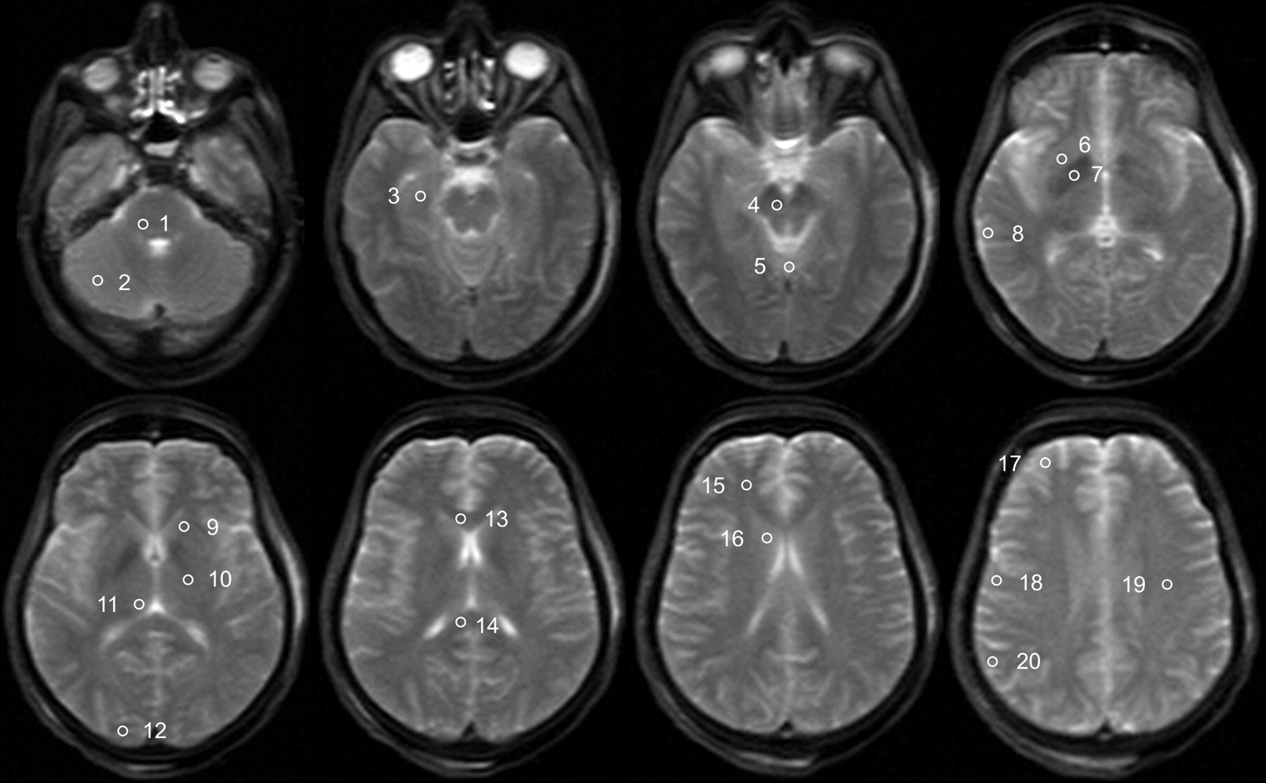
- Fig 1.
Trace-weighted image (b-value = 5 s/mm2) of a deceased subject showing the placement of ROIs for the evaluation of ADC values. ROIs are only shown unilaterally; the ROI in the medulla is not shown. The ROIs are the following: 1) pons, 2) cerebellum, 3) hippocampus, 4) mesencephalon, 5) vermis, 6) putamen, 7) pallidum, 8) temporal cortex, 9) internal capsule anterior, 10) internal capsule posterior, 11) thalamus, 12) occipital cortex, 13) corpus callosum genu, 14) corpus callosum splenium, 15) frontal WM, 16) caudate nucleus, 17) frontal cortex, 18) motor cortex, 19) centrum semiovale, and 20) parietal cortex.
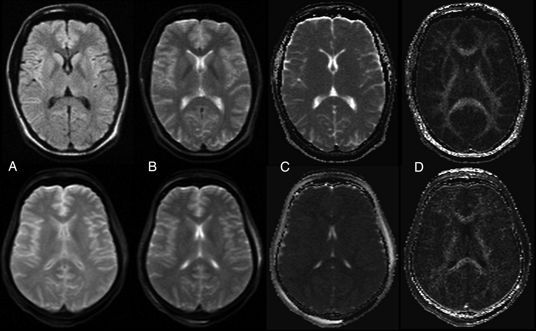
- Fig 2.
Example of a DWI image, b0 image, ADC map, and FA map of a healthy living volunteer (woman, 31 years) (top row) and a deceased subject (woman; age, 45 years; PMI, 73 hours; body temperature at acquisition, 12°C) (bottom row). A, DWI (b = 1000 s/mm2). B, Trace-weighted image with b-value = 5 s/mm2. C, ADC map. D) FA map.
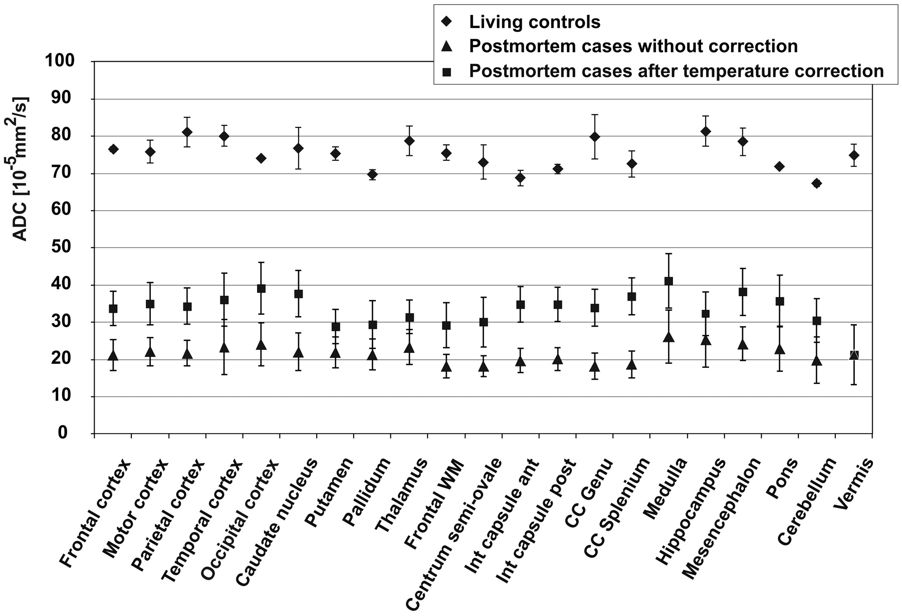
- Fig 3.
Comparison of ADC of the postmortem cases with the living controls and demonstration of the effect of temperature correction on postmortem ADC. Data points are mean values for the respective ROI; error bars show the corresponding SD.
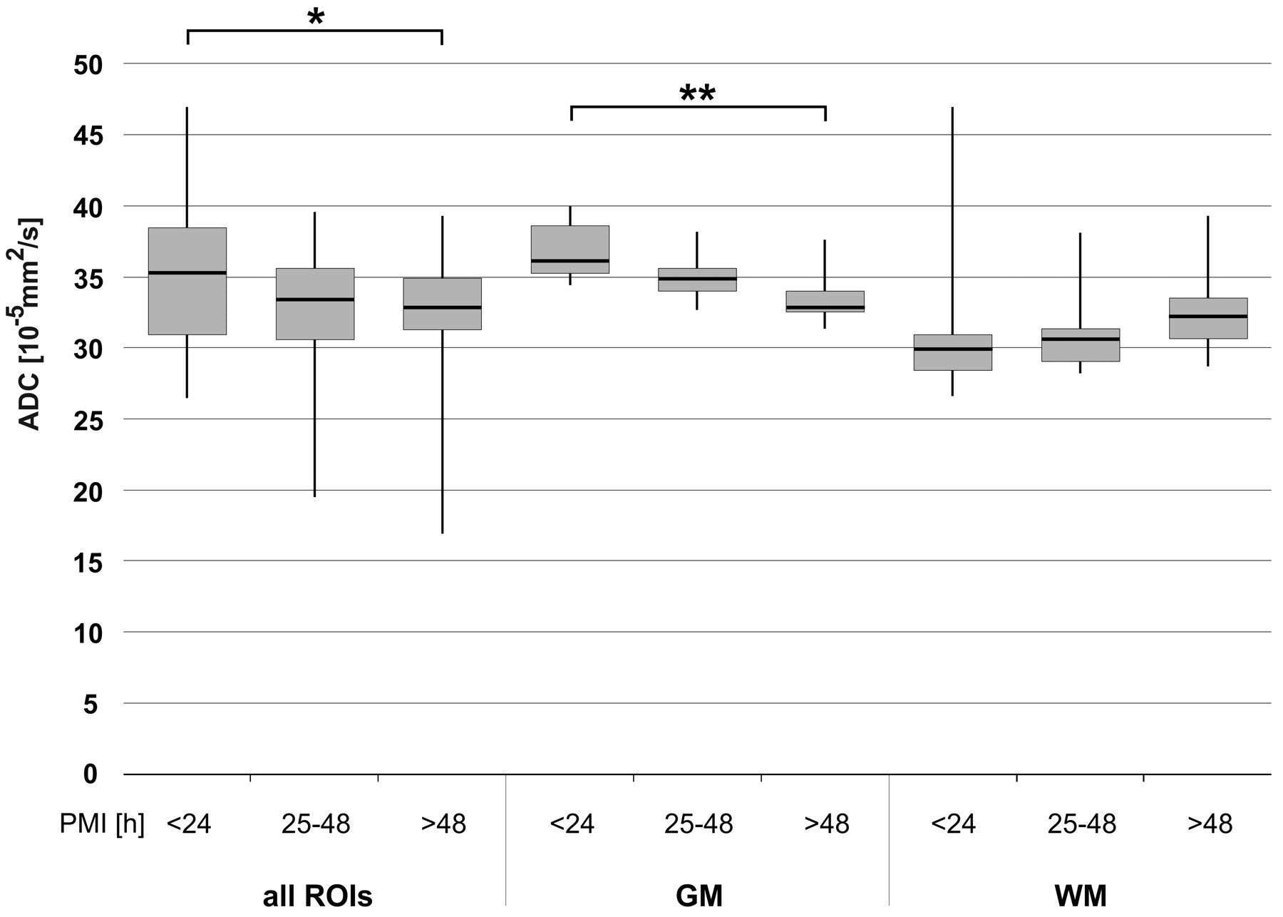
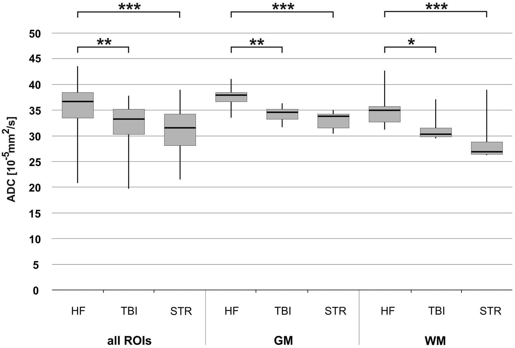
- Fig 4.
Influence of PMI on ADCTc in all ROIs, GM, and WM, respectively. Whiskers show the maximum and the minimum values. A single asterisk indicates P < .05; double asterisks, P < .01.
- Fig 5.
Influence of mechanical brain trauma and hypoxic brain injury due to strangulation on ADCTc of all ROIs, GM, and WM, respectively. A group of brain trauma cases (n = 4) and a group of strangulation cases (n = 5) are compared with a group of subjects with natural death due to cardiac failure (n = 4). Whiskers show the maximum and the minimum values. A single asterisk indicates P < .05; double asterisks, P < .005; and triple asterisks, P < .001.
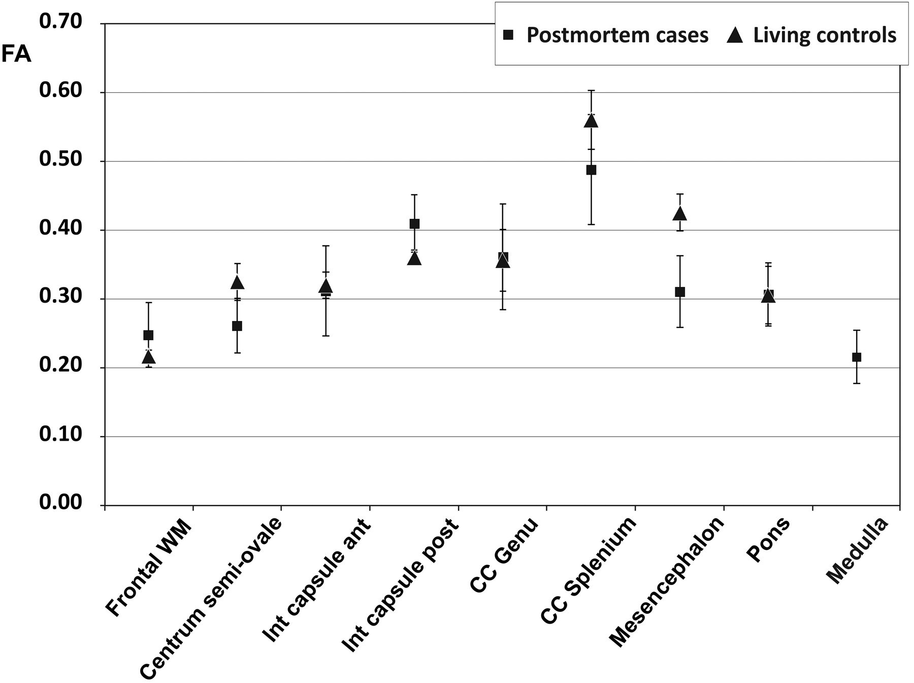
- Fig 6.
Comparison of FA of the postmortem cases with the living controls in different brain regions. Data points are mean values for the respective ROI; error bars show the corresponding SD.
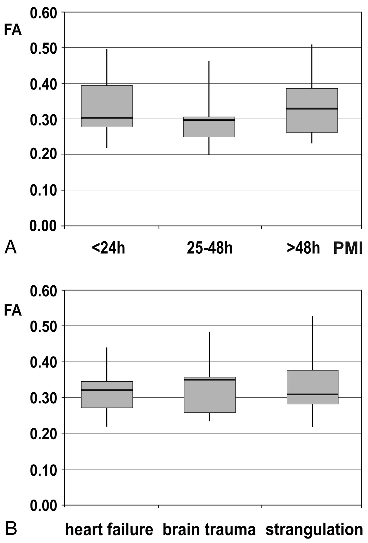
- Fig 7.
A, Influence of the PMI on FA in 3 groups of subjects (PMI < 24 hours, n = 7; PMI = 25–48 hours, n = 7; PMI > 48 hours, n = 6). Whiskers show the maximum and the minimum values. B, Effect of the cause of death—that is, mechanical brain trauma and hypoxic injury of the brain due to strangulation compared with heart failure—on FA. Whiskers show the maximum and the minimum values.


Tables
Case Age at Death [years] Incident Cause of Deatha Time of Death–MRI [hours] Tappb [°C] 1 53 Fall from great height Central respiratory arrest due to brain trauma 20 20 2 29 Natural death Heart failure 44 20 3 94 Motor vehicle crash (pedestrian) Central respiratory arrest due to brain trauma 45 10 4 63 Natural death Heart failure 49 5 5 44 Diving accident Heart failure due to gas embolism 51 5 6 45 Fall into a crevasse Organ failure due to hypothermia 73 12 7 29 Suicidal hanging Suffocation 13 30 8 79 Beating to death Heart failure due to pneumothorax and fat embolism 14 28 9 30 Suicidal hanging Suffocation 48 23 10 61 Motor vehicle crash (pedestrian) Heart & lung failure due to blood aspiration and pneumothorax 49 10 11 19 Incidental gas intoxication (propane & butane) Central respiratory arrest 14 25 12 46 Manual strangulation Suffocation 14 22 13 3 Natural death Central respiratory arrest due to suffocation (laryngitis) 19 8 14 46 Suicidal intoxication & hypothermia Central respiratory arrest and hypothermia (combined) 33 5 15 37 Natural death Heart failure 17 8 16 28 Incidental hanging Suffocation 141 5 17 58 Natural death Heart failure 25 10 18 29 Suicidal hanging Suffocation 61 15 19 58 Medical maltreatment Heart failure due to arterial air embolism 43 10 20 49 Hang-glider crash Central respiratory insufficience due to brain stem lesion 27 19 -
a In cases with combined or concurring causes of death only the most relevant are mentioned.
-
b Approximate body core temperature at the time of MR imaging
-
ROI FA Postmortem Casesa Living Controls Mean SD Median Min Max Mean SD Frontal white matter 0.25 0.05 0.25 0.15 0.34 0.22 0.01 Centrum semiovale 0.26 0.04 0.25 0.18 0.34 0.32 0.03 Internal capsule anterior 0.31 0.07 0.31 0.18 0.41 0.32 0.02 Internal capsule posterior 0.41 0.04 0.41 0.33 0.51 0.36 0.01 Corpus callosum genu 0.36 0.08 0.37 0.19 0.48 0.36 0.04 Corpus callosum splenium 0.49 0.08 0.51 0.32 0.59 0.56 0.04 Mesencephalon 0.31 0.05 0.30 0.24 0.49 0.43 0.03 Pons 0.31 0.05 0.31 0.23 0.39 0.31 0.04 Medulla 0.22 0.04 0.22 0.16 0.28 n.a. n.a. -
a Given are the mean, standard deviation, median, minimum, and maximum except case 9 (n = 19).
-


