
Article Figures & Data
Figures
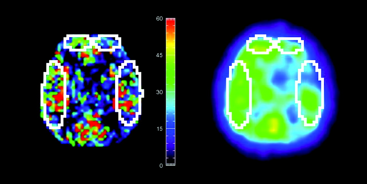
- Fig 1.
Representative region of interest sizes and locations in SPECT and ASL perfusion CBF images after registration. ROIs defined on 1 image are automatically copied to another.
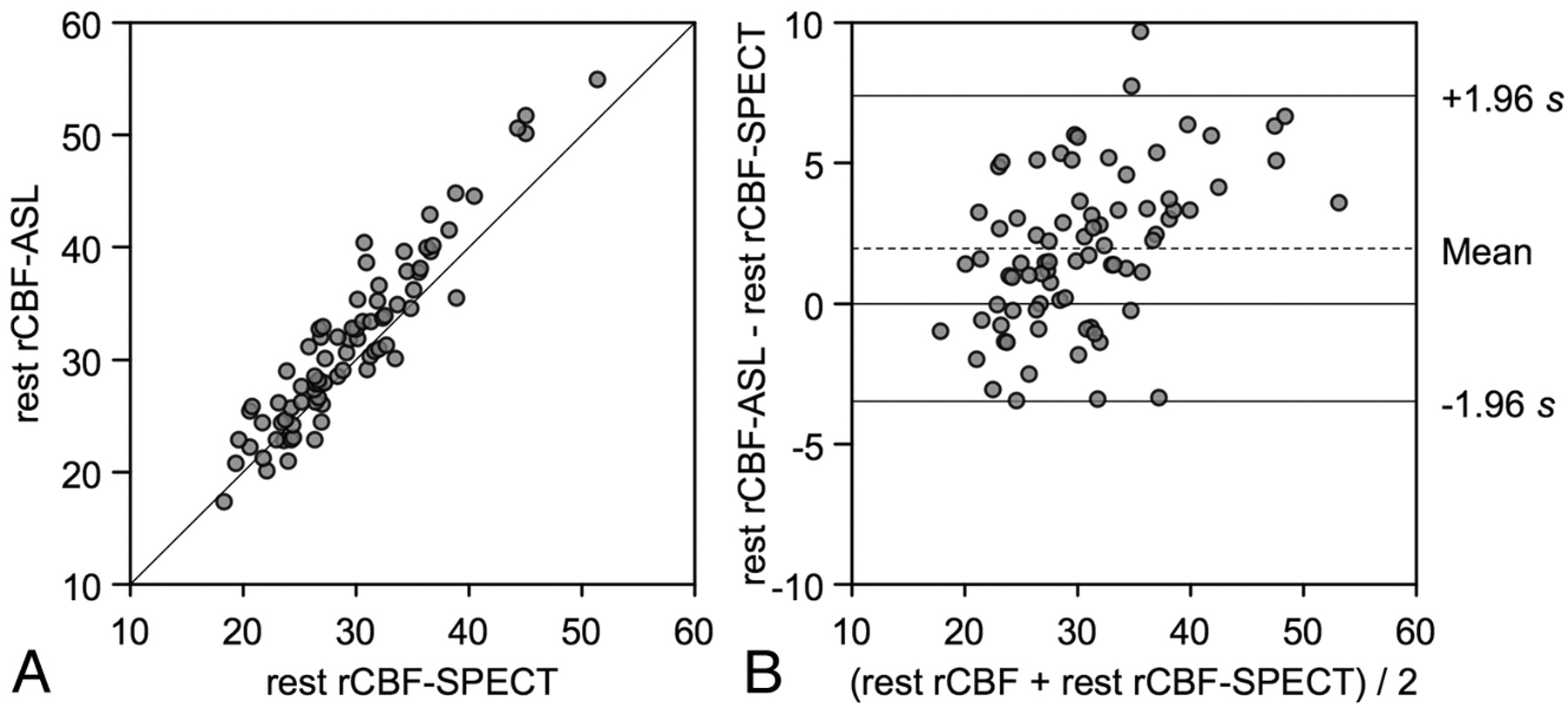
- Fig 2.
CBF assessments by ASL and SPECT in the region of interest analysis. A, Resting CBF values measured by ASL are plotted against the corresponding CBF values measured by SPECT from bilateral frontal and temporal ROIs. The solid line shows the line of equality. B, Bland-Altman plot of CBF values measured by ASL and SPECT. A plot of the differences between the methods against their means is shown. The broken line shows average of differences, and the solid line indicates ±1.96 × SD of differences.
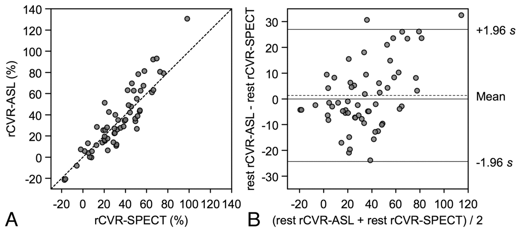
- Fig 3.
CVR assessments by ASL and SPECT in the ROI study. A, CVR values measured by ASL are plotted against the corresponding CVR values measured by SPECT from bilateral frontal and temporal ROIs. The broken line shows the line of equality. B, Bland-Altman plot of CVR values measured by ASL and SPECT. The broken line shows average of differences, and the solid line indicates ±1.96× SD of differences.
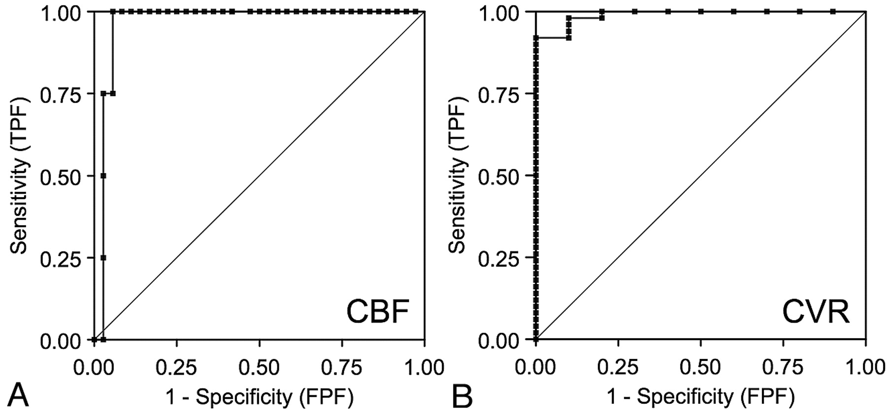
- Fig 4.
A, ROC curves of CBF by ASL for detection of CBF reduction on SPECT. Area under the curve is 0.98. B, ROC curves of CVR by ASL for detection of CVR reduction on SPECT. Area under the curve is 0.99. TPF, true-positive fraction; FPF, false-positive fraction.
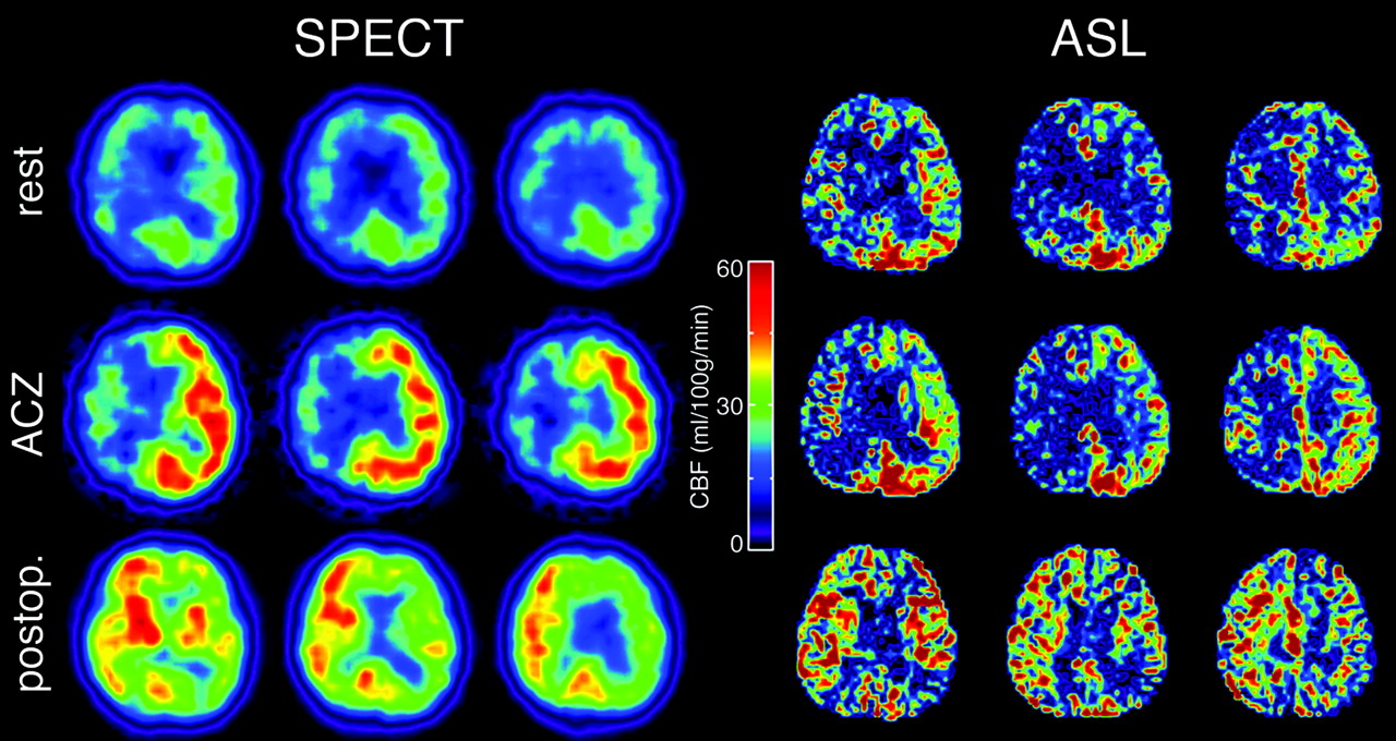
- Fig 5.
Typical CBF maps obtained by SPECT and ASL in patients with severe right carotid stenosis. Preoperative resting images (top row), preoperative images with ACZ challenge (middle row), and postoperative images (bottom row) are shown. Preoperative hypoperfusion, poor vasoreactivity, and postoperative hyperperfusion in the right internal carotid artery region can be seen in both SPECT and ASL.


Tables
Parameter n = 20 Age (yr) 70.0 ± 8.1 Men 16 (80) Symptomatic stenosis 12 (60) Right side affected 12 (60) NASCET degree of stenosis Severe (>70%) 10 (50) Moderate (50–70%) 6 (30) Mild (<50%) 4 (20) Contralateral stenosis 4 (20) Ipsilateral intracranial stenosis 3 (15) Surgery CEA 10 (50) Carotid artery stenting 2 (10) None 8 (40) A1 hypoplasia 4 (20) -
Note:—Values in parentheses are percentages.
-
ASL SPECT ROI size (no. of pixels) Frontal (n = 40) 24.9 ± 5.2 Temporal (n = 40) 66.1 ± 9.0 Avg. CBF (ml/100 g/min) All ROIs (n = 80) 31.5 ± 7.7 29.6 ± 6.4 Ipsilateral ROIs (n = 40) 31.4 ± 8.1 29.4 ± 6.7 Contralateral ROIs (n = 40) 31.6 ± 7.5 29.6 ± 6.3 Frontal ROIs (n = 40) 27.5 ± 5.5 26.8 ± 4.5 Temporal ROIs (n = 40) 35.5 ± 7.6 32.3 ± 7.0 Avg. CVR (%) All ROIs (n = 60) 35.4 ± 29.2 34.1 ± 23.1 Ipsilateral ROIs (n = 30) 32.0 ± 31.2 31.7 ± 24.1 Contralateral ROIs (n = 30) 38.4 ± 28.2 35.5 ± 23.3 Frontal ROIs (n = 30) 43.6 ± 33.0 40.6 ± 25.4 Temporal ROIs (n = 30) 27.2 ± 23.2 27.6 ± 19.3
In this issue


Jump to section
Related Articles
Cited By...
- Predicting Impaired Cerebrovascular Reactivity and Hyperperfusion Syndrome with BeamSAT MRI in Carotid Artery Stenosis
- Cerebral Blood Flow Improvement after Indirect Revascularization for Pediatric Moyamoya Disease: A Statistical Analysis of Arterial Spin-Labeling MRI
- Correlation of Asymmetry Indices Measured by Arterial Spin-Labeling MR Imaging and SPECT in Patients with Crossed Cerebellar Diaschisis
- Arterial Spin-Labeling Evaluation of Cerebrovascular Reactivity to Acetazolamide in Healthy Subjects