Advertisement

Main menu

  • Home
  • Content
    • Current Issue
    • Accepted Manuscripts
    • Article Preview
    • Past Issue Archive
    • AJNR Case Collection
    • Case of the Week Archive
    • Classic Case Archive
    • Case of the Month Archive
  • Special Collections
    • Spinal CSF Leak Articles (Jan 2020-June 2024)
    • 2024 AJNR Journal Awards
    • Most Impactful AJNR Articles
  • Multimedia
    • AJNR Podcast
    • AJNR Scantastics
    • Video Articles
  • For Authors
    • Submit a Manuscript
    • Author Policies
    • Fast publishing of Accepted Manuscripts
    • Graphical Abstract Preparation
    • Manuscript Submission Guidelines
    • Imaging Protocol Submission
    • Submit a Case for the Case Collection
  • About Us
    • About AJNR
    • Editorial Board
  • More
    • Become a Reviewer/Academy of Reviewers
    • Subscribers
    • Permissions
    • Alerts
    • Feedback
    • Advertisers
    • ASNR Home
  • Other Publications
    • ajnr

User menu

  • Alerts
  • Log in

Search

  • Advanced search
American Journal of Neuroradiology

American Journal of Neuroradiology

ASHNR American Society of Functional Neuroradiology ASHNR American Society of Pediatric Neuroradiology ASSR
  • Alerts
  • Log in

Advanced Search

  • Home
  • Content
    • Current Issue
    • Accepted Manuscripts
    • Article Preview
    • Past Issue Archive
    • AJNR Case Collection
    • Case of the Week Archive
    • Classic Case Archive
    • Case of the Month Archive
  • Special Collections
    • Spinal CSF Leak Articles (Jan 2020-June 2024)
    • 2024 AJNR Journal Awards
    • Most Impactful AJNR Articles
  • Multimedia
    • AJNR Podcast
    • AJNR Scantastics
    • Video Articles
  • For Authors
    • Submit a Manuscript
    • Author Policies
    • Fast publishing of Accepted Manuscripts
    • Graphical Abstract Preparation
    • Manuscript Submission Guidelines
    • Imaging Protocol Submission
    • Submit a Case for the Case Collection
  • About Us
    • About AJNR
    • Editorial Board
  • More
    • Become a Reviewer/Academy of Reviewers
    • Subscribers
    • Permissions
    • Alerts
    • Feedback
    • Advertisers
    • ASNR Home
  • Follow AJNR on Twitter
  • Visit AJNR on Facebook
  • Follow AJNR on Instagram
  • Join AJNR on LinkedIn
  • RSS Feeds

Welcome to the new AJNR, Updated Hall of Fame, and more. Read the full announcements.


AJNR is seeking candidates for the position of Associate Section Editor, AJNR Case Collection. Read the full announcement.


Getting new auth cookie, if you see this message a lot, tell someone!
Research ArticleHead and NeckF

Oncocytoma: The Vanishing Parotid Mass

N.D. Patel, A. van Zante, D.W. Eisele, H.R. Harnsberger and C.M. Glastonbury
American Journal of Neuroradiology October 2011, 32 (9) 1703-1706; DOI: https://doi.org/10.3174/ajnr.A2569
N.D. Patel
  • Find this author on Google Scholar
  • Find this author on PubMed
  • Search for this author on this site
A. van Zante
  • Find this author on Google Scholar
  • Find this author on PubMed
  • Search for this author on this site
D.W. Eisele
  • Find this author on Google Scholar
  • Find this author on PubMed
  • Search for this author on this site
H.R. Harnsberger
  • Find this author on Google Scholar
  • Find this author on PubMed
  • Search for this author on this site
C.M. Glastonbury
  • Find this author on Google Scholar
  • Find this author on PubMed
  • Search for this author on this site
  • Article
  • Figures & Data
  • Info & Metrics
  • Responses
  • References
  • PDF
Loading

Abstract

SUMMARY: Parotid gland oncocytoma is an uncommon, benign salivary neoplasm composed of mitochondria-rich oncocytes. The purpose of this study was to correlate MR imaging and histopathology of parotid gland oncocytomas and to define the features that may distinguish these neoplasms from other benign and malignant parotid gland tumors. The MR imaging features in 9 patients with a pathologic diagnosis of oncocytoma were retrospectively reviewed. The imaging features were strikingly similar for 8 of the 9 patients. All lesions appeared T1 hypointense but isointense to the native parotid gland on fat-saturated T2 and postcontrast T1 imaging. On MR imaging, parotid gland oncocytomas share specific imaging characteristics that have not been described for benign or malignant parotid gland tumors. Oncocytomas are isointense to native parotid gland on fat-saturated T2 and T1 postcontrast MR images. Preoperative identification of correct histology may help surgical planning.

Abbreviations

FDG
fluorodeoxyglucose
FNAB
fine needle aspiration biopsy
T1+C
postcontrast T1
T1 FS C+ WI
postcontrast T1-weighted imaging with fat saturation
T1WI
T1-weighted imaging
T2 FS WI
T2-weighted imaging with fat saturation
T2WI
T2-weighted imaging

Parotid gland oncocytoma is an uncommon, benign salivary neoplasm composed of mitochondria-rich epithelial cells called oncocytes.1 In the parotid gland, oncocytomas most commonly occur in adults in their sixth to eighth decades, with no sex predilection.1,2 Parotid gland oncocytomas have been shown to demonstrate intense FDG uptake on positron-emission tomography scans and to accumulate technetium Tc99m pertechnetate,3 which have both been attributed to their high content of metabolically active mitochondria; however, these findings are a common radiographic and histopathologic feature shared with Warthin tumors. High FDG uptake also is reported with many parotid lesions, including the significantly more common benign mixed tumor.4 A recent article favors multiphase contrast-enhanced CT to distinguish oncocytomas and suggests some potentially helpful but nonetheless nonspecific CT features.5,6 CT carries the additional risk of ionizing radiation, and many parotid gland tumors are “invisible” on CT, further minimizing its utility in assessment of parotid masses.7

Cytologic analysis of a parotid mass by FNAB can be useful. It must be understood, however, that despite excellent reported accuracy rates of FNAB, some parotid neoplasms are prone to diagnostic error. Thus, it has been proposed that the fine needle aspiration findings should contribute to, not replace, the overall clinical and imaging diagnostic impression.8 Infrequently, FNAB can alter the tumor histopathology to the point of obscuring final pathologic diagnosis. Inadequate specimen sampling also may occur. To this end, a large series by Capone et al9 reported sensitivity in the detection of oncocytomas by FNAB of 29%. Clearly, exclusive reliance on the results of FNAB findings at the expense of clinical and radiographic findings is unwarranted and potentially misleading.

MR imaging allows for markedly improved soft tissue contrast, providing information that can help to characterize parotid gland masses. Among MR imaging features, T1WI remains invaluable in localizing parotid lesions, while determining the margin contour and relationship to adjacent structures. T2 FS WI best helps to characterize the neoplastic nature of the lesion.10 Benign mixed tumors characteristically demonstrate markedly high signal intensity similar to CSF on T2WI and heterogeneous low-to-intermediate signal intensity on T1WI, with solid enhancement. In contrast, salivary gland malignant tumors are typically low signal intensity on T2 FS WI. T1 FS C+ WI is important in detecting the perineural spread of disease and intracranial extension of disease. In addition, administration of contrast is necessary to distinguish a cyst from solid mass.

The purpose of this study was to correlate MR imaging and histopathology of parotid gland oncocytomas and to define the features that distinguish these from other benign and malignant parotid gland tumors. We report the largest series reviewing the MR imaging features of parotid gland oncocytoma and histopathologic correlates to date.

Materials and Methods

After approval by our institutional review board, the departments of pathology and radiology archives were reviewed to identify all parotid gland oncocytomas that had undergone MR imaging before surgery or before fine needle aspiration. Seven such cases were identified at our institution during a period of 10 years. An additional 2 cases were similarly identified at another institution and submitted for review. Clinical presentation, diagnostic imaging, surgical findings, surgical pathology, and postoperative course were analyzed in all patients. MR imaging scans were reviewed for all 9 patients. CT scans were also available for 3 of these patients, and sonography imaging was reviewed in 6 patients. All preoperative MR images were acquired on 1.5T MR imaging systems (Signa 1.5T HDX; GE Healthcare, Waukesha, Wisconsin). Our standard imaging protocol used axial and coronal T1-weighted fast spin-echo, axial and coronal fat-suppressed gadolinium-enhanced T1-weighted fast spin-echo, and fast spin-echo T2 with fat suppression sequences in the axial plane. One certificate of added qualification–certified experienced neuroradiologist and 1 neuroradiology fellowship–trained clinical instructor together reviewed the MR images, analyzing tumor margin and location, size, and T1 and T2 signal intensity characteristics and enhancement patterns.

Results

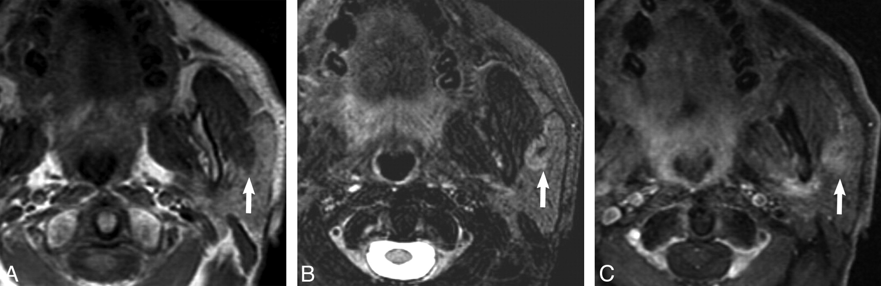
The Table illustrates our patient group and MR imaging findings. Of the 9 patients, 6 were women and 3 were men between the ages of 36 and 83 years. A painless mass was the most common presenting feature, though ipsilateral ear pain was the presenting symptom in 1 case. Imaging features among all lesions were strikingly similar. Most tumors (6/9) resided in the superficial parotid gland, with 3 also extending to the deep lobe. All oncocytomas appeared hypointense relative to native parotid tissue on T1WI, with well-demarcated margins that were lobulated in 4 cases. On T2 FS WI, 8 oncocytomas appeared isointense to the native parotid gland (Figs 1 and 2), whereas 1 mixed oncocytoma/pleomorphic adenoma was hyperintense on T2 FS WI (Fig 3 ). On T1 FS C+ WI, 8 oncocytomas appeared isointense to the native parotid gland (Figs 1 and 2), whereas 1 mixed oncocytoma/pleomorphic adenoma was heterogeneous (Fig 3).

View this table:
  • View inline
  • View popup

Presentation and MR imaging features of parotid gland oncocytomas

Fig 1.
  • Download figure
  • Open in new tab
  • Download powerpoint
Fig 1.

Case 1, 62-year-old female with 2-year history of firm, painless left parotid mass. A, Axial T1-weighted MR image shows a well-demarcated mass within the superficial lobe of the left parotid gland (white arrow) that is T1 hypointense. B, Axial T2-weighted MR image with fat saturation shows the mass (white arrow) to be isointense to the native parotid gland. C, Axial T1-weighted postgadolinium sequences illustrates the mass (white arrow) as isointense to the native parotid gland.

Fig 2.
  • Download figure
  • Open in new tab
  • Download powerpoint
Fig 2.

Case 5, 55-year-old woman with painless “lump” behind her left ear. A, Axial T1-weighted MR image shows a well-demarcated mass within the superficial and deep lobe of the left parotid gland (white arrow) that is T1 hypointense. B, Axial T2-weighted MR image with fat saturation illustrates the mass (white arrow) to be isointense to the native parotid gland. C, Axial T1-weighted postgadolinium sequence shows the mass (white arrow) as isointense to the native parotid gland.

Fig 3.
  • Download figure
  • Open in new tab
  • Download powerpoint
Fig 3.

Case 7, 75-year-old man with history of squamous cell carcinoma of the larynx status postradiation found to have an enlarging mass in the left parotid gland on surveillance imaging. A, Axial T1-weighted MR image shows a well-demarcated mass within the superficial lobe of the left parotid gland (white arrow) that is T1 hypointense. B, Axial T2-weighted MR image with fat saturation shows the mass to be hyperintense but heterogeneous to the native parotid gland. C, Axial T1-weighted postgadolinium sequence shows the mass (white arrow) to demonstrate heterogeneous enhancement to the native parotid gland.

Discussion

An oncocytoma is an uncommon benign parotid gland neoplasm, accounting for <1% of all salivary gland tumors.1 Due to their low prevalence, these lesions are more frequently reported in the pathology literature, with only a few case reports published that describe MR imaging features. These case reports all describe lesions to be hypointense on T2WI, but few of these papers present examples of this, and it is not clear to what tissue they are being compared.11,12 Interestingly, although Kasai et al13 describe multinodular oncocytomas in their case report as being hypointense on T2 FS WI, review of the images provided within the article undoubtedly demonstrates the multinodular lesions to be isointense to the native parotid gland and isointense to the native parotid gland on delayed T1 postcontrast imaging.

Although there are no reliable distinguishing features of oncocytomas on either CT or sonography, our series suggests that MR may be able to characterize oncocytomas preoperatively. Specifically, in our series, 8 of 8 proved oncocytomas demonstrated well-demarcated contours on T1WI and appeared isointense to parotid on T1 FS C+ WI and T2 FS WI, effectively “vanishing” on T1 FS C+ WI and T2 FS WI. One additional oncocytoma that did not follow this pattern on T2 FS WI and appeared more heterogeneous and hyperintense to parotid was shown to be a mixed oncocytoma and benign mixed tumor.

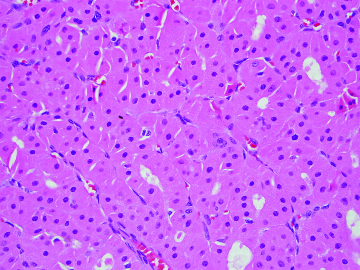
The origin of this vanishing behavior on T1 FS C+ WI and T2 FS MR is unknown. Our hypothesis is based upon the subcellular structure of normal parotid tissue and oncocytes. A normal parotid gland is equally composed of epithelial cells (acinar and ductal cells) and adipocytes (fat cells). An oncocytoma consists of a pure population of oncocytes arranged in solid nests of sheets.14 Innumerable mitochondria fill the oncocytes, producing an abundant deeply eosinophilic finely granular cytoplasm, the most striking feature by light microscopy, and resulting in a paucity of other organelles (eg, DNA, protein; Fig 4).15 The cell body of the oncocytes is otherwise composed primarily of membrane lipids. The low free water content and rich lipid membrane composition might approximate the lipid content of the normal parotid gland, thereby explaining why these lesions appeared isointense to parotid gland on T1 FS C+ WI and T2 FS WI and appeared to vanish. Were this hypothesis to be correct, however, one might expect higher intrinsic T1 signal intensity on the precontrast T1WI. Management of parotid gland oncocytoma is complete surgical resection by parotidectomy. Preoperative identification of correct histology and tumor extent may help in presurgical patient preparation and in surgical planning, and in the case of a benign neoplasm, patient reassurance.

Fig 4.
  • Download figure
  • Open in new tab
  • Download powerpoint
Fig 4.

Typical histologic features of a parotid gland oncocytoma including eosinophilic cells with finely granular cytoplasm and uniform, round, centrally placed nuclei (hematoxylin-eosin stain, original magnification, ×400).

Conclusions

Parotid gland oncocytomas seem to share unique imaging characteristics that have not been described for other benign or malignant parotid gland tumors. These imaging characteristics may help in the preoperative determination of a benign parotid tumor. We found oncocytomas to be hypointense, well-demarcated lesions on T1-weighted sequences. On T1 FS C+ WI and T2 FS WI this tumor seems to vanish or become isointense to parotid tissue. Preoperative identification of correct histology may help surgical planning. We believe this further validates the utility of MR for the preoperative evaluation of neoplastic parotid masses.

Footnotes

  • Paper previously presented at Annual Meeting of the American Society of Head and Neck Radiology, October 2, 2010; Houston, Texas.

  • Disclosures: Hugh Harnsberger, Ownership Interest: Amirsys Inc. Details: CEO, Amirsys Inc., has no relationship to this paper; Christine M. Glastonbury, Consultant: Amirsys Inc. Details: Author and editor of textbook chapters; editor and author of StatDx. Ownership Interest: Amirsys. Details: Investor in company.

References

  1. 1.↵
    1. Barnes L,
    2. Eveson JW,
    3. Reichart P,
    4. et al.
    1. Huvos AG
    .Oncocytoma. In: Barnes L, Eveson JW, Reichart P, et al. , eds. World Health Organization Classification of Tumors: Pathology and Genetics, Head and Neck Tumors. Lyon, France: IARC Press; 2005: 242–43
  2. 2.↵
    1. Brandwein MS,
    2. Huvos AG
    . Oncocytic tumors of major salivary glands: a study of 68 cases with follow-up of 44 patients. Am J Surg Pathol 1991; 15: 514–28
    PubMed
  3. 3.↵
    1. Shah VN,
    2. Branstetter BF
    . Oncocytoma of the parotid gland: a potential false-positive finding on 18F-FDG PET. AJR Am J Roentgenol 2007; 189: W212–14
    CrossRefPubMed
  4. 4.↵
    1. Branstetter BF IV.,
    2. Blodgett TM,
    3. Zimmer LA,
    4. et al
    . Head and neck malignancy: is PET/CT more accurate than PET or CT alone? Radiology 2005; 235: 580–86
    PubMed
  5. 5.↵
    1. Tan TJ,
    2. Tan TY
    . CT features of parotid gland oncocytomas: a study of 10 cases and literature review AJNR Am J Neuroradiol 2010; 31: 1413–17
    Abstract/FREE Full Text
  6. 6.↵
    1. Lev MH,
    2. Khanduja K,
    3. Morris PP,
    4. et al
    . Parotid pleomorphic adenomas: delayed CT enhancement. AJNR Am J Neuroradiol 1998; 19: 941–66
  7. 7.↵
    1. Kei PL,
    2. Tan TY
    . CT “invisible” lesion of the major salivary glands a diagnostic pitfall of contrast-enhanced CT. Clin Radiol 2009; 64: 744–46
    CrossRefPubMed
  8. 8.↵
    1. Alps HH,
    2. Eisele DW,
    3. Westra WH
    . The role of fine needle aspiration in the evaluation of parotid masses. Curr Opin Otolaryngol Head Neck Surg 2006; 14: 62–66
    CrossRefPubMed
  9. 9.↵
    1. Capone RB,
    2. Ha PK,
    3. Westra WH,
    4. et al
    . Oncocytic neoplasms of the parotid gland: a 16-year institutional review. Otolaryngol Head Neck Surg 2002; 126: 657–62
    CrossRefPubMed
  10. 10.↵
    1. Yousem DM,
    2. Kraut MA,
    3. Chalian AA
    . Major salivary gland imaging. Radiology 2000; 216: 19–29
    CrossRefPubMed
  11. 11.↵
    1. Joe VQ,
    2. Westesson PL
    . Tumors of the parotid gland: MR imaging characteristics of various histologic types. AJR Am J Roentgenol 1994; 163: 433–38
    PubMed
  12. 12.↵
    1. Sakai E,
    2. Yoda T,
    3. Shimamoto H,
    4. et al
    . Pathologic and imaging findings of an oncocytoma in the deep lobe of the left parotid gland. J Oral Maxillofac Surg 2003; 32: 563–65
  13. 13.↵
    1. Kasai T,
    2. Motoori K,
    3. Hanazawa T,
    4. et al
    . MR imaging of multinodular bilateral oncocytoma of the parotid gland. Eur J Radiol 2007; 63: 97–100
    CrossRef
  14. 14.↵
    1. Zhou CX,
    2. Gao Y
    . Oncocytoma of the salivary glands: a clinicopathologic and immunohistochemical study. Oral Oncology 2009; e232–238
  15. 15.↵
    1. Wakely PE Jr.
    . Oncocytic and oncocyte-like lesions of the head and neck. Ann Diagn Pathol 2008; 12: 222–30
    CrossRefPubMed
  • Received January 11, 2011.
  • Accepted after revision January 24, 2011.
  • © 2011 by American Journal of Neuroradiology
View Abstract
PreviousNext
Back to top

In this issue

American Journal of Neuroradiology: 32 (9)
American Journal of Neuroradiology
Vol. 32, Issue 9
1 Oct 2011
  • Table of Contents
  • Index by author
Advertisement
Print
Download PDF
Email Article

Thank you for your interest in spreading the word on American Journal of Neuroradiology.

NOTE: We only request your email address so that the person you are recommending the page to knows that you wanted them to see it, and that it is not junk mail. We do not capture any email address.

Enter multiple addresses on separate lines or separate them with commas.
Oncocytoma: The Vanishing Parotid Mass
(Your Name) has sent you a message from American Journal of Neuroradiology
(Your Name) thought you would like to see the American Journal of Neuroradiology web site.
CAPTCHA
This question is for testing whether or not you are a human visitor and to prevent automated spam submissions.
Cite this article
N.D. Patel, A. van Zante, D.W. Eisele, H.R. Harnsberger, C.M. Glastonbury
Oncocytoma: The Vanishing Parotid Mass
American Journal of Neuroradiology Oct 2011, 32 (9) 1703-1706; DOI: 10.3174/ajnr.A2569

Citation Manager Formats

  • BibTeX
  • Bookends
  • EasyBib
  • EndNote (tagged)
  • EndNote 8 (xml)
  • Medlars
  • Mendeley
  • Papers
  • RefWorks Tagged
  • Ref Manager
  • RIS
  • Zotero
0 Responses
Respond to this article
Share
Bookmark this article
Oncocytoma: The Vanishing Parotid Mass
N.D. Patel, A. van Zante, D.W. Eisele, H.R. Harnsberger, C.M. Glastonbury
American Journal of Neuroradiology Oct 2011, 32 (9) 1703-1706; DOI: 10.3174/ajnr.A2569
del.icio.us logo Twitter logo Facebook logo Mendeley logo
  • Tweet Widget
  • Facebook Like
  • Google Plus One
Purchase

Jump to section

  • Article
    • Abstract
    • Abbreviations
    • Materials and Methods
    • Results
    • Discussion
    • Conclusions
    • Footnotes
    • References
  • Figures & Data
  • Info & Metrics
  • Responses
  • References
  • PDF

Related Articles

  • No related articles found.
  • PubMed
  • Google Scholar

Cited By...

  • No citing articles found.
  • Crossref (32)
  • Google Scholar

This article has been cited by the following articles in journals that are participating in Crossref Cited-by Linking.

  • State-of-the-Art Imaging of Salivary Gland Tumors
    Ahmed Abdel Khalek Abdel Razek, Suresh K. Mukherji
    Neuroimaging Clinics of North America 2018 28 2
  • Usefulness of diffusion-weighted MR imaging for differentiating between Warthin’s tumor and oncocytoma of the parotid gland
    Hiroki Kato, Keita Fujimoto, Masayuki Matsuo, Keisuke Mizuta, Mitsuhiro Aoki
    Japanese Journal of Radiology 2017 35 2
  • Review of the Major and Minor Salivary Glands, Part 2: Neoplasms and Tumor-like Lesions
    Alexander T Kessler, Alok A Bhatt
    Journal of Clinical Imaging Science 2018 8
  • Imaging Evaluation of the Suprahyoid Neck
    Caryn Gamss, Ajay Gupta, J. Levi Chazen, C. Douglas Phillips
    Radiologic Clinics of North America 2015 53 1
  • The key radiologic and cytomorphologic features of oncocytic and oncocytoid lesions of the salivary gland
    Daniel Lubin, Sharon Song, Hanna M. Zafar, Zubair Baloch
    Diagnostic Cytopathology 2019 47 6
  • Imaging of Benign Neoplastic and Nonneoplastic Salivary Gland Tumors
    Daniel Thomas Ginat
    Neuroimaging Clinics of North America 2018 28 2
  • Bilateral parotid oncocytoma with spontaneous intratumoral hemorrhage: a rare hypervascular parotid tumor with ASL perfusion
    Etsushi Iida, Richard H. Wiggins, Yoshimi Anzai
    Clinical Imaging 2016 40 3
  • Diagnostic imaging of parotid gland oncocytoma: a pictorial review with emphasis on ultrasound assessment
    Antonio Corvino, Martina Caruso, Carlo Varelli, Francesca Di Gennaro, Saverio Pignata, Fabio Corvino, Gianfranco Vallone, Orlando Catalano
    Journal of Ultrasound 2021 24 3
  • Parotid Oncocytoma as a Manifestation of Birt-Hogg-Dubé Syndrome
    Kazuki Yoshida, Masao Miyagawa, Teruhito Kido, Kana Ide, Yoshifumi Sano, Yoshifumi Sugawara, Hiroyuki Takahata, Nobuya Monden, Mitsuko Furuya, Teruhito Mochizuki
    Case Reports in Radiology 2018 2018
  • Imaging of Major Salivary Gland Lesions and Disease
    Elliott Friedman, Yu Cai, Bo Chen
    Oral and Maxillofacial Surgery Clinics of North America 2023 35 3

More in this TOC Section

  • Correlation of Apparent Diffusion Coefficient at 3T with Prognostic Parameters of Retinoblastoma
  • Parathyroid Lesions: Characterization with Dual-Phase Arterial and Venous Enhanced CT of the Neck
  • MR Evaluation of Sinonasal Angiomatous Polyp
Show more Head and Neck

Similar Articles

Advertisement

Indexed Content

  • Current Issue
  • Accepted Manuscripts
  • Article Preview
  • Past Issues
  • Editorials
  • Editors Choice
  • Fellow Journal Club
  • Letters to the Editor

Cases

  • Case Collection
  • Archive - Case of the Week
  • Archive - Case of the Month
  • Archive - Classic Case

Special Collections

  • Special Collections

Resources

  • News and Updates
  • Turn around Times
  • Submit a Manuscript
  • Author Policies
  • Manuscript Submission Guidelines
  • Evidence-Based Medicine Level Guide
  • Publishing Checklists
  • Graphical Abstract Preparation
  • Imaging Protocol Submission
  • Submit a Case
  • Become a Reviewer/Academy of Reviewers
  • Get Peer Review Credit from Publons

Multimedia

  • AJNR Podcast
  • AJNR SCANtastic
  • Video Articles

About Us

  • About AJNR
  • Editorial Board
  • Not an AJNR Subscriber? Join Now
  • Alerts
  • Feedback
  • Advertise with us
  • Librarian Resources
  • Permissions
  • Terms and Conditions

American Society of Neuroradiology

  • Not an ASNR Member? Join Now

© 2025 by the American Society of Neuroradiology All rights, including for text and data mining, AI training, and similar technologies, are reserved.
Print ISSN: 0195-6108 Online ISSN: 1936-959X

Powered by HighWire