Advertisement

Main menu

  • Home
  • Content
    • Current Issue
    • Accepted Manuscripts
    • Article Preview
    • Past Issue Archive
    • AJNR Case Collection
    • Case of the Week Archive
    • Classic Case Archive
    • Case of the Month Archive
  • Special Collections
    • Spinal CSF Leak Articles (Jan 2020-June 2024)
    • 2024 AJNR Journal Awards
    • Most Impactful AJNR Articles
  • Multimedia
    • AJNR Podcast
    • AJNR Scantastics
    • Video Articles
  • For Authors
    • Submit a Manuscript
    • Author Policies
    • Fast publishing of Accepted Manuscripts
    • Graphical Abstract Preparation
    • Manuscript Submission Guidelines
    • Imaging Protocol Submission
    • Submit a Case for the Case Collection
  • About Us
    • About AJNR
    • Editorial Board
  • More
    • Become a Reviewer/Academy of Reviewers
    • Subscribers
    • Permissions
    • Alerts
    • Feedback
    • Advertisers
    • ASNR Home
  • Other Publications
    • ajnr

User menu

  • Alerts
  • Log in

Search

  • Advanced search
American Journal of Neuroradiology

American Journal of Neuroradiology

ASHNR American Society of Functional Neuroradiology ASHNR American Society of Pediatric Neuroradiology ASSR
  • Alerts
  • Log in

Advanced Search

  • Home
  • Content
    • Current Issue
    • Accepted Manuscripts
    • Article Preview
    • Past Issue Archive
    • AJNR Case Collection
    • Case of the Week Archive
    • Classic Case Archive
    • Case of the Month Archive
  • Special Collections
    • Spinal CSF Leak Articles (Jan 2020-June 2024)
    • 2024 AJNR Journal Awards
    • Most Impactful AJNR Articles
  • Multimedia
    • AJNR Podcast
    • AJNR Scantastics
    • Video Articles
  • For Authors
    • Submit a Manuscript
    • Author Policies
    • Fast publishing of Accepted Manuscripts
    • Graphical Abstract Preparation
    • Manuscript Submission Guidelines
    • Imaging Protocol Submission
    • Submit a Case for the Case Collection
  • About Us
    • About AJNR
    • Editorial Board
  • More
    • Become a Reviewer/Academy of Reviewers
    • Subscribers
    • Permissions
    • Alerts
    • Feedback
    • Advertisers
    • ASNR Home
  • Follow AJNR on Twitter
  • Visit AJNR on Facebook
  • Follow AJNR on Instagram
  • Join AJNR on LinkedIn
  • RSS Feeds

Welcome to the new AJNR, Updated Hall of Fame, and more. Read the full announcements.


AJNR is seeking candidates for the position of Associate Section Editor, AJNR Case Collection. Read the full announcement.


Getting new auth cookie, if you see this message a lot, tell someone!
Research ArticleMethodologic Perspectives
Open Access

An Expanded Role for Neuroimaging in the Evaluation of Memory Impairment

R.S. Desikan, M.S. Rafii, J.B. Brewer and C.P. Hess
American Journal of Neuroradiology November 2013, 34 (11) 2075-2082; DOI: https://doi.org/10.3174/ajnr.A3644
R.S. Desikan
aFrom the Departments of Radiology (R.S.D., J.B.B.)
  • Find this author on Google Scholar
  • Find this author on PubMed
  • Search for this author on this site
M.S. Rafii
bNeurosciences (M.S.R., J.B.B.), University of California, San Diego, La Jolla, California
  • Find this author on Google Scholar
  • Find this author on PubMed
  • Search for this author on this site
J.B. Brewer
aFrom the Departments of Radiology (R.S.D., J.B.B.)
bNeurosciences (M.S.R., J.B.B.), University of California, San Diego, La Jolla, California
  • Find this author on Google Scholar
  • Find this author on PubMed
  • Search for this author on this site
C.P. Hess
cNeuroradiology Section, Department of Radiology and Biomedical Imaging (C.P.H.), University of California, San Francisco, San Francisco, California.
  • Find this author on Google Scholar
  • Find this author on PubMed
  • Search for this author on this site
  • Article
  • Figures & Data
  • Supplemental
  • Info & Metrics
  • Responses
  • References
  • PDF
Loading

Article Figures & Data

Figures

  • Tables
  • Fig 1.
    • Download figure
    • Open in new tab
    • Download powerpoint
    Fig 1.

    Brain MR imaging evaluation of a patient with amnestic MCI by use of a volumetric technique (NeuroQuant, http://www.cortechslabs.com). The top panel illustrates subcortical regions, such as the hippocampus (dark yellow), automatically classified on axial, coronal, and sagittal T1-weighted MR images. The middle and bottom panel demonstrate volumes and normative percentiles for the hippocampus and ventricles. Analyses of the baseline MR imaging scan demonstrated hippocampal volumes that were at the < 1 normative percentile, lending objective support to an impression of medial temporal lobe atrophy. At the time of volumetric assessment, the patient's Mini-Mental Status Examination score was 29 of 30, yet memory impairment was suggested by more detailed neuropsychological testing. Three years later, his Mini-Mental Status Examination score was 22 of 30, and he had clinically progressed to dementia with high biomarker probability of AD, as supported by evidence of neuronal injury on structural MR imaging and elevated amyloid levels on a florbetapir scan (Fig 3).
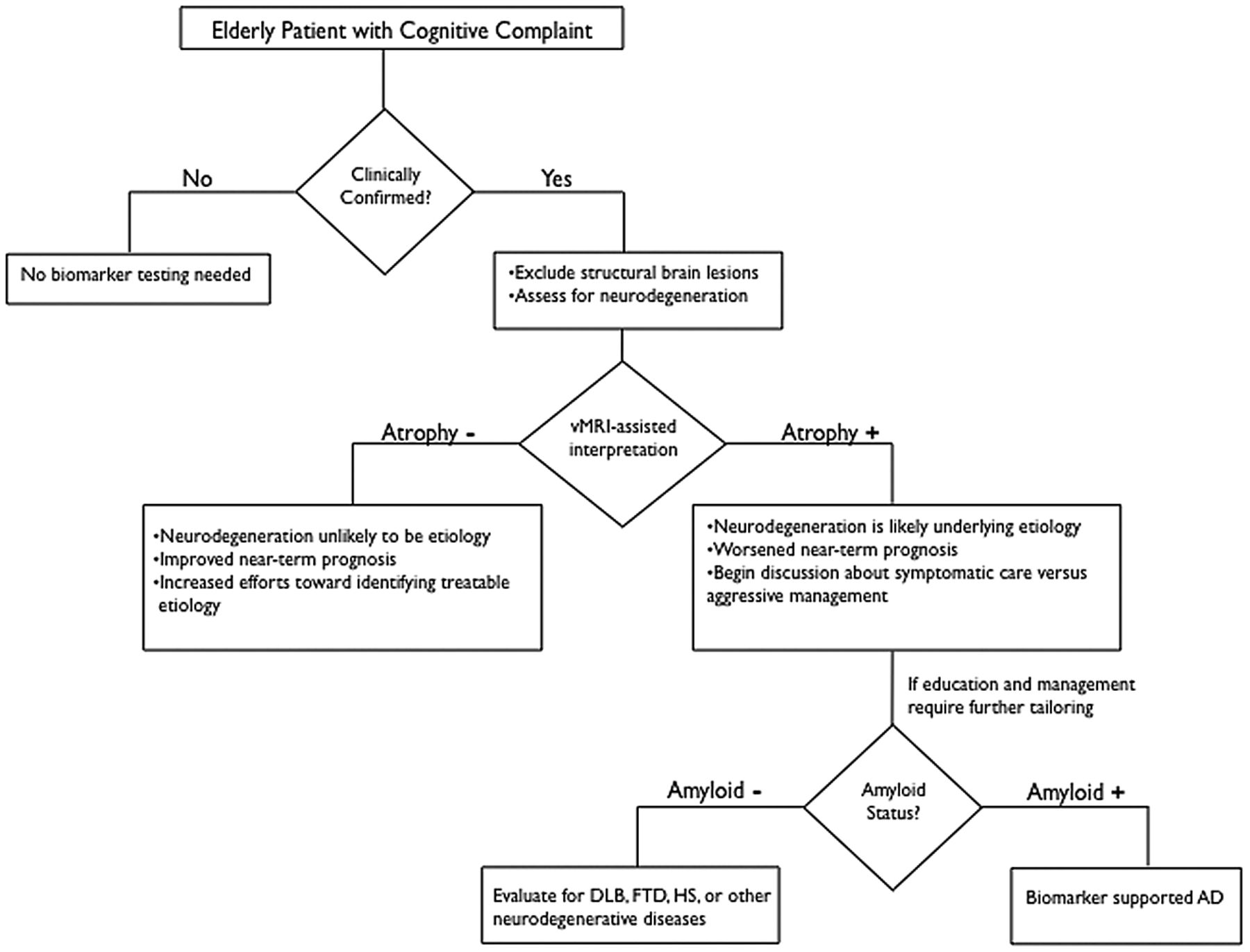
  • Fig 2.
    • Download figure
    • Open in new tab
    • Download powerpoint
    Fig 2.

    Recommended decision tree for evaluating the elderly patient with a cognitive complaint. DLB = Dementia with Lewy bodies; HS = hippocampal sclerosis. Figure adapted from McEvoy and Brewer.48

  • Fig 3.
    • Download figure
    • Open in new tab
    • Download powerpoint
    Fig 3.

    Assessment of amyloid deposition by use of florbetapir (Amyvid). The axial PET image on the left shows normal preserved gray-white contrast with the cortical radioactivity less than the adjacent white matter (amyloid-“negative” scan). The axial PET image on the right demonstrates areas of decreased gray-white contrast with increased cortical radioactivity that is comparable to the radioactivity in the adjacent white matter (amyloid-“positive” scan). The florbetapir scan on the right was acquired on a patient with MCI who clinically progressed to dementia with a high biomarker probability of AD, as supported by this amyloid-positive scan and evidence of neuronal injury on structural MR imaging (Fig 1).

Tables

  • Figures
    • View popup
    Table 1:

    Clinical features in amnestic patients with MCI

    Clinical Characteristics
    Memory ImpairmentEpisodic Memory Dysfunction
    Nonmemory cognitive impairmentExecutive dysfunction, apraxia, aphasia, and/or visuospatial dysfunction may be present in amnestic MCI multidomain
    Functional impairmentNo change in ability to perform activities of daily living
    Behavioral impairmentDepression and anxiety may be present
    Annual rate of progression to dementiaVariable (range, 3%–15%)
    • View popup
    Table 2:

    Disease progression markers in amnestic patients with MCI

    Markers of Disease ProgressionCharacteristicsProcedure(s)aApproximate Cost (in US dollars)b
    Structural neuroimaging with vMRIMedial temporal lobe and/or neocortical atrophy; white matter abnormalities may also be present1) Noncontrast MRI brain CPT 705511) 437.20 (365.75f+71.45g)
    2) 3D quantitative segmental volume reporting and assessmentc CPT 763772) 82.68 (44.57f+38.11g)
    FDG-PETTemporoparietal hypometabolismBrain imaging (PET) metabolic evaluation CPT 786081266.40 (1041.99f+150i+74.41g)
    Amyloid imagingIncreased uptake in frontal, parietal, and/or temporal regionsPET imaging limited area CPT 788112721.83 (1041.99f+1600i+79.84g)
    CSF amyloidDecreased1) CSF lumbar puncture CPT 622701) 242.58 (78.93h+163.65g)
    CSF tau (total tau)Increased2) CSF analysis and interpretationd CPT 835202) 1080
    APOE ϵ4 carrier statusDose-dependent effect (risk for AD: ϵ4/ϵ4 > ϵ3/ϵ4 > ϵ3/ϵ3 > ϵ3/ϵ2 > ϵ2/ϵ2)1) Buccal swab or routine venipuncture CPT 364151) 3
    2) APOE genotype analysis and interpretatione CPT 814012) 500
    • Note:—APOE ϵ4 indicates apolipoprotein E4; CPT, Current Procedural Terminology; vMRI, volumetric-based MR imaging.

    • ↵a Determined using data from the Centers for Medicare and Medicaid Services (www.cms.gov). For informational purposes only. Selected CPT code may vary.

    • ↵b Determined, when possible, using National Payment Amount data from the Centers for Medicare and Medicaid Services (www.cms.gov). For informational purposes only. Payment amount varies by location.

    • ↵c Using NeuroQuant (http://www.cortechs.net/products/neuroquant.php).

    • ↵d Using the ADmark Phospho-Tau/Total-Tau/Ab42 CSF Analysis & Interpretation (Symptomatic) test (http://www.athenadiagnostics.com/content/test-catalog/find-test/service-detail/q/id/311).

    • ↵e Using the ADmark ApoE Genotype Analysis & Interpretation (Symptomatic) (http://www.athenadiagnostics.com/content/test-catalog/find-test/service-detail/q/id/35).

    • ↵f Approximate technical charge.

    • ↵g Approximate professional charge.

    • ↵h Approximate facility price.

    • ↵i Approximate ligand price.

PreviousNext
Back to top

In this issue

American Journal of Neuroradiology: 34 (11)
American Journal of Neuroradiology
Vol. 34, Issue 11
1 Nov 2013
  • Table of Contents
  • Index by author
  • Complete Issue (PDF)
Advertisement
Print
Download PDF
Email Article

Thank you for your interest in spreading the word on American Journal of Neuroradiology.

NOTE: We only request your email address so that the person you are recommending the page to knows that you wanted them to see it, and that it is not junk mail. We do not capture any email address.

Enter multiple addresses on separate lines or separate them with commas.
An Expanded Role for Neuroimaging in the Evaluation of Memory Impairment
(Your Name) has sent you a message from American Journal of Neuroradiology
(Your Name) thought you would like to see the American Journal of Neuroradiology web site.
CAPTCHA
This question is for testing whether or not you are a human visitor and to prevent automated spam submissions.
Cite this article
R.S. Desikan, M.S. Rafii, J.B. Brewer, C.P. Hess
An Expanded Role for Neuroimaging in the Evaluation of Memory Impairment
American Journal of Neuroradiology Nov 2013, 34 (11) 2075-2082; DOI: 10.3174/ajnr.A3644

Citation Manager Formats

  • BibTeX
  • Bookends
  • EasyBib
  • EndNote (tagged)
  • EndNote 8 (xml)
  • Medlars
  • Mendeley
  • Papers
  • RefWorks Tagged
  • Ref Manager
  • RIS
  • Zotero
0 Responses
Respond to this article
Share
Bookmark this article
An Expanded Role for Neuroimaging in the Evaluation of Memory Impairment
R.S. Desikan, M.S. Rafii, J.B. Brewer, C.P. Hess
American Journal of Neuroradiology Nov 2013, 34 (11) 2075-2082; DOI: 10.3174/ajnr.A3644
del.icio.us logo Twitter logo Facebook logo Mendeley logo
  • Tweet Widget
  • Facebook Like
  • Google Plus One
Purchase

Jump to section

  • Article
    • Abstract
    • ABBREVIATIONS:
    • Acknowledgments
    • Footnotes
    • REFERENCES
  • Figures & Data
  • Supplemental
  • Info & Metrics
  • Responses
  • References
  • PDF

Related Articles

  • PubMed
  • Google Scholar

Cited By...

  • Automated Segmentation of Hippocampal Volume: The Next Step in Neuroradiologic Diagnosis of Mesial Temporal Sclerosis
  • Polygenic hazard score: an enrichment marker for Alzheimers associated amyloid and tau deposition
  • Entorhinal Cortex: Antemortem Cortical Thickness and Postmortem Neurofibrillary Tangles and Amyloid Pathology
  • Crossref (23)
  • Google Scholar

This article has been cited by the following articles in journals that are participating in Crossref Cited-by Linking.

  • Polygenic hazard score: an enrichment marker for Alzheimer’s associated amyloid and tau deposition
    Chin Hong Tan, Chun Chieh Fan, Elizabeth C. Mormino, Leo P. Sugrue, Iris J. Broce, Christopher P. Hess, William P. Dillon, Luke W. Bonham, Jennifer S. Yokoyama, Celeste M. Karch, James B. Brewer, Gil D. Rabinovici, Bruce L. Miller, Gerard D. Schellenberg, Karolina Kauppi, Howard A. Feldman, Dominic Holland, Linda K. McEvoy, Bradley T. Hyman, David A. Bennett, Ole A. Andreassen, Anders M. Dale, Rahul S. Desikan
    Acta Neuropathologica 2018 135 1
  • Comparison of Automated Brain Volume Measures obtained with NeuroQuant® and FreeSurfer
    Alfred L. Ochs, David E. Ross, Megan D. Zannoni, Tracy J. Abildskov, Erin D. Bigler
    Journal of Neuroimaging 2015 25 5
  • Late-onset Alzheimer Disease
    Gil D. Rabinovici
    CONTINUUM: Lifelong Learning in Neurology 2019 25 1
  • Quantitative Neuroimaging Software for Clinical Assessment of Hippocampal Volumes on MR Imaging
    Jamila Ahdidan, Cyrus A. Raji, Edgar A. DeYoe, Jedidiah Mathis, Karsten Ø. Noe, Jens Rimestad, Thomas K. Kjeldsen, Jesper Mosegaard, James T. Becker, Oscar Lopez
    Journal of Alzheimer's Disease 2015 49 3
  • Entorhinal Cortex: Antemortem Cortical Thickness and Postmortem Neurofibrillary Tangles and Amyloid Pathology
    A.A. Thaker, B.D. Weinberg, W.P. Dillon, C.P. Hess, H.J. Cabral, D.A. Fleischman, S.E. Leurgans, D.A. Bennett, B.T. Hyman, M.S. Albert, R.J. Killiany, B. Fischl, A.M. Dale, R.S. Desikan
    American Journal of Neuroradiology 2017 38 5
  • Diagnostic Efficacy of Structural MRI in Patients With Mild-to-Moderate Alzheimer Disease: Automated Volumetric Assessment Versus Visual Assessment
    Jeeyoung Min, Won-Jin Moon, Ji Young Jeon, Jin Woo Choi, Yeon-Sil Moon, Seol-Heui Han
    American Journal of Roentgenology 2017 208 3
  • PET and Single-Photon Emission Computed Tomography in Brain Concussion
    Cyrus A. Raji, Theodore A. Henderson
    Neuroimaging Clinics of North America 2018 28 1
  • Handgrip Strength Is Related to Hippocampal and Lobar Brain Volumes in a Cohort of Cognitively Impaired Older Adults with Confirmed Amyloid Burden
    Somayeh Meysami, Cyrus A. Raji, Ryan M. Glatt, Emily S. Popa, Aarthi S. Ganapathi, Tess Bookheimer, Colby B. Slyapich, Kyron P. Pierce, Casey J. Richards, Melanie G. Lampa, Jaya M. Gill, Molly K. Rapozo, John F. Hodes, Ynez M. Tongson, Claudia L. Wong, Mihae Kim, Verna R. Porter, Scott A. Kaiser, Stella E. Panos, Richelin V. Dye, Karen J. Miller, Susan Y. Bookheimer, Neil A. Martin, Santosh Kesari, Daniel F. Kelly, Jennifer E. Bramen, Prabha Siddarth, David A. Merrill
    Journal of Alzheimer's Disease 2023 91 3
  • Neuroimaging Part I
    Mykol Larvie, Bruce Fischl
    2016 135
  • Brain Networks, Clinical Manifestations, and Neuroimaging of Cognitive Disorders
    Borna Bonakdarpour, Clara Takarabe
    Clinics in Geriatric Medicine 2023 39 1

More in this TOC Section

  • A Meta-Analysis on the Diagnostic Performance of 18F-FDG and 11C-Methionine PET for Differentiating Brain Tumors
  • Multivariate Classification of Blood Oxygen Level–Dependent fMRI Data with Diagnostic Intention: A Clinical Perspective
  • Guidelines for the Ethical Use of Neuroimages in Medical Testimony: Report of a Multidisciplinary Consensus Conference
Show more Methodologic Perspectives

Similar Articles

Advertisement

Indexed Content

  • Current Issue
  • Accepted Manuscripts
  • Article Preview
  • Past Issues
  • Editorials
  • Editors Choice
  • Fellow Journal Club
  • Letters to the Editor

Cases

  • Case Collection
  • Archive - Case of the Week
  • Archive - Case of the Month
  • Archive - Classic Case

Special Collections

  • Special Collections

Resources

  • News and Updates
  • Turn around Times
  • Submit a Manuscript
  • Author Policies
  • Manuscript Submission Guidelines
  • Evidence-Based Medicine Level Guide
  • Publishing Checklists
  • Graphical Abstract Preparation
  • Imaging Protocol Submission
  • Submit a Case
  • Become a Reviewer/Academy of Reviewers
  • Get Peer Review Credit from Publons

Multimedia

  • AJNR Podcast
  • AJNR SCANtastic
  • Video Articles

About Us

  • About AJNR
  • Editorial Board
  • Not an AJNR Subscriber? Join Now
  • Alerts
  • Feedback
  • Advertise with us
  • Librarian Resources
  • Permissions
  • Terms and Conditions

American Society of Neuroradiology

  • Not an ASNR Member? Join Now

© 2025 by the American Society of Neuroradiology All rights, including for text and data mining, AI training, and similar technologies, are reserved.
Print ISSN: 0195-6108 Online ISSN: 1936-959X

Powered by HighWire