Advertisement

Main menu

  • Home
  • Content
    • Current Issue
    • Accepted Manuscripts
    • Article Preview
    • Past Issue Archive
    • AJNR Case Collection
    • Case of the Week Archive
    • Classic Case Archive
    • Case of the Month Archive
  • Special Collections
    • Spinal CSF Leak Articles (Jan 2020-June 2024)
    • 2024 AJNR Journal Awards
    • Most Impactful AJNR Articles
  • Multimedia
    • AJNR Podcast
    • AJNR Scantastics
    • Video Articles
  • For Authors
    • Submit a Manuscript
    • Author Policies
    • Fast publishing of Accepted Manuscripts
    • Graphical Abstract Preparation
    • Manuscript Submission Guidelines
    • Imaging Protocol Submission
    • Submit a Case for the Case Collection
  • About Us
    • About AJNR
    • Editorial Board
  • More
    • Become a Reviewer/Academy of Reviewers
    • Subscribers
    • Permissions
    • Alerts
    • Feedback
    • Advertisers
    • ASNR Home
  • Other Publications
    • ajnr

User menu

  • Alerts
  • Log in

Search

  • Advanced search
American Journal of Neuroradiology

American Journal of Neuroradiology

ASHNR American Society of Functional Neuroradiology ASHNR American Society of Pediatric Neuroradiology ASSR
  • Alerts
  • Log in

Advanced Search

  • Home
  • Content
    • Current Issue
    • Accepted Manuscripts
    • Article Preview
    • Past Issue Archive
    • AJNR Case Collection
    • Case of the Week Archive
    • Classic Case Archive
    • Case of the Month Archive
  • Special Collections
    • Spinal CSF Leak Articles (Jan 2020-June 2024)
    • 2024 AJNR Journal Awards
    • Most Impactful AJNR Articles
  • Multimedia
    • AJNR Podcast
    • AJNR Scantastics
    • Video Articles
  • For Authors
    • Submit a Manuscript
    • Author Policies
    • Fast publishing of Accepted Manuscripts
    • Graphical Abstract Preparation
    • Manuscript Submission Guidelines
    • Imaging Protocol Submission
    • Submit a Case for the Case Collection
  • About Us
    • About AJNR
    • Editorial Board
  • More
    • Become a Reviewer/Academy of Reviewers
    • Subscribers
    • Permissions
    • Alerts
    • Feedback
    • Advertisers
    • ASNR Home
  • Follow AJNR on Twitter
  • Visit AJNR on Facebook
  • Follow AJNR on Instagram
  • Join AJNR on LinkedIn
  • RSS Feeds

Welcome to the new AJNR, Updated Hall of Fame, and more. Read the full announcements.


AJNR is seeking candidates for the position of Associate Section Editor, AJNR Case Collection. Read the full announcement.


Research ArticlePediatrics
Open Access

Development of the Fetal Cerebral Cortex in the Second Trimester: Assessment with 7T Postmortem MR Imaging

Z. Zhang, Z. Hou, X. Lin, G. Teng, H. Meng, F. Zang, F. Fang and S. Liu
American Journal of Neuroradiology July 2013, 34 (7) 1462-1467; DOI: https://doi.org/10.3174/ajnr.A3406
Z. Zhang
aFrom the Research Center for Sectional and Imaging Anatomy (Z.Z., Z.H., X.L., H.M., S.L.), Shandong University School of Medicine, Shandong, China
bDepartment of Medical Imaging (Z.Z., Z.H.), Provincial Hospital Affiliated with Shandong University, Shandong, China
  • Find this author on Google Scholar
  • Find this author on PubMed
  • Search for this author on this site
Z. Hou
aFrom the Research Center for Sectional and Imaging Anatomy (Z.Z., Z.H., X.L., H.M., S.L.), Shandong University School of Medicine, Shandong, China
bDepartment of Medical Imaging (Z.Z., Z.H.), Provincial Hospital Affiliated with Shandong University, Shandong, China
  • Find this author on Google Scholar
  • Find this author on PubMed
  • Search for this author on this site
X. Lin
aFrom the Research Center for Sectional and Imaging Anatomy (Z.Z., Z.H., X.L., H.M., S.L.), Shandong University School of Medicine, Shandong, China
cDepartment of MR (X.L.), Shandong Medical Imaging Research Institute, Shandong, China
  • Find this author on Google Scholar
  • Find this author on PubMed
  • Search for this author on this site
G. Teng
dDepartment of Radiology (G.T., F.Z., F.F.), Zhong Da Hospital, Southeast University School of Clinical Medicine, Jiangsu, China.
  • Find this author on Google Scholar
  • Find this author on PubMed
  • Search for this author on this site
H. Meng
aFrom the Research Center for Sectional and Imaging Anatomy (Z.Z., Z.H., X.L., H.M., S.L.), Shandong University School of Medicine, Shandong, China
  • Find this author on Google Scholar
  • Find this author on PubMed
  • Search for this author on this site
F. Zang
dDepartment of Radiology (G.T., F.Z., F.F.), Zhong Da Hospital, Southeast University School of Clinical Medicine, Jiangsu, China.
  • Find this author on Google Scholar
  • Find this author on PubMed
  • Search for this author on this site
F. Fang
dDepartment of Radiology (G.T., F.Z., F.F.), Zhong Da Hospital, Southeast University School of Clinical Medicine, Jiangsu, China.
  • Find this author on Google Scholar
  • Find this author on PubMed
  • Search for this author on this site
S. Liu
aFrom the Research Center for Sectional and Imaging Anatomy (Z.Z., Z.H., X.L., H.M., S.L.), Shandong University School of Medicine, Shandong, China
  • Find this author on Google Scholar
  • Find this author on PubMed
  • Search for this author on this site
  • Article
  • Figures & Data
  • Supplemental
  • Info & Metrics
  • Responses
  • References
  • PDF
Loading

Article Figures & Data

Figures

  • Tables
  • Fig 1.
    • Download figure
    • Open in new tab
    • Download powerpoint
    Fig 1.

    Methods of image segmentation with Amira 4.1. A, The original image. B, Segmentation of the hemisphere. C, Different colors are filled in the structures after segmentation. D, 3D visualization model and measurements of brain length, width, and height.

  • Fig 2.
    • Download figure
    • Open in new tab
    • Download powerpoint
    Fig 2.

    Transverse T2-weighted 7T MR images of 12 (A, E), 16 (B, F), 20 (C, G), and 22 (D, H) weeks GA. Sagittal T2-weighted 7T MR images of 13 (I), 16 (J), 18 (K), and 20 (L) weeks GA. The lateral sulcus and the interhemispheric fissure can be distinguished at 12 weeks GA (A, E). At 16 weeks GA, more sulci can be observed (B, F), such as the central sulcus and the superior frontal sulcus. At 20–22 weeks GA, the sulci are more clearly delineated (E–H). On the sagittal images, development of the calcarine fissure, the parieto-occipital sulcus, the central sulcus, and the superior frontal sulcus can be clearly distinguished. The bar in each figure represents 1 cm; if, interhemisphaeric fissure; cas, callosal sulcus; cis, cingular sulcus; cf, calcarine fissure; pof, parieto-occipital sulcus; ots, occipitotemporal sulcus; las, lateral sulcus; sfs, superior frontal sulcus; its, inferior temporal sulcus; pcs, precentral sulcus; cns, central sulcus.
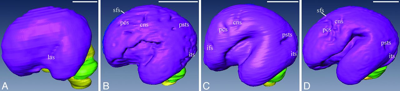
  • Fig 3.
    • Download figure
    • Open in new tab
    • Download powerpoint
    Fig 3.

    3D visualization model of the telencephalon, cerebellum, and brain stem of 12 (A), 16 (B), 20 (C), and 22 (D) weeks GA. Sulci on the brain surface are visible. The tiny lateral sulcus can be observed at 12 weeks GA (A), then it becomes wider and deeper (B–D). A cluster of shallow “sulcal roots” is delineated at 16 weeks GA (B), then the roots merge together and become deeper (C–D). ifs indicates inferior frontal sulcus; psts, posterior part of the superior temporal sulcus.

  • Fig 4.
    • Download figure
    • Open in new tab
    • Download powerpoint
    Fig 4.

    Quantitative measurements of fetal brain surface area and volume. The measurements increase linearly with GA. The surface area increases faster. Surface area is in square centimeters, and volume is in cubic centimeters. BA indicates brain surface area; BV, brain volume. The blue points represent the males, and the green represent the females.

  • Fig 5.
    • Download figure
    • Open in new tab
    • Download powerpoint
    Fig 5.

    Quantitative measurements of fetal brain length (A), width (B), and height (C). All of the measurements increase linearly with GA. Brain length increases the fastest, and height increases the slowest. Length, width, and height are in centimeters. BL indicates brain length; BW, brain width; BH, brain height.

Tables

  • Figures
    • View popup
    Table 1:

    GA dispositions and numbers of chosen specimens (n = 69)

    GANumber
    123
    133
    143
    154
    165
    177
    184
    196
    2016
    2112
    226
    • View popup
    Table 2:

    Chronology of sulcal development during 12–22 weeks GAa

    SulciObserved 25%–75%Present ≥ 75%SulciObserved 25%–75%Present ≥ 75%
    Medial Cerebral SurfaceLateral Cerebral Surface
    Interhemispheric fissure−b12Lateral sulcus1214
    Callosal sulcus1214Superior frontal sulcus1516
    Cingular sulcus1214Inferior frontal sulcus1822
    Calcarine fissure1315Posterior part of superior temporal sulcus1618
    Parieto-occipital sulcus1516Inferior temporal sulcus1516
    Ventral Cerebral SurfaceVertex
    Hippocampic fissure1214Precentral sulcus1618
    Orbital sulcus1516Central sulcus1516
    Collateral sulcus1618
    Occipitotemporal sulcus2022
    Olfactory sulcus1516
    • ↵a Sulci were present on 7T postmortem MR imaging scans.

    • ↵b No cases were studied.

PreviousNext
Back to top

In this issue

American Journal of Neuroradiology: 34 (7)
American Journal of Neuroradiology
Vol. 34, Issue 7
1 Jul 2013
  • Table of Contents
  • Index by author
Advertisement
Print
Download PDF
Email Article

Thank you for your interest in spreading the word on American Journal of Neuroradiology.

NOTE: We only request your email address so that the person you are recommending the page to knows that you wanted them to see it, and that it is not junk mail. We do not capture any email address.

Enter multiple addresses on separate lines or separate them with commas.
Development of the Fetal Cerebral Cortex in the Second Trimester: Assessment with 7T Postmortem MR Imaging
(Your Name) has sent you a message from American Journal of Neuroradiology
(Your Name) thought you would like to see the American Journal of Neuroradiology web site.
CAPTCHA
This question is for testing whether or not you are a human visitor and to prevent automated spam submissions.
Cite this article
Z. Zhang, Z. Hou, X. Lin, G. Teng, H. Meng, F. Zang, F. Fang, S. Liu
Development of the Fetal Cerebral Cortex in the Second Trimester: Assessment with 7T Postmortem MR Imaging
American Journal of Neuroradiology Jul 2013, 34 (7) 1462-1467; DOI: 10.3174/ajnr.A3406

Citation Manager Formats

  • BibTeX
  • Bookends
  • EasyBib
  • EndNote (tagged)
  • EndNote 8 (xml)
  • Medlars
  • Mendeley
  • Papers
  • RefWorks Tagged
  • Ref Manager
  • RIS
  • Zotero
0 Responses
Respond to this article
Share
Bookmark this article
Development of the Fetal Cerebral Cortex in the Second Trimester: Assessment with 7T Postmortem MR Imaging
Z. Zhang, Z. Hou, X. Lin, G. Teng, H. Meng, F. Zang, F. Fang, S. Liu
American Journal of Neuroradiology Jul 2013, 34 (7) 1462-1467; DOI: 10.3174/ajnr.A3406
del.icio.us logo Twitter logo Facebook logo Mendeley logo
  • Tweet Widget
  • Facebook Like
  • Google Plus One
Purchase

Jump to section

  • Article
    • Abstract
    • ABBREVIATIONS:
    • Materials and Methods
    • Results
    • Discussion
    • Conclusions
    • Acknowledgments
    • Footnotes
    • REFERENCES
  • Figures & Data
  • Supplemental
  • Info & Metrics
  • Responses
  • References
  • PDF

Related Articles

  • PubMed
  • Google Scholar

Cited By...

  • Mapping Fetal Brain Development of 10 Weeks Gestational Age with 9.4T Postmortem MRI and Histologic Sections
  • Characterization of dynamic patterns of human fetal to neonatal brain asymmetry with deformation-based morphometry
  • Association of Isolated Congenital Heart Disease with Fetal Brain Maturation
  • Morphologic Evolution and Coordinated Development of the Fetal Lateral Ventricles in the Second and Third Trimesters
  • Sulcal Depth-Position Profile Is a Genetically Mediated Neuroscientific Trait: Description and Characterization in the Central Sulcus
  • Crossref (43)
  • Google Scholar

This article has been cited by the following articles in journals that are participating in Crossref Cited-by Linking.

  • Learning-based prediction of gestational age from ultrasound images of the fetal brain
    Ana I.L. Namburete, Richard V. Stebbing, Bryn Kemp, Mohammad Yaqub, Aris T. Papageorghiou, J. Alison Noble
    Medical Image Analysis 2015 21 1
  • Abnormalities in structural covariance of cortical gyrification in schizophrenia
    Lena Palaniyappan, Bert Park, Vijender Balain, Raj Dangi, Peter Liddle
    Brain Structure and Function 2015 220 4
  • Premature Birth and Developmental Programming: Mechanisms of Resilience and Vulnerability
    Femke Lammertink, Christiaan H. Vinkers, Maria L. Tataranno, Manon J. N. L. Benders
    Frontiers in Psychiatry 2021 11
  • Brain microstructural development at near-term age in very-low-birth-weight preterm infants: An atlas-based diffusion imaging study
    Jessica Rose, Rachel Vassar, Katelyn Cahill-Rowley, Ximena Stecher Guzman, David K. Stevenson, Naama Barnea-Goraly
    NeuroImage 2014 86
  • NR2F1 regulates regional progenitor dynamics in the mouse neocortex and cortical gyrification in BBSOAS patients
    Michele Bertacchi, Anna Lisa Romano, Agnès Loubat, Frederic Tran Mau‐Them, Marjolaine Willems, Laurence Faivre, Philippe Khau van Kien, Laurence Perrin, Françoise Devillard, Arthur Sorlin, Paul Kuentz, Christophe Philippe, Aurore Garde, Francesco Neri, Rossella Di Giaimo, Salvatore Oliviero, Silvia Cappello, Ludovico D'Incerti, Carolina Frassoni, Michèle Studer
    The EMBO Journal 2020 39 13
  • Quantitative In vivo MRI Assessment of Structural Asymmetries and Sexual Dimorphism of Transient Fetal Compartments in the Human Brain
    Lana Vasung, Caitlin K Rollins, Hyuk Jin Yun, Clemente Velasco-Annis, Jennings Zhang, Konrad Wagstyl, Alan Evans, Simon K Warfield, Henry A Feldman, P Ellen Grant, Ali Gholipour
    Cerebral Cortex 2020 30 3
  • Sulcal Depth-Position Profile Is a Genetically Mediated Neuroscientific Trait: Description and Characterization in the Central Sulcus
    D. Reese McKay, Peter Kochunov, Matthew D. Cykowski, Jack W. Kent, Angela R. Laird, Jack L. Lancaster, John Blangero, David C. Glahn, Peter T. Fox
    The Journal of Neuroscience 2013 33 39
  • Association of Isolated Congenital Heart Disease with Fetal Brain Maturation
    C. Jaimes, V. Rofeberg, C. Stopp, C.M. Ortinau, A. Gholipour, K.G. Friedman, W. Tworetzky, J. Estroff, J.W. Newburger, D. Wypij, S.K. Warfield, E. Yang, C.K. Rollins
    American Journal of Neuroradiology 2020 41 8
  • Morphometric development of the human fetal cerebellum during the early second trimester
    Feifei Xu, Xinting Ge, Yonggang Shi, Zhonghe Zhang, Yuchun Tang, Xiangtao Lin, Gaojun Teng, Fengchao Zang, Nuonan Gao, Haihong Liu, Arthur W. Toga, Shuwei Liu
    NeuroImage 2020 207
  • Mimicking Cortex Convolutions Through the Wrinkling of Growing Soft Bilayers
    Martine Ben Amar, Adrien Bordner
    Journal of Elasticity 2017 129 1-2

More in this TOC Section

  • Comparison of Image Quality and Radiation Dose in Pediatric Temporal Bone CT Using Photon-Counting Detector CT and Energy-Integrating Detector CT
  • SyMRI & MR Fingerprinting in Brainstem Myelination
  • Pons&Vermis Localization on Fetal MRI Using U-Net
Show more Pediatrics

Similar Articles

Advertisement

Indexed Content

  • Current Issue
  • Accepted Manuscripts
  • Article Preview
  • Past Issues
  • Editorials
  • Editors Choice
  • Fellow Journal Club
  • Letters to the Editor

Cases

  • Case Collection
  • Archive - Case of the Week
  • Archive - Case of the Month
  • Archive - Classic Case

Special Collections

  • Special Collections

Resources

  • News and Updates
  • Turn around Times
  • Submit a Manuscript
  • Author Policies
  • Manuscript Submission Guidelines
  • Evidence-Based Medicine Level Guide
  • Publishing Checklists
  • Graphical Abstract Preparation
  • Imaging Protocol Submission
  • Submit a Case
  • Become a Reviewer/Academy of Reviewers
  • Get Peer Review Credit from Publons

Multimedia

  • AJNR Podcast
  • AJNR SCANtastic
  • Video Articles

About Us

  • About AJNR
  • Editorial Board
  • Not an AJNR Subscriber? Join Now
  • Alerts
  • Feedback
  • Advertise with us
  • Librarian Resources
  • Permissions
  • Terms and Conditions

American Society of Neuroradiology

  • Not an ASNR Member? Join Now

© 2025 by the American Society of Neuroradiology All rights, including for text and data mining, AI training, and similar technologies, are reserved.
Print ISSN: 0195-6108 Online ISSN: 1936-959X

Powered by HighWire