Advertisement

Main menu

  • Home
  • Content
    • Current Issue
    • Accepted Manuscripts
    • Article Preview
    • Past Issue Archive
    • AJNR Case Collection
    • Case of the Week Archive
    • Classic Case Archive
    • Case of the Month Archive
  • Special Collections
    • Spinal CSF Leak Articles (Jan 2020-June 2024)
    • 2024 AJNR Journal Awards
    • Most Impactful AJNR Articles
  • Multimedia
    • AJNR Podcast
    • AJNR Scantastics
    • Video Articles
  • For Authors
    • Submit a Manuscript
    • Author Policies
    • Fast publishing of Accepted Manuscripts
    • Graphical Abstract Preparation
    • Manuscript Submission Guidelines
    • Imaging Protocol Submission
    • Submit a Case for the Case Collection
  • About Us
    • About AJNR
    • Editorial Board
  • More
    • Become a Reviewer/Academy of Reviewers
    • Subscribers
    • Permissions
    • Alerts
    • Feedback
    • Advertisers
    • ASNR Home
  • Other Publications
    • ajnr

User menu

  • Alerts
  • Log in

Search

  • Advanced search
American Journal of Neuroradiology

American Journal of Neuroradiology

ASHNR American Society of Functional Neuroradiology ASHNR American Society of Pediatric Neuroradiology ASSR
  • Alerts
  • Log in

Advanced Search

  • Home
  • Content
    • Current Issue
    • Accepted Manuscripts
    • Article Preview
    • Past Issue Archive
    • AJNR Case Collection
    • Case of the Week Archive
    • Classic Case Archive
    • Case of the Month Archive
  • Special Collections
    • Spinal CSF Leak Articles (Jan 2020-June 2024)
    • 2024 AJNR Journal Awards
    • Most Impactful AJNR Articles
  • Multimedia
    • AJNR Podcast
    • AJNR Scantastics
    • Video Articles
  • For Authors
    • Submit a Manuscript
    • Author Policies
    • Fast publishing of Accepted Manuscripts
    • Graphical Abstract Preparation
    • Manuscript Submission Guidelines
    • Imaging Protocol Submission
    • Submit a Case for the Case Collection
  • About Us
    • About AJNR
    • Editorial Board
  • More
    • Become a Reviewer/Academy of Reviewers
    • Subscribers
    • Permissions
    • Alerts
    • Feedback
    • Advertisers
    • ASNR Home
  • Follow AJNR on Twitter
  • Visit AJNR on Facebook
  • Follow AJNR on Instagram
  • Join AJNR on LinkedIn
  • RSS Feeds

Welcome to the new AJNR, Updated Hall of Fame, and more. Read the full announcements.


AJNR is seeking candidates for the position of Associate Section Editor, AJNR Case Collection. Read the full announcement.


Research ArticleBrain
Open Access

Discrimination of Tumorous Intracerebral Hemorrhage from Benign Causes Using CT Densitometry

Y.S. Choi, T.H. Rim, S.S. Ahn and S.-K. Lee
American Journal of Neuroradiology May 2015, 36 (5) 886-892; DOI: https://doi.org/10.3174/ajnr.A4233
Y.S. Choi
aFrom the Department of Radiology and Research Institute of Radiological Science (Y.S.C., S.S.A., S.-K.L.)
  • Find this author on Google Scholar
  • Find this author on PubMed
  • Search for this author on this site
  • ORCID record for Y.S. Choi
T.H. Rim
bDepartment of Ophthalmology, Institute of Vision Research (T.H.R.), College of Medicine, Yonsei University, Seoul, Korea.
  • Find this author on Google Scholar
  • Find this author on PubMed
  • Search for this author on this site
S.S. Ahn
aFrom the Department of Radiology and Research Institute of Radiological Science (Y.S.C., S.S.A., S.-K.L.)
  • Find this author on Google Scholar
  • Find this author on PubMed
  • Search for this author on this site
S.-K. Lee
aFrom the Department of Radiology and Research Institute of Radiological Science (Y.S.C., S.S.A., S.-K.L.)
  • Find this author on Google Scholar
  • Find this author on PubMed
  • Search for this author on this site
  • Article
  • Figures & Data
  • Info & Metrics
  • Responses
  • References
  • PDF
Loading

Article Figures & Data

Figures

  • Tables
  • Fig 1.
    • Download figure
    • Open in new tab
    • Download powerpoint
    Fig 1.

    Flowchart of study population enrollment. IVH indicates intraventricular hemorrhage.
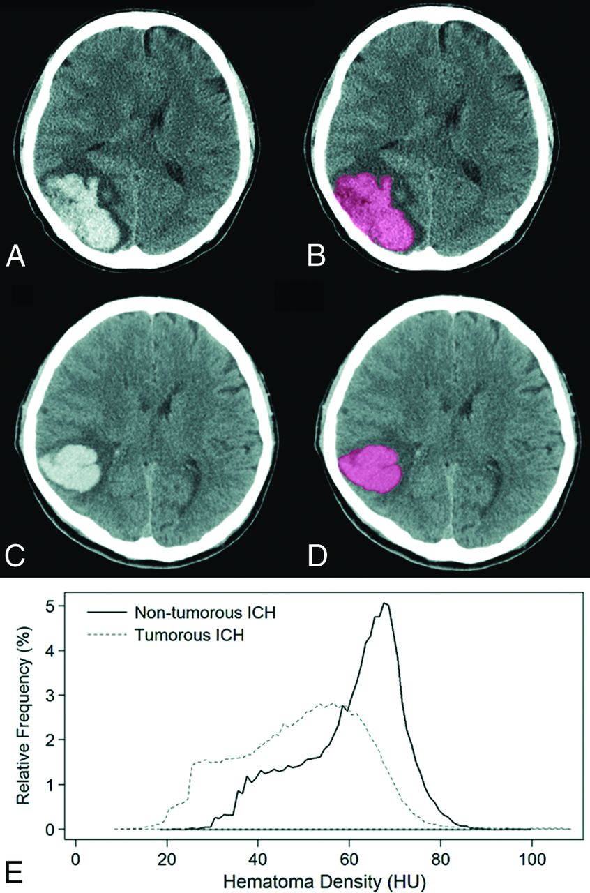
  • Fig 2.
    • Download figure
    • Open in new tab
    • Download powerpoint
    Fig 2.

    Representative cases of semiautomatic segmentation of tumorous (A and B) and nontumorous (C and D) ICHs and their relative frequency histogram of hematoma attenuation (E). ICH was segmented with a semiautomatic method based on a voxel-intensity threshold of 40–130 HU. On histogram analysis, the 5th and 25th percentile values were 26 HU and 38 HU in tumorous ICHs and 40 HU and 65 HU in nontumorous ICHs, respectively, and were discriminated correctly by using our histogram analysis.

  • Fig 3.
    • Download figure
    • Open in new tab
    • Download powerpoint
    Fig 3.

    Differences among the 5 groups in histogram parameters of ICH attenuation are list on the x-axis of each boxplot: 1) primary ICH without antithrombotics, 2) primary ICH with antithrombotics, 3) secondary ICH due to vascular malformation, 4) secondary ICH due to brain metastasis, and 5) secondary ICH due to primary brain tumor.
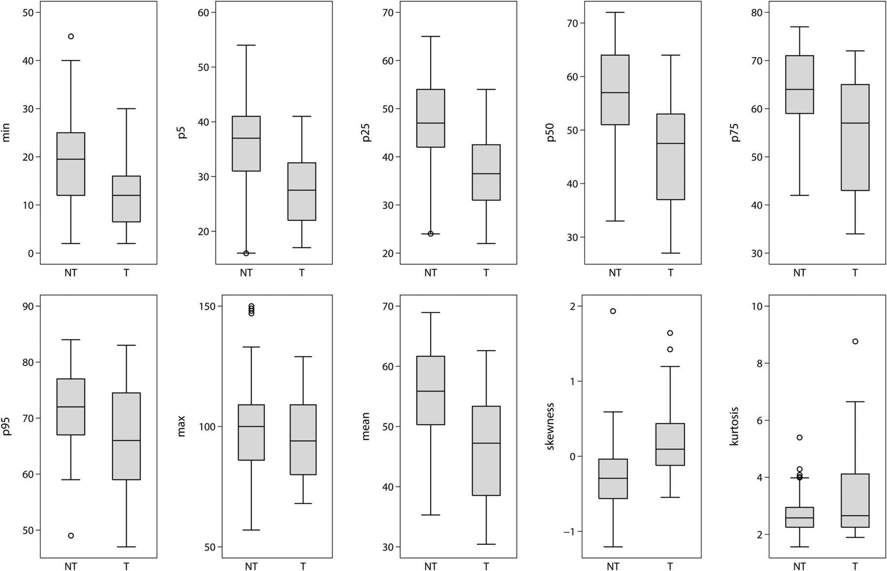
  • Fig 4.
    • Download figure
    • Open in new tab
    • Download powerpoint
    Fig 4.

    Differences in histogram parameters of ICH attenuation in tumor and nontumorous ICHs. All histogram parameters except maximum value and kurtosis were significantly different between tumorous and nontumorous ICHs. Tumors had lower cumulative histogram parameters and positive skewness. NT indicates nontumorous ICHs; T, tumorous ICHs.

  • Fig 5.
    • Download figure
    • Open in new tab
    • Download powerpoint
    Fig 5.

    Receiver operating characteristic curves of histogram parameters for discrimination of tumor and nontumorous ICHs.

Tables

  • Figures
    • View popup
    Table 1:

    Demographic data of study population (n = 110)

    ParameterNontumorous ICH (n = 90)Tumorous ICH (n = 20)P Valuea
    Primary ICH (n = 64)Secondary ICH (n = 46)
    Without Antithrombotics (n = 51)With Antithrombotics (n = 13)Vascular Malformation (n = 26)Metastasis (n = 12)Primary Brain Tumor (n = 8)
    Age (year)57.0 ± 20.466.8 ± 11.731.0 ± 20.956.8 ± 14.947.9 ± 20.9<.01
    Gender (M:F)29:228:514:129:34:4.75
    Onset–CT interval time (hours)10.0 ± 8.215.2 ± 7.18.5 ± 7.87.0 ± 6.49.5 ± 7.1.08
    • ↵a Difference among the 5 groups tested by 1-way analysis of variance or χ2 test.

    • View popup
    Table 2:

    Differences in histogram parameters for ICH attenuation among the 5 groups

    ParameterPrimary ICH without Antithrombotics (n = 51)Primary ICH with Antithrombotics (n = 13)Vascular Malformation (n = 26)Metastasis (n = 12)Primary Brain Tumor (n = 8)P Valuea
    Mean57.31 ± 7.4052.64 ± 7.3554.12 ± 6.9646.94 ± 9.0745.08 ± 10.34<.01
    Minimum21.65 ± 9.6511.69 ± 9.5218.62 ± 7.0514.83 ± 8.509.00 ± 5.29<.01
    Percentiles
        5th37.73 ± 6.7030.38 ± 7.3835.56 ± 5.5328.50 ± 6.2626.38 ± 6.50<.01
        25th49.47 ± 8.3642.77 ± 9.1445.85 ± 7.0538.58 ± 8.5034.38 ± 8.00<.01
        50th58.78 ± 8.4654.00 ± 8.7155.35 ± 8.3147.42 ± 10.3244.38 ± 11.65<.01
        75th65.73 ± 7.4262.92 ± 6.7162.65 ± 7.9555.00 ± 10.5754.13 ± 14.88<.01
        95th73.02 ± 7.0271.15 ± 5.5170.00 ± 7.3864.58 ± 9.0267.25 ± 11.65<.01
    Maximum99.33 ± 16.46104.69 ± 17.6794.31 ± 18.4792.08 ± 14.05122.00 ± 65.95.29
    Skewness−0.32 ± 0.35−0.18 ± 0.50−0.13 ± 0.560.05 ± 0.470.63 ± 0.66<.01
    Kurtosis2.64 ± 0.522.95 ± 0.963.00 ± 2.112.96 ± 1.214.11 ± 2.47<.01
    • ↵a Difference among the 5 groups tested using 1-way analysis of variance before pair-wise comparison.

    • View popup
    Table 3:

    Diagnostic performance of ICH attenuation histogram parameters for discriminating tumor and nontumorous ICHs

    ParameterAz Value95% CICutoff ValueaSensitivity (%)Specificity (%)
    Minimum0.710.59–0.841762.285.0
    Percentiles
        5th0.810.72–0.913465.685.0
        25th0.810.71–0.924470.080.0
        50th0.780.66–0.895365.675.0
        75th0.740.60–0.876072.265.0
        95th0.690.54–0.846390.045.0
    Mean0.780.66–0.9049.1181.165.0
    Skewness0.780.67–0.88−0.0275.025.0
    • Note:—Az indicates area under the receiver operating characteristic curve.

    • ↵a Determined by maximizing the Youden index.

PreviousNext
Back to top

In this issue

American Journal of Neuroradiology: 36 (5)
American Journal of Neuroradiology
Vol. 36, Issue 5
1 May 2015
  • Table of Contents
  • Index by author
  • Complete Issue (PDF)
Advertisement
Print
Download PDF
Email Article

Thank you for your interest in spreading the word on American Journal of Neuroradiology.

NOTE: We only request your email address so that the person you are recommending the page to knows that you wanted them to see it, and that it is not junk mail. We do not capture any email address.

Enter multiple addresses on separate lines or separate them with commas.
Discrimination of Tumorous Intracerebral Hemorrhage from Benign Causes Using CT Densitometry
(Your Name) has sent you a message from American Journal of Neuroradiology
(Your Name) thought you would like to see the American Journal of Neuroradiology web site.
CAPTCHA
This question is for testing whether or not you are a human visitor and to prevent automated spam submissions.
Cite this article
Y.S. Choi, T.H. Rim, S.S. Ahn, S.-K. Lee
Discrimination of Tumorous Intracerebral Hemorrhage from Benign Causes Using CT Densitometry
American Journal of Neuroradiology May 2015, 36 (5) 886-892; DOI: 10.3174/ajnr.A4233

Citation Manager Formats

  • BibTeX
  • Bookends
  • EasyBib
  • EndNote (tagged)
  • EndNote 8 (xml)
  • Medlars
  • Mendeley
  • Papers
  • RefWorks Tagged
  • Ref Manager
  • RIS
  • Zotero
0 Responses
Respond to this article
Share
Bookmark this article
Discrimination of Tumorous Intracerebral Hemorrhage from Benign Causes Using CT Densitometry
Y.S. Choi, T.H. Rim, S.S. Ahn, S.-K. Lee
American Journal of Neuroradiology May 2015, 36 (5) 886-892; DOI: 10.3174/ajnr.A4233
del.icio.us logo Twitter logo Facebook logo Mendeley logo
  • Tweet Widget
  • Facebook Like
  • Google Plus One
Purchase

Jump to section

  • Article
    • Abstract
    • ABBREVIATIONS:
    • Materials and Methods
    • Results
    • Discussion
    • Conclusions
    • REFERENCES
  • Figures & Data
  • Info & Metrics
  • Responses
  • References
  • PDF

Related Articles

  • No related articles found.
  • PubMed
  • Google Scholar

Cited By...

  • No citing articles found.
  • Crossref (16)
  • Google Scholar

This article has been cited by the following articles in journals that are participating in Crossref Cited-by Linking.

  • Hemorrhage in brain tumor – An unresolved issue
    Robert P. Ostrowski, Zhaohui He, Emanuela B. Pucko, Ewa Matyja
    Brain Hemorrhages 2022 3 2
  • Neoplastic and Non-neoplastic Acute Intracerebral Hemorrhage in CT Brain Scans: Machine Learning-Based Prediction Using Radiomic Image Features
    Jawed Nawabi, Helge Kniep, Reza Kabiri, Gabriel Broocks, Tobias D. Faizy, Christian Thaler, Gerhard Schön, Jens Fiehler, Uta Hanning
    Frontiers in Neurology 2020 11
  • Seizures, Edema, Thrombosis, and Hemorrhages: An Update Review on the Medical Management of Gliomas
    Marco Zoccarato, Lucia Nardetto, Anna Maria Basile, Bruno Giometto, Vittorina Zagonel, Giuseppe Lombardi
    Frontiers in Oncology 2021 11
  • Machine Learning-Based Perihematomal Tissue Features to Predict Clinical Outcome after Spontaneous Intracerebral Hemorrhage
    Xin Qi, Guorui Hu, Haiyan Sun, Zhigeng Chen, Chao Yang
    Journal of Stroke and Cerebrovascular Diseases 2022 31 6
  • Neoplastic and Non-Neoplastic Causes of Acute Intracerebral Hemorrhage on CT
    Jawed Nawabi, Uta Hanning, Gabriel Broocks, Gerhard Schön, Tanja Schneider, Jens Fiehler, Christian Thaler, Susanne Gellissen
    Clinical Neuroradiology 2020 30 2
  • Machine learning-based CT radiomics model to discriminate the primary and secondary intracranial hemorrhage
    Jianbo Lyu, Zhaohui Xu, HaiYan Sun, Fangbing Zhai, Xiaofeng Qu
    Scientific Reports 2023 13 1
  • A Radiomics Nomogram for Classifying Hematoma Entities in Acute Spontaneous Intracerebral Hemorrhage on Non-contrast-Enhanced Computed Tomography
    Jia Wang, Xing Xiong, Jing Ye, Yang Yang, Jie He, Juan Liu, Yi-Li Yin
    Frontiers in Neuroscience 2022 16
  • Air Pollution and Intracranial Hemorrhage
    Mervyn Lim Jun Rui, Jaclyn Tan, Benjamin Yong-Qiang Tan, Tseng Tsai Yeo, Vijay K. Sharma
    Annals of Indian Academy of Neurology 2022 25 Suppl 1
  • External validation of the diagnostic value of perihematomal edema characteristics in neoplastic and non‐neoplastic intracerebral hemorrhage
    Jawed Nawabi, Tobias Orth, Sophia Schulze‐Weddige, Georg Lukas Baumgaertner, Anna Tietze, Christian Thaler, Tobias Penzkofer
    European Journal of Neurology 2023 30 6
  • Building nonenhanced CT based radiomics model in discriminating arteriovenous malformation related hematomas from hypertensive intracerebral hematomas
    Huanhuan Xie, Fei Dong, Ruiting Zhang, Xinfeng Yu, Peng Xu, Yinshan Tang, Peiyu Huang, Chao Wang
    Frontiers in Neuroscience 2023 17

More in this TOC Section

  • Multimodal CT Provides Improved Performance for Lacunar Infarct Detection
  • Statin Therapy Does Not Affect the Radiographic and Clinical Profile of Patients with TIA and Minor Stroke
  • Optimal MRI Sequence for Identifying Occlusion Location in Acute Stroke: Which Value of Time-Resolved Contrast-Enhanced MRA?
Show more Brain

Similar Articles

Advertisement

Indexed Content

  • Current Issue
  • Accepted Manuscripts
  • Article Preview
  • Past Issues
  • Editorials
  • Editors Choice
  • Fellow Journal Club
  • Letters to the Editor

Cases

  • Case Collection
  • Archive - Case of the Week
  • Archive - Case of the Month
  • Archive - Classic Case

Special Collections

  • Special Collections

Resources

  • News and Updates
  • Turn around Times
  • Submit a Manuscript
  • Author Policies
  • Manuscript Submission Guidelines
  • Evidence-Based Medicine Level Guide
  • Publishing Checklists
  • Graphical Abstract Preparation
  • Imaging Protocol Submission
  • Submit a Case
  • Become a Reviewer/Academy of Reviewers
  • Get Peer Review Credit from Publons

Multimedia

  • AJNR Podcast
  • AJNR SCANtastic
  • Video Articles

About Us

  • About AJNR
  • Editorial Board
  • Not an AJNR Subscriber? Join Now
  • Alerts
  • Feedback
  • Advertise with us
  • Librarian Resources
  • Permissions
  • Terms and Conditions

American Society of Neuroradiology

  • Not an ASNR Member? Join Now

© 2025 by the American Society of Neuroradiology All rights, including for text and data mining, AI training, and similar technologies, are reserved.
Print ISSN: 0195-6108 Online ISSN: 1936-959X

Powered by HighWire