
Article Figures & Data
Figures
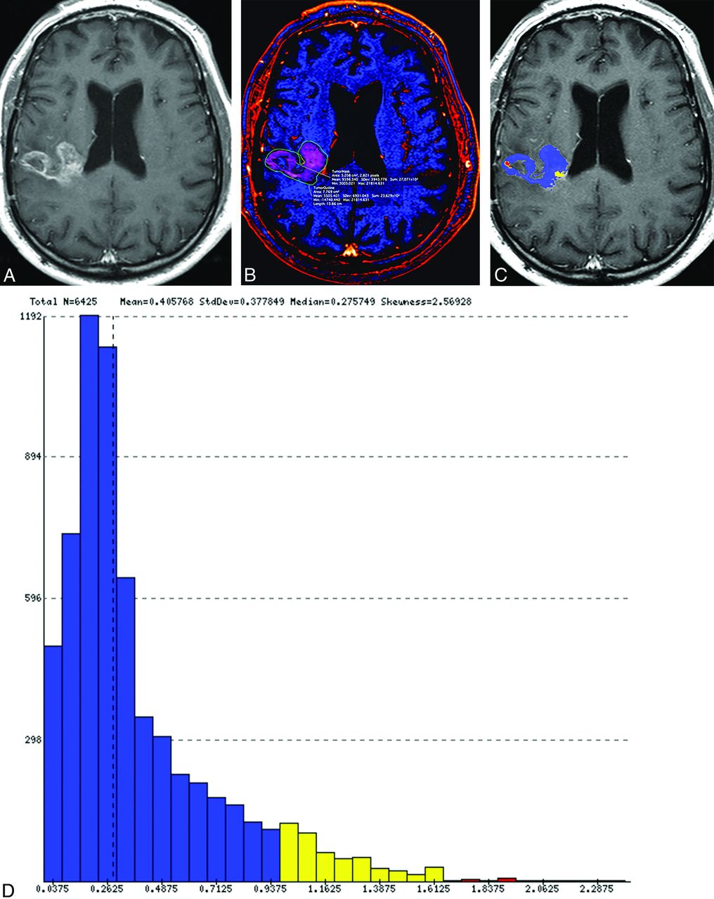
- Fig 1.
Representative examples of treatment effect (upper row) and recurrent tumor (lower row) in 2 patients with previously resected and irradiated glioblastomas. Contrast-enhancing lesions on postcontrast T1-weighted (A and E) and ΔT1 (B and F) images. Output FTB maps superimposed on the contrast-enhanced T1-weighted images (C and G). Blue represents areas of low blood volume (FTBlow), and red represents areas of high blood volume (FTBhigh). Histograms (D and H) show all voxels of the contrast-enhancing volume classified into the respective FTBlow, FTBmid (yellow), and FTBhigh classes, which is based on the rCBV thresholds of 1.0 and 1.75.
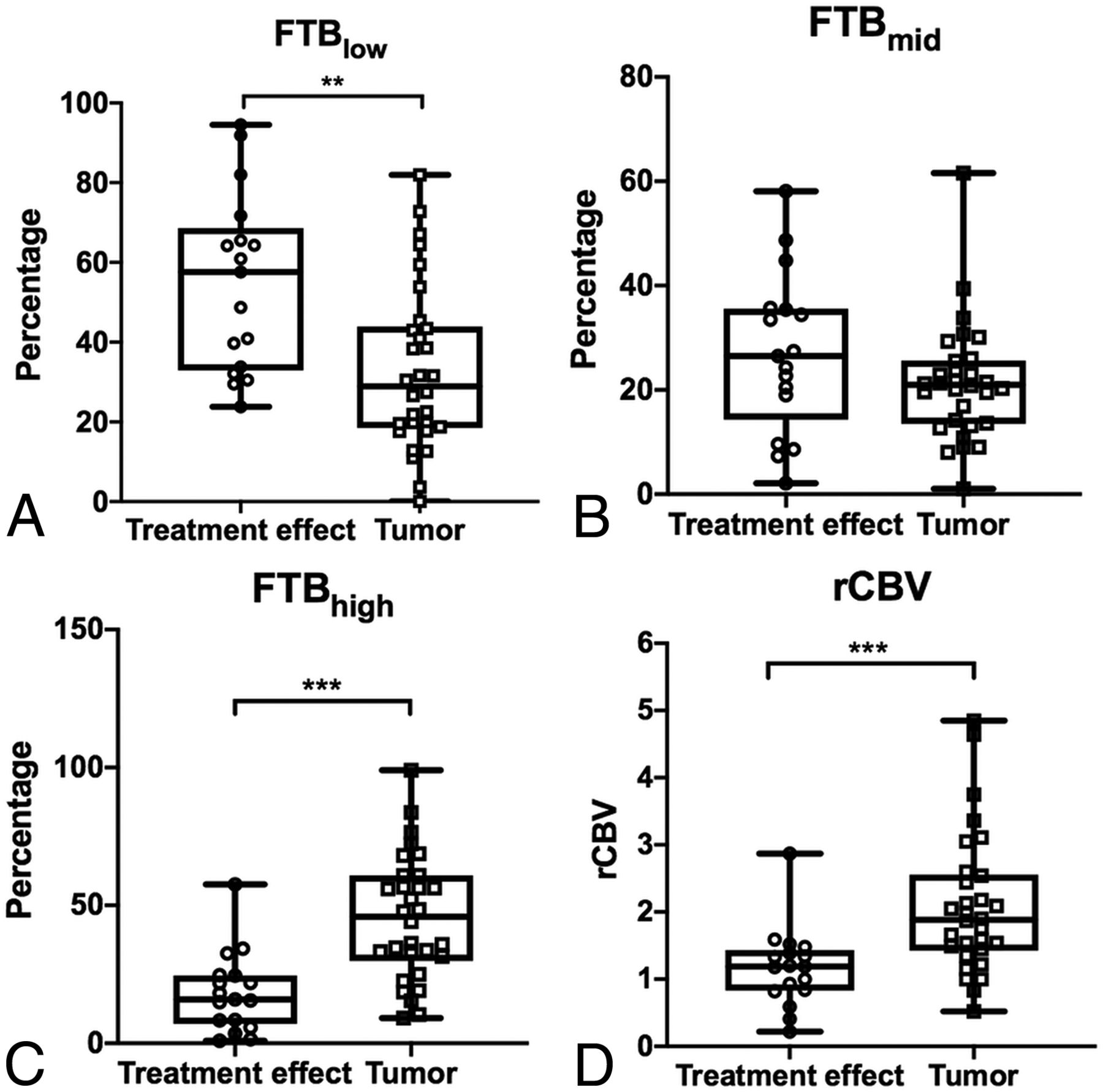
- Fig 2.
Boxplots of the relationship between FTB and normalized rCBV in 2 histopathologically defined groups: treatment effect and recurrent tumor. Open circles and squares represent individual measurements. The upper and lower limits of the whiskers represent the minimum and maximum of all of the data. Double asterisks indicate P < .01; triple asterisks, P < .001.
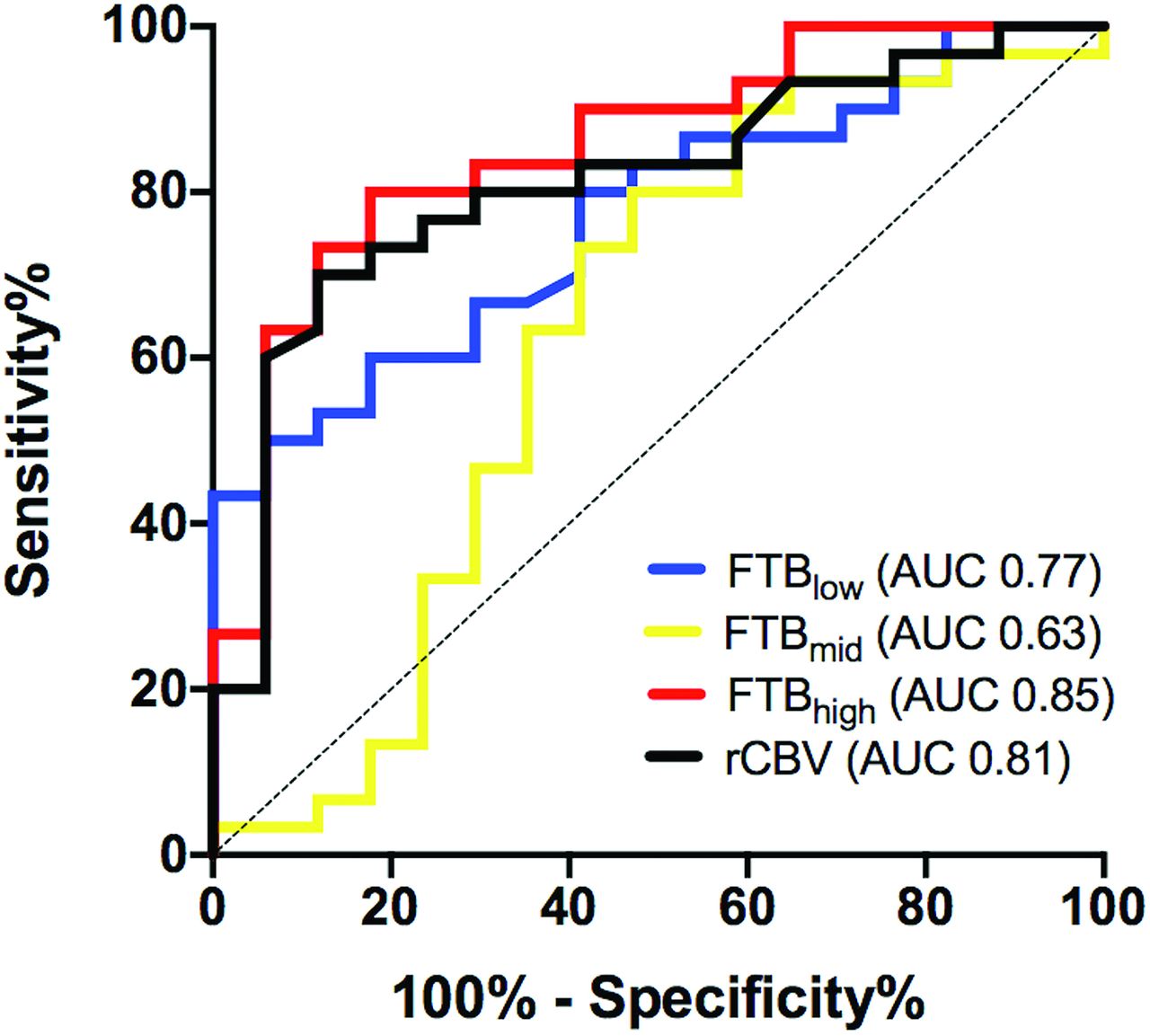
- Fig 3.
Receiver operating characteristic curves for the use of fractional tumor burden classes and normalized rCBV to differentiate tumor from treatment effect.
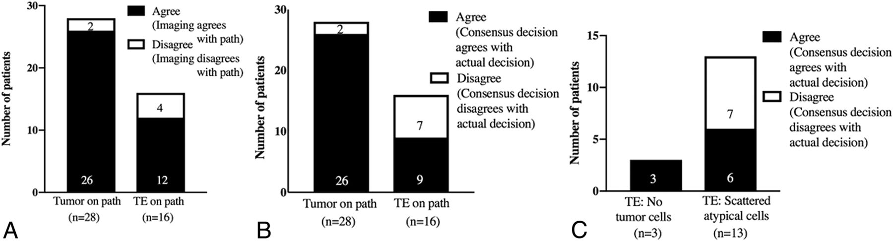
- Fig 4.
Agreement between the consensus (among 5 physician raters) qualitative interpretation of imaging and the actual histopathologic diagnosis (A). Agreement between the hypothetic consensus decision to change treatment plans and the actual (real-time) management plans (B and C).


Tables
TE (n = 17) Tumor (n = 30) Total (n = 47) Age (yr) Mean (SD) 56 (10) 55 (13) 55 (12) Range 38-77 20-80 20-80 Sex Male 11 (65%) 18 (60%) 29 (62%) Female 6 (35%) 12 (40%) 18 (38%) Interval time between end or radiation therapy and surgery (mo) Median (range) 11.4 (0.6-60.4) 10.7 (1.3-101.5) 10.9 (0.6-101.5) Bevacizumab at time of surgeryb 2 (12%) 3 (10%) 5 (11%) Surgical procedure Biopsy 3 (18%) 4 (13%) 7 (15%) >90% resection 3 (18%) 9 (30%) 12 (25%) Gross total resection 11 (64%) 17 (57%) 28 (60%) HGG histopathology Anaplastic astrocytoma, WHO grade III 2 (12%) 0 2 (4%) Glioblastoma, WHO grade IV 15 (88%) 29 (97%) 44 (94%) Gliosarcoma, WHO grade IV 0 1 (3%) 1 (2%) HGG molecular features IDH wild type 11 (65%) 11 (37%) 22 (47%) IDH mutant 0 2 (7%) 2 (4%) Unknown IDH status 6 (35%) 17 (56%) 23 (49%) MGMT-unmethylated 6 (35%) 15 (50%) 21 (45%) MGMT-methylated 6 (35%) 8 (27%) 14 (30%) Unknown MGMT status 5 (30%) 7 (23%) 12 (25%) Note:—IDH indicates isocitrate dehydrogenase; MGMT, O-6-methylguanine-DNA methyltransferase; WHO, World Health Organization.
↵a Percentage values in parentheses for sex, bevacizumab at time of the operation, surgical procedure, HGG histopathology, and HGG molecular features are percentages relative to the number of patients in each column.
↵b Patient received a dose of bevacizumab within 1 month of the surgical procedure for suspected recurrence.
- Table 2:
Mean values of FTB classes and normalized rCBV in histopathologically defined treatment effect and tumor groupsa
TE Tumor P Values FTBlow 54.8 (22.3) 33.1 (20.8) .002 FTBmid 27.0 (15.4) 21.3 (11.3) .16 FTBhigh 18.2 (14.4) 45.5 (22.6) <.001 rCBV 1.2 (0.6) 2.1 (1.0) <.001 ↵a Values are reported as mean (SD), except for P values.


Jump to section
Related Articles
Cited By...
- Multisite Benchmark Study for Standardized Relative CBV in Untreated Brain Metastases Using the DSC-MRI Consensus Acquisition Protocol
- Identification of a Single-Dose, Low-Flip-Angle-Based CBV Threshold for Fractional Tumor Burden Mapping in Recurrent Glioblastoma
- Arterial Spin-Labeling and DSC Perfusion Metrics Improve Agreement in Neuroradiologists Clinical Interpretations of Posttreatment High-Grade Glioma Surveillance MR Imaging--An Institutional Experience
- DSC Perfusion MRI-Derived Fractional Tumor Burden and Relative CBV Differentiate Tumor Progression and Radiation Necrosis in Brain Metastases Treated with Stereotactic Radiosurgery
- Performance of Standardized Relative CBV for Quantifying Regional Histologic Tumor Burden in Recurrent High-Grade Glioma: Comparison against Normalized Relative CBV Using Image-Localized Stereotactic Biopsies