
Article Figures & Data
Figures
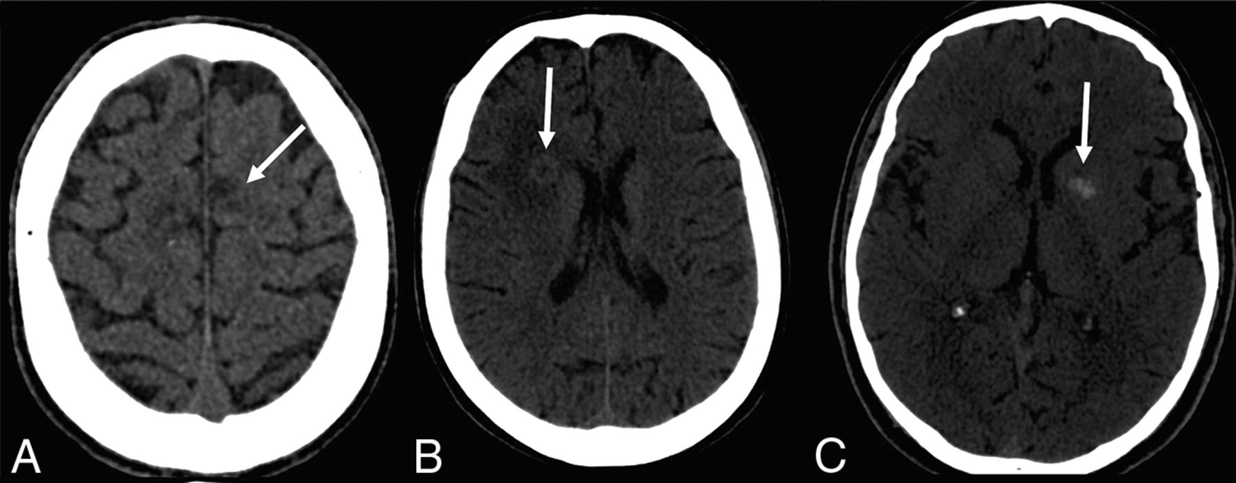
- Fig 1.
Brain CT showing different presentations of the lesions. A, Hypoattenuating lesions. B, Hypoattenuating lesions with a hyperattenuating halo. C, Hyperattenuating lesions.
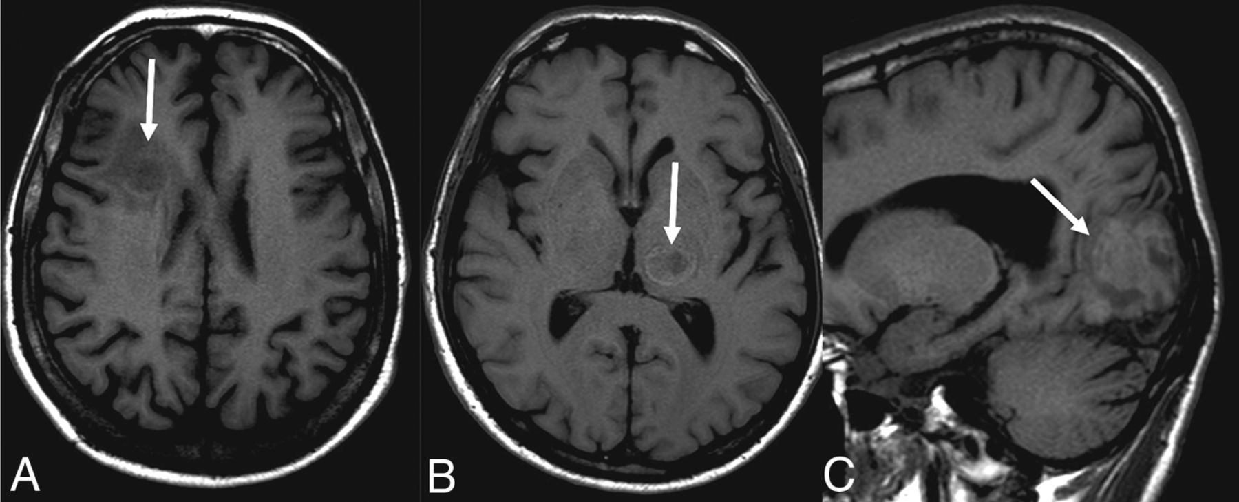
- Fig 2.
Signal on the T1-weighted images: hypointense lesion (A), lesion with hypointense center and hyperintense halo (B), and hyperintense lesion (C).
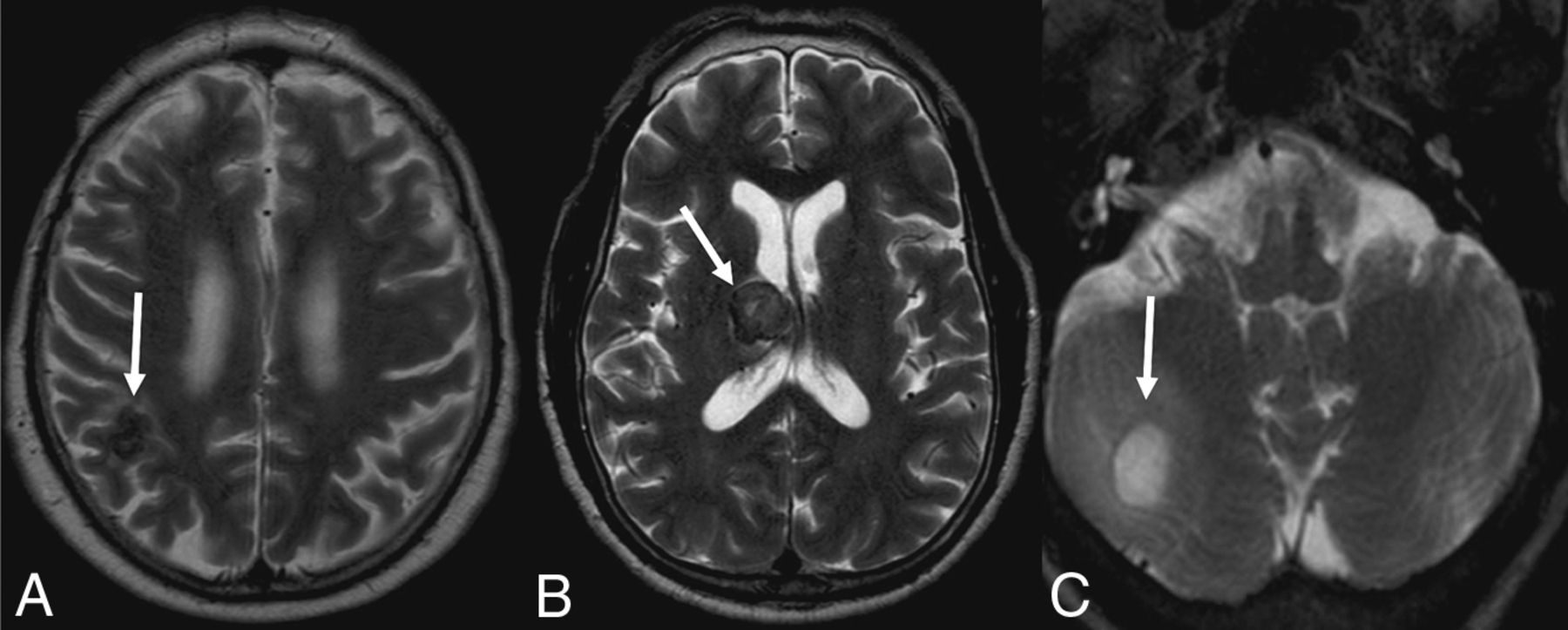
- Fig 3.
T2-weighted images show a hypointense lesion (A), a heterogeneous signal (B), and a hyperintense lesion (C).
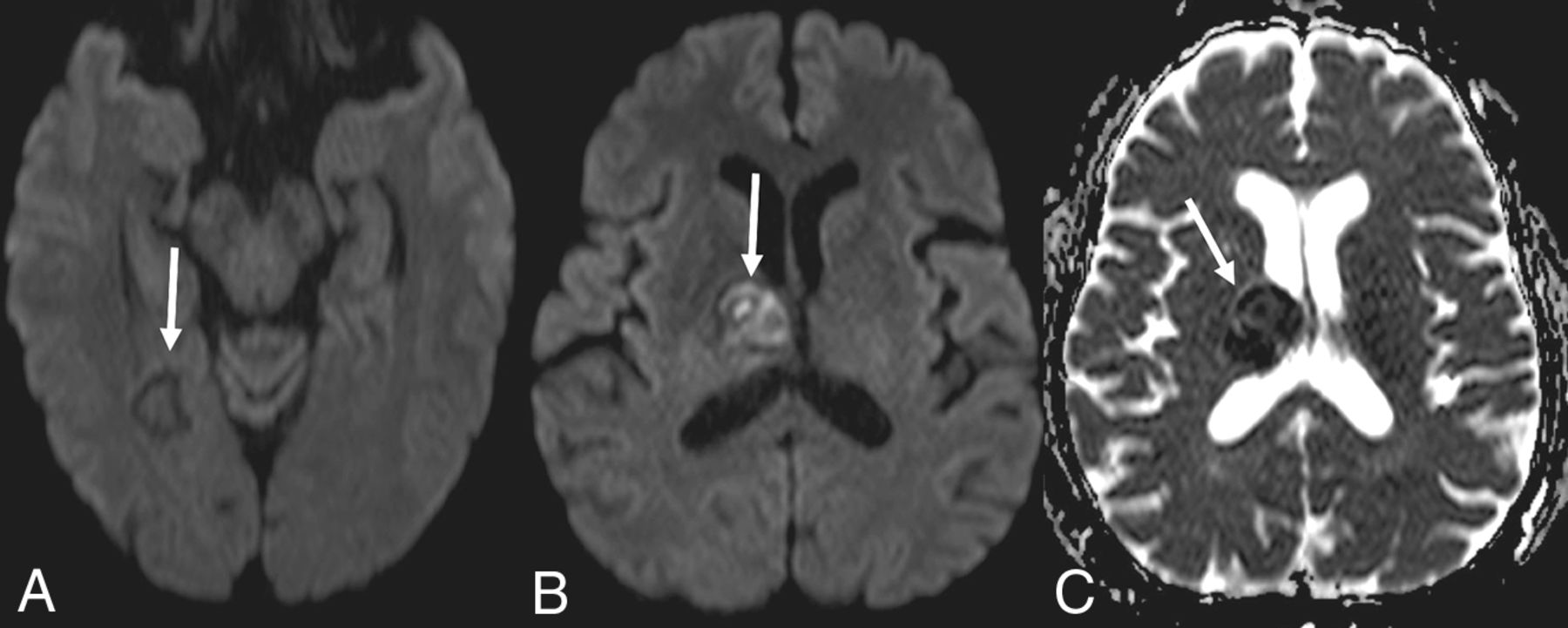
- Fig 4.
Demonstration that the lesions may not present with diffusion restriction (A) or diffusion restriction (B) with a low signal on the ADC map (C).
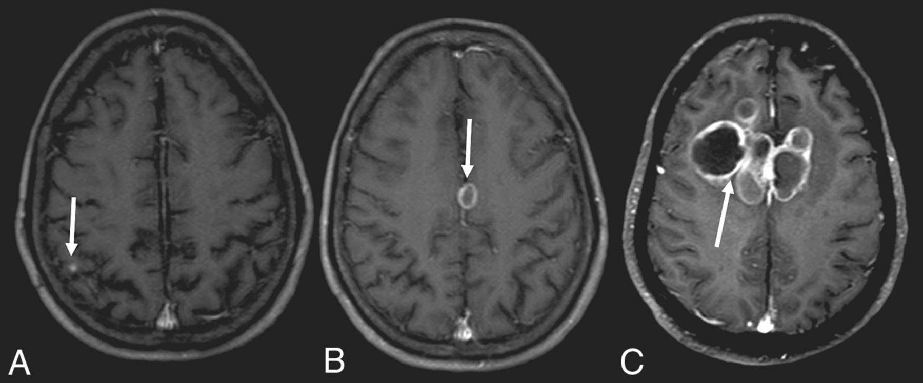
- Fig 5.
T1-weighted images after contrast administration demonstrating a small nodule in the cortical-subcortical transition (A), nodular lesion with annular enhancement (B), and multiple nodular lesions with annular enhancement and “daughter cysts” in a complex heterogeneous mass (C).
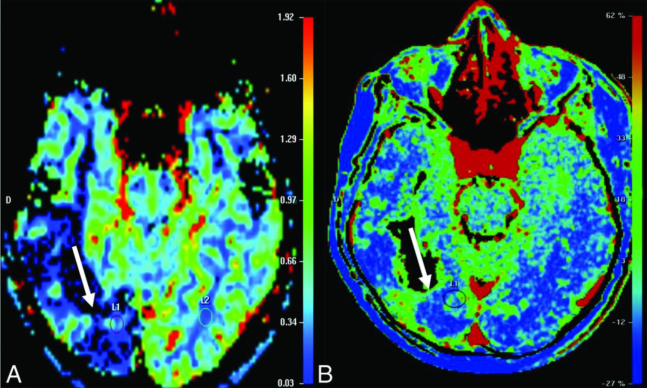
- Fig 6.
T2-weighted perfusion shows a lesion with low perfusion (A). T1-weighted perfusion shows the blood-brain barrier breakdown (B).
- Fig 7.
Proton spectroscopy with a TE = 144 ms showing increased lipid and choline peaks, with decreased NAA peaks.
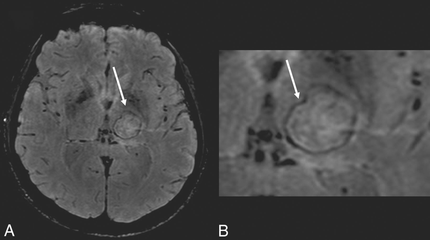
- Fig 8.
SWI sequence showing the dual rim sign. Note the external hypointense halo with an internal hyperintense halo.
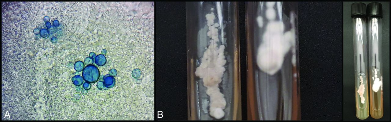
- Fig 9.
Laboratory diagnosis of paracoccidioidomycosis. Fresh examination in Parker-KOH stain shows yeast cells with multiple buds (A). Cultivation of Paracoccidioides spp. Left, yeast colonies to 37°C; and right, filamentous colonies to 25°C (B).


Tables
Characteristic Value Age Median (range) (yr) 54 (19–66) Subgroup (No. of patients) (%) 0–19 1 (4.1) 20–39 6 (25.0) 40–59 11 (45.8) ≥60 6 (25.0) Male sex (No.) (%) 24 (100) Neurologic symptoms (No. of patients) (%) Headache 8 (33.3) Epilepsy 7 (29.1) Focal neurologic signs 6 (25.0) Paresis 5 (20.8) Paresthesia 4 (16.6) Plegia 2 (8.3) Dysarthria 2 (8.3) Mental confusion 2 (8.3) Head lump 2 (8.3) Diplopia 1 (4.1) Chorea 1 (4.1) Vertigo 1 (4.1) Absence of neurologic symptom 2 (8.3) Characteristic Value Patterns (No. of patients) (%) Pseudotumoral 21 (87.5) Meningeal 1 (4.1) Pseudotumoral + meningeal (combined) 2 (8.3) Calcifications (No. of patients) (%) 0 (0) Perilesional edema (No. of patients) (%)a 19 (90.4) Hydrocephalus (No. of patients) (%) 6 (25.0) Hemorrhage (No. of patients) (%)b 3 (13.0) No. of lesionsb Mean (range) 3.0 (1–11) Subgroup (No. of patients) (%) Single lesions 9 (39.1) 2–5 11 (47.8) 6–10 2 (8.6) >10 1 (4.3) Lesion size (No. of patients) (%)b >2 cm 15 (65.2) Larger axial diameter of the major lesionb Mean (range) (cm) 2.6 (0.3–5.6) CT attenuation (No. of patients) (%)c Hyperattenuating lesions 11 (20.7) Hypoattenuating lesions 30 (56.6) Hypoattenuating center and hyperattenuating margin 12 (22.6) CT or MR imaging contrast enhancement (No. of patients) (%) Ring enhancement 17 (70.8) Nodular enhancement 3 (12.5) Ring and nodular enhancement 3 (12.5) Leptomeningeal enhancement 1 (4.1) Characteristic Value Localization (No. of patients) (%) Basal meningitis 1 (4.1) Pachymeningitis 1 (4.1) Skull 2 (8.3) Parietal lobe 7 (29.1) Occipital lobe 6 (25.0) Frontal lobe 9 (37.5) Temporal lobe 5 (20.8) Cerebellum 8 (33.3) Cingulate gyrus 3 (12.5) Thalamus 5 (20.8) Basal ganglia 3 (12.5) Striatum 1 (4.1) Globus pallidus 1 (4.1) Caudate nucleus 1 (4.1) Putamen 1(4.1) Corpus callosum 2 (8.3) Hippocampus 1 (4.1) Hypothalamus 1 (4.1) Pons 2 (8.3) Characteristic Value MR imaging scans (No. of patients) (%)a T1-weightedb Hyperintense lesions 21 (34.4) Hypointense lesions 31 (50.8) Hypointense center and hyperintense margin T2-weightedb Hyperintense lesions 6 (9.8) Hypointense lesions 36 (59.0) Heterogeneous lesions (hyperintense + hypointense) 19 (31.1) T1, perfusionc Slow and progressive ascending 3 (100) T2, perfusionc Decreased rCBV 3 (100) Spectroscopyc Decrease of NAA 2 (100) Increase of choline 2 (100) Increase of lipids 2 (100) Diffusion-weighted (No. of patients) (%)d Restricted diffusion 9 (47.3) Target sign 1 (5.2) Note:—rCBV indicates relative CBV.
↵a Five patients did not have MR imaging scans and were eliminated from the calculations.
↵b The patient with the meningeal form was eliminated from the calculations.
↵c Only 3 patients underwent perfusion, and only 2 underwent spectroscopy.
↵d Five patients did not have diffusion and were eliminated from the calculations.


Jump to section
Related Articles
Cited By...
- No citing articles found.