Advertisement

Main menu

  • Home
  • Content
    • Current Issue
    • Accepted Manuscripts
    • Article Preview
    • Past Issue Archive
    • AJNR Case Collection
    • Case of the Week Archive
    • Classic Case Archive
    • Case of the Month Archive
  • Special Collections
    • Spinal CSF Leak Articles (Jan 2020-June 2024)
    • 2024 AJNR Journal Awards
    • Most Impactful AJNR Articles
  • Multimedia
    • AJNR Podcast
    • AJNR Scantastics
    • Video Articles
  • For Authors
    • Submit a Manuscript
    • Author Policies
    • Fast publishing of Accepted Manuscripts
    • Graphical Abstract Preparation
    • Manuscript Submission Guidelines
    • Imaging Protocol Submission
    • Submit a Case for the Case Collection
  • About Us
    • About AJNR
    • Editorial Board
  • More
    • Become a Reviewer/Academy of Reviewers
    • Subscribers
    • Permissions
    • Alerts
    • Feedback
    • Advertisers
    • ASNR Home
  • Other Publications
    • ajnr

User menu

  • Alerts
  • Log in

Search

  • Advanced search
American Journal of Neuroradiology

American Journal of Neuroradiology

ASHNR American Society of Functional Neuroradiology ASHNR American Society of Pediatric Neuroradiology ASSR
  • Alerts
  • Log in

Advanced Search

  • Home
  • Content
    • Current Issue
    • Accepted Manuscripts
    • Article Preview
    • Past Issue Archive
    • AJNR Case Collection
    • Case of the Week Archive
    • Classic Case Archive
    • Case of the Month Archive
  • Special Collections
    • Spinal CSF Leak Articles (Jan 2020-June 2024)
    • 2024 AJNR Journal Awards
    • Most Impactful AJNR Articles
  • Multimedia
    • AJNR Podcast
    • AJNR Scantastics
    • Video Articles
  • For Authors
    • Submit a Manuscript
    • Author Policies
    • Fast publishing of Accepted Manuscripts
    • Graphical Abstract Preparation
    • Manuscript Submission Guidelines
    • Imaging Protocol Submission
    • Submit a Case for the Case Collection
  • About Us
    • About AJNR
    • Editorial Board
  • More
    • Become a Reviewer/Academy of Reviewers
    • Subscribers
    • Permissions
    • Alerts
    • Feedback
    • Advertisers
    • ASNR Home
  • Follow AJNR on Twitter
  • Visit AJNR on Facebook
  • Follow AJNR on Instagram
  • Join AJNR on LinkedIn
  • RSS Feeds

Welcome to the new AJNR, Updated Hall of Fame, and more. Read the full announcements.


AJNR is seeking candidates for the position of Associate Section Editor, AJNR Case Collection. Read the full announcement.


Getting new auth cookie, if you see this message a lot, tell someone!
Research ArticleExtracranial Vascular

Carotid Intraplaque-Hemorrhage Volume and Its Association with Cerebrovascular Events

L. Saba, G. Micheletti, W. Brinjikji, P. Garofalo, R. Montisci, A. Balestrieri, J.S. Suri, J.K. DeMarco, G. Lanzino and R. Sanfilippo
American Journal of Neuroradiology October 2019, 40 (10) 1731-1737; DOI: https://doi.org/10.3174/ajnr.A6189
L. Saba
aFrom the Departments of Radiology (L.S., G.M., P.G., A.B.)
  • Find this author on Google Scholar
  • Find this author on PubMed
  • Search for this author on this site
  • ORCID record for L. Saba
G. Micheletti
aFrom the Departments of Radiology (L.S., G.M., P.G., A.B.)
  • Find this author on Google Scholar
  • Find this author on PubMed
  • Search for this author on this site
  • ORCID record for G. Micheletti
W. Brinjikji
cDepartments of Radiology (W.B.)
  • Find this author on Google Scholar
  • Find this author on PubMed
  • Search for this author on this site
  • ORCID record for W. Brinjikji
P. Garofalo
aFrom the Departments of Radiology (L.S., G.M., P.G., A.B.)
  • Find this author on Google Scholar
  • Find this author on PubMed
  • Search for this author on this site
  • ORCID record for P. Garofalo
R. Montisci
bVascular Surgery (R.M., R.S.), Azienda Ospedaliero Universitaria, Monserrato (Cagliari), Italy
  • Find this author on Google Scholar
  • Find this author on PubMed
  • Search for this author on this site
  • ORCID record for R. Montisci
A. Balestrieri
aFrom the Departments of Radiology (L.S., G.M., P.G., A.B.)
  • Find this author on Google Scholar
  • Find this author on PubMed
  • Search for this author on this site
  • ORCID record for A. Balestrieri
J.S. Suri
eStroke Monitoring and Diagnostic Division (J.S.S.), AtheroPoint, Roseville, California
fPoint-of-Care Devices (J.S.S.), Global Biomedical Technologies, Roseville, California
gDepartment of Electrical Engineering (J.S.S.), University of Idaho, Moscow, Idaho (Affiliated)
  • Find this author on Google Scholar
  • Find this author on PubMed
  • Search for this author on this site
  • ORCID record for J.S. Suri
J.K. DeMarco
hDepartment of Radiology (J.K.D.), Walter Reed Medical Center, Bethesda, Maryland.
  • Find this author on Google Scholar
  • Find this author on PubMed
  • Search for this author on this site
  • ORCID record for J.K. DeMarco
G. Lanzino
dNeurosurgery (G.L.), Mayo Clinic, Rochester, Minnesota
  • Find this author on Google Scholar
  • Find this author on PubMed
  • Search for this author on this site
  • ORCID record for G. Lanzino
R. Sanfilippo
bVascular Surgery (R.M., R.S.), Azienda Ospedaliero Universitaria, Monserrato (Cagliari), Italy
  • Find this author on Google Scholar
  • Find this author on PubMed
  • Search for this author on this site
  • ORCID record for R. Sanfilippo
  • Article
  • Figures & Data
  • Info & Metrics
  • Responses
  • References
  • PDF
Loading

Article Figures & Data

Figures

  • Tables
  • Fig 1.
    • Download figure
    • Open in new tab
    • Download powerpoint
    Fig 1.

    The first case is a 69-year-old male patient with right stroke who underwent CTA that showed a large IPH component of 107 mm3 (A–C). A, The coronal view of the carotid CTA is given with the segmentation of the software (white open arrow). B, A coronal cut of the postprocessed carotid arteries is shown (white arrowhead). C, The white arrow indicates the internal carotid artery in the axial selected section. The legend of the chromatic scale is the following: red = IPH; yellow = lipid-IPH component; blue = mixed component; green = calcified component. The second case is a 73-year-old male patient with left MCA stroke with an IPH/lipid ratio of 0.93 (D–F). D–F, Three axial slices from the bifurcation upward were selected showing the presence of a large IPH and the small amount of lipid-IPH (white arrowheads).
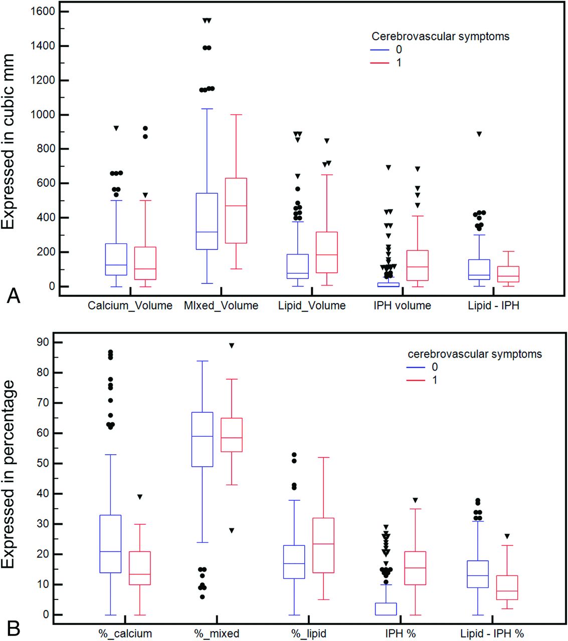
  • Fig 2.
    • Download figure
    • Open in new tab
    • Download powerpoint
    Fig 2.

    Boxplot of the volume components of the carotid artery plaque according to the presence or absence of cerebral symptoms (A) and boxplot of the percentages of the components according to the presence or absence of cerebrovascular symptoms (B).

  • Fig 3.
    • Download figure
    • Open in new tab
    • Download powerpoint
    Fig 3.

    ROC curve analysis of the volume components of the carotid artery plaque according to the presence or absence of cerebral symptoms (A) and ROC curve analysis of the percentages of the components according to the presence or absence of cerebral symptoms (B).

Tables

  • Figures
    • View popup
    Table 1:

    Demographic and plaque characteristic summary table

    Cerebrovascular SymptomsYesNoP ValueTest
    Demographics
        Age (mean) (95% CI) (yr)70 (66–74)68 (65–71).37Paired Student t
        Sex (male = 93)80% (37/46)74% (57/77).55χ2
        Hypertension26% (12/46)27% (42/154).98χ2
        CAD50% (23/46)45% (70/154).71χ2
        Smoking status43% (20/46)30% (46/154).12χ2
        Diabetes7% (3/43)7% (11/154).85χ2
        Dyslipidemia28% (13/46)32% (49/154).79χ2
    Plaque composition
        Total plaque volume (mm3)793 (565–984)560 (503–669).105Mann-Whitney
        Lipid volume (mm3)187 (132–240)79 (63–101).002aMann-Whitney
        Mixed volume (mm3)471 (400–544)318 (290–367).062Mann-Whitney
        Calcified volume (mm3)103 (70–150)128 (109–156).16Mann-Whitney
        IPH volume (mm3)115 (74–160)2 (0–9).001aMann-Whitney
        Lipid-IPH volume (mm3)61 (37–110)67 (60–87).14Mann-Whitney
        % of lipid23 (21–30)17 (14–18).002aMann-Whitney
        % of mixed58 (56–63)59 (56–61).51Mann-Whitney
        % of calcium13 (12–17)21 (18–22).001aMann-Whitney
        % of IPH15 (12–18)1 (0–3).001aMann-Whitney
        % of lipid-IPH8 (6–11)13 (12–14).001aMann-Whitney
        IPH/lipid ratio0.69 (0.59–0.73)0.019 (0–0.064).001aMann-Whitney
    • Note:—CAD indicates coronary artery disease.

    • ↵a Significant.

    • View popup
    Table 2:

    ROC curve analysis between symptoms and volume/percentage of plaque tissues

    AUCSE95% CIP Value
    Plaque volume0.5790.0490.507–0.648.108
    IPH volume0.7930.04210.730–0.847.001
    Lipid minus IPH0.5740.04860.502–0.643.129
    Lipid volume0.6480.04860.577–0.714.002
    Mixed volume0.5940.04650.523–0.663.043
    Calcium volume0.5680.05170.497–0.638.186
    % of lipid0.6790.04760.609–0.743.001
    % of mixed0.5320.04590.460–0.602.489
    % of calcium0.5910.04260.522–0.654.135
    % of IPH0.8120.04130.751–0.863.001
    % of lipid minus IPH0.7020.04280.633–0.764.001
    IPH/lipid radio0.8110.04240.751–0.863.001
    • Note:—SE indicates standard error.

    • View popup
    Table 3:

    χ2 test between symptoms and different volume thresholds of IPH

    χ2Contingency CoefficientP Value
    IPH, 10 mm312.5270.243.001
    IPH, 50 mm343.9130.424.001
    IPH, 100 mm337.4780.397.001
    IPH, 150 mm334.7630.385.001
    IPH, 200 mm314.9350.264.001
    IPH, 250 mm37.9440.195.048
PreviousNext
Back to top

In this issue

American Journal of Neuroradiology: 40 (10)
American Journal of Neuroradiology
Vol. 40, Issue 10
1 Oct 2019
  • Table of Contents
  • Index by author
  • Complete Issue (PDF)
Advertisement
Print
Download PDF
Email Article

Thank you for your interest in spreading the word on American Journal of Neuroradiology.

NOTE: We only request your email address so that the person you are recommending the page to knows that you wanted them to see it, and that it is not junk mail. We do not capture any email address.

Enter multiple addresses on separate lines or separate them with commas.
Carotid Intraplaque-Hemorrhage Volume and Its Association with Cerebrovascular Events
(Your Name) has sent you a message from American Journal of Neuroradiology
(Your Name) thought you would like to see the American Journal of Neuroradiology web site.
CAPTCHA
This question is for testing whether or not you are a human visitor and to prevent automated spam submissions.
Cite this article
L. Saba, G. Micheletti, W. Brinjikji, P. Garofalo, R. Montisci, A. Balestrieri, J.S. Suri, J.K. DeMarco, G. Lanzino, R. Sanfilippo
Carotid Intraplaque-Hemorrhage Volume and Its Association with Cerebrovascular Events
American Journal of Neuroradiology Oct 2019, 40 (10) 1731-1737; DOI: 10.3174/ajnr.A6189

Citation Manager Formats

  • BibTeX
  • Bookends
  • EasyBib
  • EndNote (tagged)
  • EndNote 8 (xml)
  • Medlars
  • Mendeley
  • Papers
  • RefWorks Tagged
  • Ref Manager
  • RIS
  • Zotero
0 Responses
Respond to this article
Share
Bookmark this article
Carotid Intraplaque-Hemorrhage Volume and Its Association with Cerebrovascular Events
L. Saba, G. Micheletti, W. Brinjikji, P. Garofalo, R. Montisci, A. Balestrieri, J.S. Suri, J.K. DeMarco, G. Lanzino, R. Sanfilippo
American Journal of Neuroradiology Oct 2019, 40 (10) 1731-1737; DOI: 10.3174/ajnr.A6189
del.icio.us logo Twitter logo Facebook logo Mendeley logo
  • Tweet Widget
  • Facebook Like
  • Google Plus One
Purchase

Jump to section

  • Article
    • Abstract
    • ABBREVIATIONS:
    • Materials and Methods
    • Results
    • Discussion
    • Conclusions
    • Footnotes
    • References
  • Figures & Data
  • Info & Metrics
  • Responses
  • References
  • PDF

Related Articles

  • No related articles found.
  • PubMed
  • Google Scholar

Cited By...

  • Carotid Plaque Calcification Attenuation Characteristics are Associated with Intraplaque Hemorrhage Volumes: A 3D Segmentation-Based Analysis
  • Assessment of Attenuation in Pericarotid Fat among Patients with Carotid Plaque and Spontaneous Carotid Dissection
  • Explainable machine-learning model to classify culprit calcified carotid plaque in embolic stroke of undetermined source
  • Impact Analysis of Different CT Configurations of Carotid Artery Plaque Calcifications on Cerebrovascular Events
  • Perivascular Fat Density and Contrast Plaque Enhancement: Does a Correlation Exist?
  • Crossref (12)
  • Google Scholar

This article has been cited by the following articles in journals that are participating in Crossref Cited-by Linking.

  • Perivascular Fat Density and Contrast Plaque Enhancement: Does a Correlation Exist?
    L. Saba, S. Zucca, A. Gupta, G. Micheletti, J.S. Suri, A. Balestrieri, M. Porcu, P. Crivelli, G. Lanzino, Y. Qi, V. Nardi, G. Faa, R. Montisci
    American Journal of Neuroradiology 2020 41 8
  • Impact Analysis of Different CT Configurations of Carotid Artery Plaque Calcifications on Cerebrovascular Events
    L. Saba, H. Chen, R. Cau, G.D. Rubeis, G. Zhu, F. Pisu, B. Jang, G. Lanzino, J.S. Suri, Y. Qi, M. Wintermark
    American Journal of Neuroradiology 2022 43 2
  • Carotid Plaques From Symptomatic Patients With Mild Stenosis Is Associated With Intraplaque Hemorrhage
    Valentina Nardi, John Benson, Melanie C. Bois, Luca Saba, Anthony S. Larson, Ilke Özcan, Ali Ahmad, David W. Morse, Fredric B. Meyer, Waleed Brinjikji, Giuseppe Lanzino, Lilach O. Lerman, Luis E. Savastano, Amir Lerman
    Hypertension 2022 79 1
  • Differences in Stable and Unstable Atherosclerotic Plaque
    Kenji Kawai, Rika Kawakami, Aloke V. Finn, Renu Virmani
    Arteriosclerosis, Thrombosis, and Vascular Biology 2024 44 7
  • Studying the Factors of Human Carotid Atherosclerotic Plaque Rupture, by Calculating Stress/Strain in the Plaque, Based on CEUS Images: A Numerical Study
    Zhenzhou Li, Yongfeng Wang, Xinyin Wu, Xin Liu, Shanshan Huang, Yi He, Shuyu Liu, Lijie Ren
    Frontiers in Neuroinformatics 2020 14
  • Quantitative and qualitative features of carotid and coronary atherosclerotic plaque among men and women
    Carlotta Onnis, Christian Cadeddu Dessalvi, Filippo Cademartiri, Giuseppe Muscogiuri, Simone Angius, Francesca Contini, Jasjit S. Suri, Sandro Sironi, Rodrigo Salgado, Antonio Esposito, Luca Saba
    Frontiers in Cardiovascular Medicine 2022 9
  • Semiautomated carotid artery plaque composition: are intraplaque CT imaging features associated with cardiovascular risk factors?
    John C. Benson, Giuseppe Lanzino, Valentina Nardi, Luis Savastano, Amir Lerman, Waleed Brinjikji
    Neuroradiology 2021 63 10
  • Carotid Artery Tortuosity and Internal Carotid Artery Plaque Composition
    John C. Benson, Adnan Shahid, Anthony Larson, Waleed Brinjikji, Deena Nasr, Luca Saba, Giuseppe Lanzino, Luis E. Savastano
    Clinical Neuroradiology 2023 33 4
  • CT and MR Imaging of Carotid Wall and Plaque
    Luca Saba, Lorenzo Mannelli, Antonella Balestrieri, Alessandra Serra, PierPaolo Bassareo, Alessandro Murgia, Carola Politi, Michele Porcu, Paola Crivelli, Giulio Micheletti, Maurizio Conti, Jasjit S Suri
    Journal of Neurosonology and Neuroimaging 2019 11 2
  • Prevalence of High-Risk CTA-Based Carotid Plaque-RADS Subtypes in Patients With Embolic Stroke of Undetermined Source
    Jae W. Song, Huy Q. Phi, Manisha Koneru, Quy Cao, Jeremy Rubin, Yu Sakai, Lamya Ibrahim, Sonya E. Zhou, John H. Woo, Scott E. Kasner, Luca Saba, Brett L. Cucchiara
    Stroke 2025 56 3

More in this TOC Section

  • Proximal Vertebral Artery Variants and Embryology
  • High-Risk Plaque Features in Carotid MRI
  • Nonstenotic Carotid Plaques and Stroke Review
Show more Extracranial Vascular

Similar Articles

Advertisement

Indexed Content

  • Current Issue
  • Accepted Manuscripts
  • Article Preview
  • Past Issues
  • Editorials
  • Editors Choice
  • Fellow Journal Club
  • Letters to the Editor

Cases

  • Case Collection
  • Archive - Case of the Week
  • Archive - Case of the Month
  • Archive - Classic Case

Special Collections

  • Special Collections

Resources

  • News and Updates
  • Turn around Times
  • Submit a Manuscript
  • Author Policies
  • Manuscript Submission Guidelines
  • Evidence-Based Medicine Level Guide
  • Publishing Checklists
  • Graphical Abstract Preparation
  • Imaging Protocol Submission
  • Submit a Case
  • Become a Reviewer/Academy of Reviewers
  • Get Peer Review Credit from Publons

Multimedia

  • AJNR Podcast
  • AJNR SCANtastic
  • Video Articles

About Us

  • About AJNR
  • Editorial Board
  • Not an AJNR Subscriber? Join Now
  • Alerts
  • Feedback
  • Advertise with us
  • Librarian Resources
  • Permissions
  • Terms and Conditions

American Society of Neuroradiology

  • Not an ASNR Member? Join Now

© 2025 by the American Society of Neuroradiology All rights, including for text and data mining, AI training, and similar technologies, are reserved.
Print ISSN: 0195-6108 Online ISSN: 1936-959X

Powered by HighWire