Advertisement

Main menu

  • Home
  • Content
    • Current Issue
    • Accepted Manuscripts
    • Article Preview
    • Past Issue Archive
    • AJNR Case Collection
    • Case of the Week Archive
    • Classic Case Archive
    • Case of the Month Archive
  • Special Collections
    • Spinal CSF Leak Articles (Jan 2020-June 2024)
    • 2024 AJNR Journal Awards
    • Most Impactful AJNR Articles
  • Multimedia
    • AJNR Podcast
    • AJNR Scantastics
    • Video Articles
  • For Authors
    • Submit a Manuscript
    • Author Policies
    • Fast publishing of Accepted Manuscripts
    • Graphical Abstract Preparation
    • Manuscript Submission Guidelines
    • Imaging Protocol Submission
    • Submit a Case for the Case Collection
  • About Us
    • About AJNR
    • Editorial Board
  • More
    • Become a Reviewer/Academy of Reviewers
    • Subscribers
    • Permissions
    • Alerts
    • Feedback
    • Advertisers
    • ASNR Home
  • Other Publications
    • ajnr

User menu

  • Alerts
  • Log in

Search

  • Advanced search
American Journal of Neuroradiology

American Journal of Neuroradiology

ASHNR American Society of Functional Neuroradiology ASHNR American Society of Pediatric Neuroradiology ASSR
  • Alerts
  • Log in

Advanced Search

  • Home
  • Content
    • Current Issue
    • Accepted Manuscripts
    • Article Preview
    • Past Issue Archive
    • AJNR Case Collection
    • Case of the Week Archive
    • Classic Case Archive
    • Case of the Month Archive
  • Special Collections
    • Spinal CSF Leak Articles (Jan 2020-June 2024)
    • 2024 AJNR Journal Awards
    • Most Impactful AJNR Articles
  • Multimedia
    • AJNR Podcast
    • AJNR Scantastics
    • Video Articles
  • For Authors
    • Submit a Manuscript
    • Author Policies
    • Fast publishing of Accepted Manuscripts
    • Graphical Abstract Preparation
    • Manuscript Submission Guidelines
    • Imaging Protocol Submission
    • Submit a Case for the Case Collection
  • About Us
    • About AJNR
    • Editorial Board
  • More
    • Become a Reviewer/Academy of Reviewers
    • Subscribers
    • Permissions
    • Alerts
    • Feedback
    • Advertisers
    • ASNR Home
  • Follow AJNR on Twitter
  • Visit AJNR on Facebook
  • Follow AJNR on Instagram
  • Join AJNR on LinkedIn
  • RSS Feeds

Welcome to the new AJNR, Updated Hall of Fame, and more. Read the full announcements.


AJNR is seeking candidates for the position of Associate Section Editor, AJNR Case Collection. Read the full announcement.


Getting new auth cookie, if you see this message a lot, tell someone!
Research ArticlePediatrics

Growth Curves of Subependymal Giant Cell Tumors in Tuberous Sclerosis Complex

J.R. Barnett, J.H. Freedman, H. Zheng, E.A. Thiele and P. Caruso
American Journal of Neuroradiology October 2021, 42 (10) 1891-1897; DOI: https://doi.org/10.3174/ajnr.A7231
J.R. Barnett
aFrom the Carol and James Herscot Center for Tuberous Sclerosis Complex (J.R.B., J.H.F., E.A.T.), Massachusetts General Hospital, Boston, Massachusetts
  • Find this author on Google Scholar
  • Find this author on PubMed
  • Search for this author on this site
  • ORCID record for J.R. Barnett
J.H. Freedman
aFrom the Carol and James Herscot Center for Tuberous Sclerosis Complex (J.R.B., J.H.F., E.A.T.), Massachusetts General Hospital, Boston, Massachusetts
  • Find this author on Google Scholar
  • Find this author on PubMed
  • Search for this author on this site
  • ORCID record for J.H. Freedman
H. Zheng
bBiostatistics Center (H.Z.), Massachusetts General Hospital, Boston, Massachusetts
  • Find this author on Google Scholar
  • Find this author on PubMed
  • Search for this author on this site
  • ORCID record for H. Zheng
E.A. Thiele
aFrom the Carol and James Herscot Center for Tuberous Sclerosis Complex (J.R.B., J.H.F., E.A.T.), Massachusetts General Hospital, Boston, Massachusetts
  • Find this author on Google Scholar
  • Find this author on PubMed
  • Search for this author on this site
  • ORCID record for E.A. Thiele
P. Caruso
cPediatric Neuroimaging (P.C.), Lenox Hill Radiology & Medical Imaging Associates, New York
  • Find this author on Google Scholar
  • Find this author on PubMed
  • Search for this author on this site
  • ORCID record for P. Caruso
  • Article
  • Figures & Data
  • Info & Metrics
  • Responses
  • References
  • PDF
Loading

Article Figures & Data

Figures

  • Tables
  • FIG 1.
    • Download figure
    • Open in new tab
    • Download powerpoint
    FIG 1.

    Serial images of patient 11's resected lesion located in the region of the right foramen of Monro (A–C) and patient 2's indeterminate lesion located in the region of left foramen of Monro (D–F). Patient 11's lesion was resected at 16 years of age. The arrows point to the lesion of interest. Cubic measurements are as follows: (AP × TV × SI), A, 9.6 mm × 8.8 mm × 10.6 mm; B, 16.1 mm × 15.9 mm × 16.3 mm; C, 21.5 mm × 16.5 mm × 19.3 mm; D, 10.4 mm × 9.2 mm × 11.2 mm; E, 10.5 mm × 9.5 mm × 11.7 mm; F, 10.4 mm × 9.5 mm × 11.9 mm.

  • FIG 2.
    • Download figure
    • Open in new tab
    • Download powerpoint
    FIG 2.

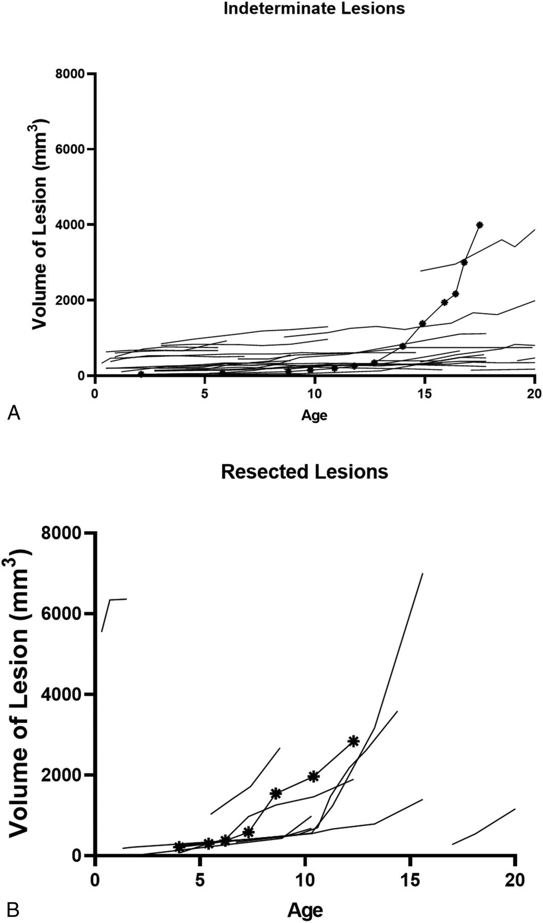
    A, Volume of indeterminate lesions by age at scan. Each line indicates 1 indeterminate nodule. The marked line indicates that an indeterminate nodule in patient 23 shows rapid growth. There is currently no evidence of obstructive hydrocephalus. The patient is being closely monitored. B, Volume of resected lesions by age at scan. The marked line indicates patient 4's resected lesion. Patient 4's resected lesion had an average growth of 531.3 mm3/year between 6.2 and 7.3 years of age and a period of decelerated growth between 7.3 and 8.7 and 8.7–10.4 years of age, in which the average growth rates were 204.7 mm3/year and 118.9 mm3/year, respectively. At 12.7 years of age, the patient underwent surgical resection for obstructive hydrocephalus.

  • FIG 3.
    • Download figure
    • Open in new tab
    • Download powerpoint
    FIG 3.

    A, Median and IQR of lesion volumes of small lesions (small), indeterminate nodules (inde), and resected lesions (resected). The median volume and IQR at the earliest scan: small lesions, 25.22 mm3 (IQR = 50.8 mm3, n = 126); indeterminate lesions, 251.9 mm3 (IQR = 319.4 mm3, n = 27), and resected lesions, 245.6 mm3 (IQR = 486.5 mm3, n = 10). The median volume and IQR at most the recent scan: small lesions, 33.18 mm3 (IQR = 61.8 mm3, n = 126); indeterminate lesions, 615.6 mm3 (IQR = 581.1 mm3, n = 27); and resected lesions, 2283.0 mm3 (IQR = 3162.0 mm3; n = 10). Indeterminate lesion volume and resected lesion volume are compared at earliest scan and most recent scan using the Mann-Whitney U test. There is no significant difference in volume at the earliest scan (P = 1.0); however, there is a significant difference at the most recent scan. The asterisk indicates P < .001. B, Median lesion volume of each lesion type by age. Each point is the median volume of ≥2 lesions at a given age. NS indicates not significant.

  • FIG 4.
    • Download figure
    • Open in new tab
    • Download powerpoint
    FIG 4.

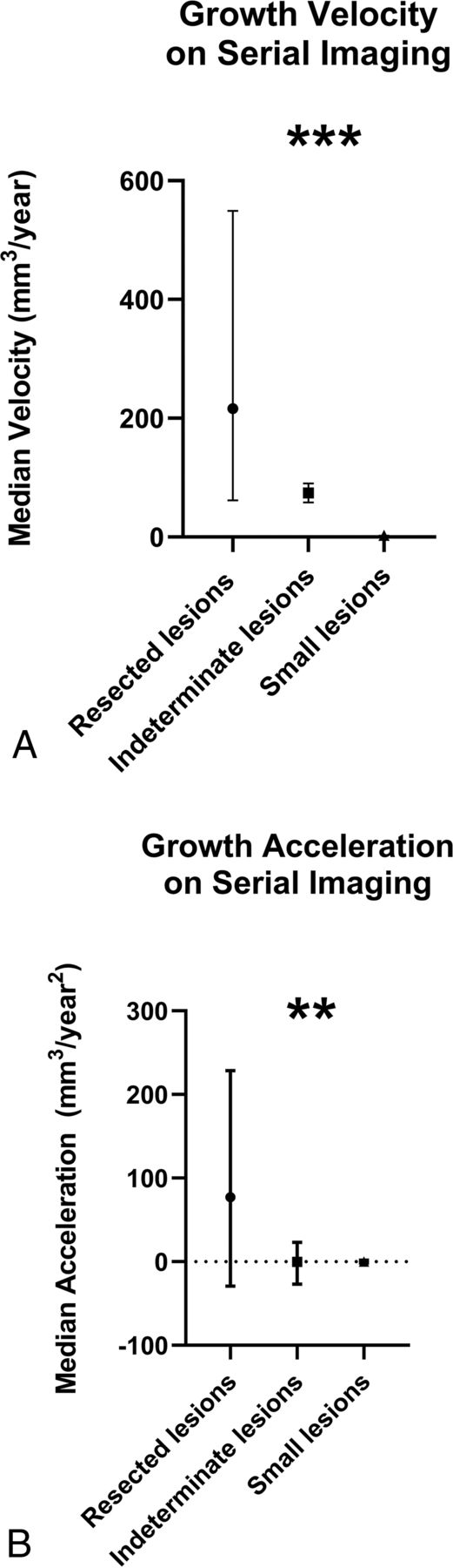
    A, Median growth velocity between scans for each lesion type: small lesions, 0.45 mm3/year (IQR = 3.2 mm3/year, n = 789); indeterminate lesions, 21.95 mm3/year (IQR = 71.6 mm3/year; n = 167); resected lesions, 216.1 mm3/year (IQR = 487.7 mm3/year; n = 38). A Mann-Whitney U test with a Bonferroni correction was used to compare the velocity of growth between lesion types. The triple asterisks indicate a significant difference in velocity comparing resected lesions and indeterminate lesions (P <.001), resected lesions and small lesions (P <.001), and indeterminate lesions and small lesions (P <.001). B, Median acceleration of growth of all lesion types: small lesions, 0.00 mm3/year2 (IQR = 3.4 mm3/year2, n = 673); indeterminate lesions, −0.18 mm3/year2 (IQR = 70.63 mm3/year2, n = 141); resected lesions, 77.01 mm3/year2 (IQR = 257.7 mm3/year2; n = 30). A Mann-Whitney U test with a Bonferroni correction was used to compare the acceleration of growth among lesion types. The double asterisks indicate significant differences between resected lesions and indeterminate lesions (P = .01) and resected lesions and small lesions (P = .004). There is not a significant difference in the rate of acceleration between indeterminate lesions and small lesions (P = 1.0).

Tables

  • Figures
    • View popup
    Table 1:

    Median growth velocitiesa

    0–5 Years of Age6–10 Years of Age11–15 Years of Age16–20 Years of Age
    Resected lesions333.2 mm3/year
    58.1–608.23b
    n = 2
    351. mm3/year
    IQR = 344.5
    n = 4
    460.3 mm3/year
    IQR = 647.7
    n = 5
    292.0 mm3/year
    NA
    n = 1
    Indeterminate lesions32.5 mm3/year
    IQR = 58.1 mm3/year
    n = 11
    18.8 mm3/year
    IQR = 37.0 mm3/year
    n = 16
    15.8 mm3/year
    IQR = 43.9 mm3/year
    n = 13
    15.7 mm3/year
    IQR = 103.6 mm3/year
    n = 13
    Small lesions0.6 mm3/year
    IQR = 4.2 mm3/year
    n = 42
    0.3 mm3/year
    IQR = 1.6 mm3/year
    n = 59
    0.3 mm3/year
    IQR = 3.0 mm3/year
    n = 39
    0.6 mm3/year
    IQR = 3.1 mm3/year
    n = 32
    • Note:—NA indicates not applicable.

    • ↵a Listed are the median growth and IQR for each lesion type for each age period.

    • ↵b Range and not IQR are reported for resected lesions at 0–5 years of age.

    • View popup
    Table 2:

    Growth velocity comparisonsa

    0-5 Years of Age6-10 Years of Age11-15 Years of Age16-20 Years of Age
    Resected vs indeP = .62P = .007bP = .006bNA
    Resected vs smallP = .01bP < .001bP < .001bNA
    Resected vs inde + smallP = .03bP < .001bP < .001bNA
    Inde vs smallP < .001bP < .001bP = .053P = .034b
    • Note:—Inde indicates indeterminate lesions; small, small lesions; resected, resected lesions; NA, not applicable.

    • ↵a P-values calculated using Mann-Whitney U test and Bonferroni correction.

    • ↵b Statistical significance.

    • View popup
    Table 3:

    Percentage of lesions with growth rates > 4.2 mm3/yeara

    Baseline to Most Recent Scan0–5 Years of Age6–10 Years of Age11–15 Years of Age16–20 Years of Age
    Small lesions14%21%12%10%16%
    Indeterminate lesions96%100%81%62%69%
    Resected lesions100%100%b100%100%100%c
    All lesions34%41%35%30%33%
    • ↵a 4.2 mm3 was chosen to reflect an increase in growth of 2 mm in the AP, TV, and SI dimensions using an ellipsoid volume formula because a 2-mm increase would be an appreciable measurement on MR imaging.

    • ↵b Of the 3 patients with symptomatic SGCTs in this age group, only 2/3 MRIs were considered diagnostic.

    • ↵c Only 1 lesion was available for measurement.

PreviousNext
Back to top

In this issue

American Journal of Neuroradiology: 42 (10)
American Journal of Neuroradiology
Vol. 42, Issue 10
1 Oct 2021
  • Table of Contents
  • Index by author
  • Complete Issue (PDF)
Advertisement
Print
Download PDF
Email Article

Thank you for your interest in spreading the word on American Journal of Neuroradiology.

NOTE: We only request your email address so that the person you are recommending the page to knows that you wanted them to see it, and that it is not junk mail. We do not capture any email address.

Enter multiple addresses on separate lines or separate them with commas.
Growth Curves of Subependymal Giant Cell Tumors in Tuberous Sclerosis Complex
(Your Name) has sent you a message from American Journal of Neuroradiology
(Your Name) thought you would like to see the American Journal of Neuroradiology web site.
CAPTCHA
This question is for testing whether or not you are a human visitor and to prevent automated spam submissions.
Cite this article
J.R. Barnett, J.H. Freedman, H. Zheng, E.A. Thiele, P. Caruso
Growth Curves of Subependymal Giant Cell Tumors in Tuberous Sclerosis Complex
American Journal of Neuroradiology Oct 2021, 42 (10) 1891-1897; DOI: 10.3174/ajnr.A7231

Citation Manager Formats

  • BibTeX
  • Bookends
  • EasyBib
  • EndNote (tagged)
  • EndNote 8 (xml)
  • Medlars
  • Mendeley
  • Papers
  • RefWorks Tagged
  • Ref Manager
  • RIS
  • Zotero
0 Responses
Respond to this article
Share
Bookmark this article
Growth Curves of Subependymal Giant Cell Tumors in Tuberous Sclerosis Complex
J.R. Barnett, J.H. Freedman, H. Zheng, E.A. Thiele, P. Caruso
American Journal of Neuroradiology Oct 2021, 42 (10) 1891-1897; DOI: 10.3174/ajnr.A7231
del.icio.us logo Twitter logo Facebook logo Mendeley logo
  • Tweet Widget
  • Facebook Like
  • Google Plus One
Purchase

Jump to section

  • Article
    • Abstract
    • ABBREVIATIONS:
    • MATERIALS AND METHODS
    • RESULTS
    • DISCUSSION
    • CONCLUSIONS
    • ACKNOWLEDGMENTS
    • Footnotes
    • References
  • Figures & Data
  • Info & Metrics
  • Responses
  • References
  • PDF

Related Articles

  • No related articles found.
  • PubMed
  • Google Scholar

Cited By...

  • No citing articles found.
  • Crossref
  • Google Scholar

This article has not yet been cited by articles in journals that are participating in Crossref Cited-by Linking.

More in this TOC Section

  • Comparison of Image Quality and Radiation Dose in Pediatric Temporal Bone CT Using Photon-Counting Detector CT and Energy-Integrating Detector CT
  • SyMRI & MR Fingerprinting in Brainstem Myelination
  • Venous Sinus Stenosis in Mucopolysaccharidosis I
Show more Pediatrics

Similar Articles

Advertisement

Indexed Content

  • Current Issue
  • Accepted Manuscripts
  • Article Preview
  • Past Issues
  • Editorials
  • Editors Choice
  • Fellow Journal Club
  • Letters to the Editor

Cases

  • Case Collection
  • Archive - Case of the Week
  • Archive - Case of the Month
  • Archive - Classic Case

Special Collections

  • Special Collections

Resources

  • News and Updates
  • Turn around Times
  • Submit a Manuscript
  • Author Policies
  • Manuscript Submission Guidelines
  • Evidence-Based Medicine Level Guide
  • Publishing Checklists
  • Graphical Abstract Preparation
  • Imaging Protocol Submission
  • Submit a Case
  • Become a Reviewer/Academy of Reviewers
  • Get Peer Review Credit from Publons

Multimedia

  • AJNR Podcast
  • AJNR SCANtastic
  • Video Articles

About Us

  • About AJNR
  • Editorial Board
  • Not an AJNR Subscriber? Join Now
  • Alerts
  • Feedback
  • Advertise with us
  • Librarian Resources
  • Permissions
  • Terms and Conditions

American Society of Neuroradiology

  • Not an ASNR Member? Join Now

© 2025 by the American Society of Neuroradiology All rights, including for text and data mining, AI training, and similar technologies, are reserved.
Print ISSN: 0195-6108 Online ISSN: 1936-959X

Powered by HighWire