Advertisement

Main menu

  • Home
  • Content
    • Current Issue
    • Accepted Manuscripts
    • Article Preview
    • Past Issue Archive
    • AJNR Case Collection
    • Case of the Week Archive
    • Classic Case Archive
    • Case of the Month Archive
  • Special Collections
    • Spinal CSF Leak Articles (Jan 2020-June 2024)
    • 2024 AJNR Journal Awards
    • Most Impactful AJNR Articles
  • Multimedia
    • AJNR Podcast
    • AJNR Scantastics
    • Video Articles
  • For Authors
    • Submit a Manuscript
    • Author Policies
    • Fast publishing of Accepted Manuscripts
    • Graphical Abstract Preparation
    • Manuscript Submission Guidelines
    • Imaging Protocol Submission
    • Submit a Case for the Case Collection
  • About Us
    • About AJNR
    • Editorial Board
  • More
    • Become a Reviewer/Academy of Reviewers
    • Subscribers
    • Permissions
    • Alerts
    • Feedback
    • Advertisers
    • ASNR Home
  • Other Publications
    • ajnr

User menu

  • Alerts
  • Log in

Search

  • Advanced search
American Journal of Neuroradiology

American Journal of Neuroradiology

ASHNR American Society of Functional Neuroradiology ASHNR American Society of Pediatric Neuroradiology ASSR
  • Alerts
  • Log in

Advanced Search

  • Home
  • Content
    • Current Issue
    • Accepted Manuscripts
    • Article Preview
    • Past Issue Archive
    • AJNR Case Collection
    • Case of the Week Archive
    • Classic Case Archive
    • Case of the Month Archive
  • Special Collections
    • Spinal CSF Leak Articles (Jan 2020-June 2024)
    • 2024 AJNR Journal Awards
    • Most Impactful AJNR Articles
  • Multimedia
    • AJNR Podcast
    • AJNR Scantastics
    • Video Articles
  • For Authors
    • Submit a Manuscript
    • Author Policies
    • Fast publishing of Accepted Manuscripts
    • Graphical Abstract Preparation
    • Manuscript Submission Guidelines
    • Imaging Protocol Submission
    • Submit a Case for the Case Collection
  • About Us
    • About AJNR
    • Editorial Board
  • More
    • Become a Reviewer/Academy of Reviewers
    • Subscribers
    • Permissions
    • Alerts
    • Feedback
    • Advertisers
    • ASNR Home
  • Follow AJNR on Twitter
  • Visit AJNR on Facebook
  • Follow AJNR on Instagram
  • Join AJNR on LinkedIn
  • RSS Feeds

Welcome to the new AJNR, Updated Hall of Fame, and more. Read the full announcements.


AJNR is seeking candidates for the position of Associate Section Editor, AJNR Case Collection. Read the full announcement.


  • Getting new auth cookie, if you see this message a lot, tell someone!
  • Getting new auth cookie, if you see this message a lot, tell someone!
Research ArticleHead & Neck

Cone-beam CT versus Multidetector CT in Postoperative Cochlear Implant Imaging: Evaluation of Image Quality and Radiation Dose

R.A. Helal, R. Jacob, M.A. Elshinnawy, A.I. Othman, I.M. Al-Dhamari, D.W. Paulus and T.T. Abdelaziz
American Journal of Neuroradiology February 2021, 42 (2) 362-367; DOI: https://doi.org/10.3174/ajnr.A6894
R.A. Helal
aFrom the Radiodiagnosis Department (R.A.H., M.A.E., A.I.O., T.T.A.), Ain Shams University, Cairo, Egypt
  • Find this author on Google Scholar
  • Find this author on PubMed
  • Search for this author on this site
  • ORCID record for R.A. Helal
R. Jacob
bHNOplus (R.J.), Höhr-Grenzhausen, Germany
  • Find this author on Google Scholar
  • Find this author on PubMed
  • Search for this author on this site
  • ORCID record for R. Jacob
M.A. Elshinnawy
aFrom the Radiodiagnosis Department (R.A.H., M.A.E., A.I.O., T.T.A.), Ain Shams University, Cairo, Egypt
  • Find this author on Google Scholar
  • Find this author on PubMed
  • Search for this author on this site
  • ORCID record for M.A. Elshinnawy
A.I. Othman
aFrom the Radiodiagnosis Department (R.A.H., M.A.E., A.I.O., T.T.A.), Ain Shams University, Cairo, Egypt
  • Find this author on Google Scholar
  • Find this author on PubMed
  • Search for this author on this site
  • ORCID record for A.I. Othman
I.M. Al-Dhamari
cInstitute for computational visualistics (I.M.A.-D., D.W.P.), Koblenz University, Koblenz, Germany
  • Find this author on Google Scholar
  • Find this author on PubMed
  • Search for this author on this site
  • ORCID record for I.M. Al-Dhamari
D.W. Paulus
cInstitute for computational visualistics (I.M.A.-D., D.W.P.), Koblenz University, Koblenz, Germany
  • Find this author on Google Scholar
  • Find this author on PubMed
  • Search for this author on this site
  • ORCID record for D.W. Paulus
T.T. Abdelaziz
aFrom the Radiodiagnosis Department (R.A.H., M.A.E., A.I.O., T.T.A.), Ain Shams University, Cairo, Egypt
  • Find this author on Google Scholar
  • Find this author on PubMed
  • Search for this author on this site
  • ORCID record for T.T. Abdelaziz
  • Article
  • Figures & Data
  • Info & Metrics
  • Responses
  • References
  • PDF
Loading

Article Figures & Data

Figures

  • Tables
  • FIG 1.
    • Download figure
    • Open in new tab
    • Download powerpoint
    FIG 1.

    Coronal oblique (cochlear view with thick MPR = 5 mm) CBCT image showing the measurement of the angle of insertion between the deepest electrode and the reference line joining the insertion point (round window/cochleostomy center) (a) and the center of a circle formed by the 3 most apical electrodes (b). The insertion angle = 360°- (c). The cochlear lateral wall is also well-visualized in this thick MPR coronal view (white arrows).
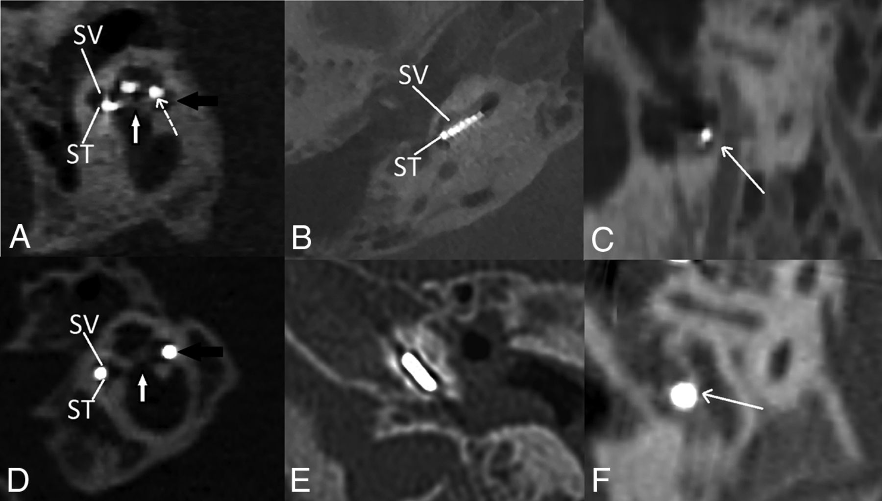
  • FIG 2.
    • Download figure
    • Open in new tab
    • Download powerpoint
    FIG 2.

    CBCT group. A, Midmodiolar view (cochlea) shows the modiolus (white arrow), the perfectly visualized cochlear lateral wall (black arrow), and the scalar translocation from ST to SV at the pars ascendens (dashed arrow). B, Axial view (cochlea) shows good visualization of single electrode contacts, C, Sagittal oblique view shows good visualization of the facial nerve canal wall (arrow). D, In the MDCT group, the midmodiolar view (cochlea) shows the modiolus (white arrow), nonvisualized cochlear lateral wall (artifacts) (black arrow), and the difficult scalar localization of the electrode (mostly ST). E, Axial view (cochlea) shows difficult identification of single electrode contacts and the osseous spiral lamina. F, Sagittal oblique view shows difficult identification of the facial nerve canal wall (arrow). ST indicates scala tympani; SV, scala vestibuli.

Tables

  • Figures
    • View popup
    Table 1:

    Different electrodes with intercontact distancing

    ElectrodesIntercontact Distancing (mm)CBCT Group (n = 32)MDCT Group (n = 19)
    Widely spaced contacts
     FLEX28a2.130
     FLEX-Synchrony-Mediuma1.922
     HiRes MIDSCALAb0.97531
     CI422*c0.85–0.9510
    Narrowly spaced contacts
     CI24RE-CAcNonuniform 0.4–0.890
     CI512-CAcNonuniform 0.4–0.830
     CI532-CAc0.6111
     CI24RE-STc0.75015
    • Note:—CA indicates contour advance; ST, straight electrode.

    • ↵a MED-EL.

    • ↵b Advanced Bionics.

    • ↵c Cochlear.

    • All distances were obtained from the portfolios of the electrodes (* except for CI422 cited from Bennink et al.29)

    • View popup
    Table 2:

    Qualitative image scoring results for fine anatomic structures and metallic artifacts

    Scale PointsCBCT Group (n = 32)MDCT Group (n = 19)P Value
    Cochlear inner wall
     Not visualized (0)1 (3%)0 (0%)>.99
     Barely visualized (1)3 (9%)2 (10.5%)
     Well-visualized (2)6 (19%)3 (15.8%)
     Perfectly visualized (3)22 (69%)14 (73.7%)
    Cochlear lateral wall
     Not visualized (0)0 (0%)1 (5%).001
     Barely visualized (1)0 (0%)0 (0%)
     Well-visualized-(2)2 (6%)8 (42%)
     Perfectly visualized (3)30 (94%)10 (53%)
    Modiolus
     Not visualized (0)0 (0%)0 (0%).37
     Barely visualized (1)2 (6%)0 (0%)
     Well-visualized (2)5 (16%)1 (5%)
     Perfectly visualized (3)25 (78%)18 (95%)
    Osseous spiral, lamina
     Not visualized (0)18 (56%)19 (100%).002
     Barely visualized (1)10 (31%)0 (0%)
     Well -visualized (2)4 (13%)0 (0%)
     Perfectly visualized (3)0 (0%)0 (0%)
    Mastoid facial, canal wall
     Not visualized (0)4 (13%)4 (21%).07
     Barely visualized (1)10 (31%)6 (32%)
     Well-visualized (2)9 (28%)9 (47%)
     Perfectly visualized (3)9 (28%)0 (0%)
    Metallic artifacts
     Very strong artifact (0)0 (0%)0 (0%)<.001
     Strong artifacts (1)6 (18.8%)14 (74%)
     Moderate artifacts (2)22 (68.8%)5 (26%)
     Weak artifacts (3)4 (12.5%)0 (0%)
    • View popup
    Table 3:

    Qualitative image scoring results for electrode evaluation

    Scale PointsCBCT Group (n = 32)MDCT Group (n = 19)P Value
    Electrode scalar position
     Not visualized (0)0 (0%)0 (0%).046
     Barely visualized (1)4 (13%)3 (16%)
     Well-visualized (2)3 (9%)7 (37%)
     Perfectly visualized (3)25 (78%)9 (47%)
    Single electrode contact  visibility
     Not visualized (0)1 (3%)10 (52.6%)<.001
     Barely visualized (1)13 (41%)6 (31.6%)
     Well-visualized (2)11 (34%)0 (0%)
     Perfectly visualized (3)7 (22%)3 (15.8%)
    Insertion angle of electrode
     Not measurable (0)0 (0%)0 (0%)>.99
     Measurable (1)32 (100%)19 (100%)
    Mean, 437.7° [SD, 120.8°]Mean, 329.4° [SD, 80°]
    • View popup
    Table 4:

    SNR comparison

    CBCT GroupMDCT GroupP Valuea
    SNR in bone (median) (IQR)8.31 (3.8)10.77 (4.25).02
    SNR in air (median) (IQR)6.58 (3.85)11.97 (5.53).001
    • Note:—IQR indicates interquartile range.

    • ↵a Mann-Whitney U test.

PreviousNext
Back to top

In this issue

American Journal of Neuroradiology: 42 (2)
American Journal of Neuroradiology
Vol. 42, Issue 2
1 Feb 2021
  • Table of Contents
  • Index by author
  • Complete Issue (PDF)
Advertisement
Print
Download PDF
Email Article

Thank you for your interest in spreading the word on American Journal of Neuroradiology.

NOTE: We only request your email address so that the person you are recommending the page to knows that you wanted them to see it, and that it is not junk mail. We do not capture any email address.

Enter multiple addresses on separate lines or separate them with commas.
Cone-beam CT versus Multidetector CT in Postoperative Cochlear Implant Imaging: Evaluation of Image Quality and Radiation Dose
(Your Name) has sent you a message from American Journal of Neuroradiology
(Your Name) thought you would like to see the American Journal of Neuroradiology web site.
CAPTCHA
This question is for testing whether or not you are a human visitor and to prevent automated spam submissions.
Cite this article
R.A. Helal, R. Jacob, M.A. Elshinnawy, A.I. Othman, I.M. Al-Dhamari, D.W. Paulus, T.T. Abdelaziz
Cone-beam CT versus Multidetector CT in Postoperative Cochlear Implant Imaging: Evaluation of Image Quality and Radiation Dose
American Journal of Neuroradiology Feb 2021, 42 (2) 362-367; DOI: 10.3174/ajnr.A6894

Citation Manager Formats

  • BibTeX
  • Bookends
  • EasyBib
  • EndNote (tagged)
  • EndNote 8 (xml)
  • Medlars
  • Mendeley
  • Papers
  • RefWorks Tagged
  • Ref Manager
  • RIS
  • Zotero
0 Responses
Respond to this article
Share
Bookmark this article
Cone-beam CT versus Multidetector CT in Postoperative Cochlear Implant Imaging: Evaluation of Image Quality and Radiation Dose
R.A. Helal, R. Jacob, M.A. Elshinnawy, A.I. Othman, I.M. Al-Dhamari, D.W. Paulus, T.T. Abdelaziz
American Journal of Neuroradiology Feb 2021, 42 (2) 362-367; DOI: 10.3174/ajnr.A6894
del.icio.us logo Twitter logo Facebook logo Mendeley logo
  • Tweet Widget
  • Facebook Like
  • Google Plus One
Purchase

Jump to section

  • Article
    • Abstract
    • ABBREVIATIONS:
    • MATERIALS AND METHODS
    • RESULTS
    • DISCUSSION
    • CONCLUSIONS
    • Footnotes
    • References
  • Figures & Data
  • Info & Metrics
  • Responses
  • References
  • PDF

Related Articles

  • No related articles found.
  • PubMed
  • Google Scholar

Cited By...

  • Measuring 3D Cochlear Duct Length on MRI: Is It Accurate and Reliable?
  • Crossref (14)
  • Google Scholar

This article has been cited by the following articles in journals that are participating in Crossref Cited-by Linking.

  • Measuring 3D Cochlear Duct Length on MRI: Is It Accurate and Reliable?
    M.B. Eser, B. Atalay, M.B. Dogan, N. Gündüz, M.T. Kalcioglu
    American Journal of Neuroradiology 2021 42 11
  • Visualization of Different Types of Cochlear Implants in Postoperative Cone-Beam CT Imaging
    Iris Burck, Franziska Drath, Moritz H. Albrecht, Tommaso D´Angelo, Hanns Ackermann, Jan-Erik Scholtz, Ibrahim Yel, Timo Stöver, Silke Helbig, Thomas J. Vogl, Benjamin Kaltenbach, Jan-Erik Scholtz
    Academic Radiology 2022 29
  • High Spatial-Resolution Skull Base Imaging With Photon-Counting Computed Tomography and Energy-Integrating Computed Tomography: A Comparative Phantom Study
    Jayasai R. Rajagopal, Fides R. Schwartz, Justin B. Solomon, David S. Enterline, Ehsan Samei
    Journal of Computer Assisted Tomography 2023 47 4
  • International Consensus Statements on Intraoperative Testing for Cochlear Implantation Surgery
    Farid Alzhrani, Isra Aljazeeri, Yassin Abdelsamad, Abdulrahman Alsanosi, Ana H. Kim, Angel Ramos-Macias, Angel Ramos-de-Miguel, Anja Kurz, Artur Lorens, Bruce Gantz, Craig A. Buchman, Dayse Távora-Vieira, Georg Sprinzl, Griet Mertens, James E. Saunders, Julie Kosaner, Laila M. Telmesani, Luis Lassaletta, Manohar Bance, Medhat Yousef, Meredith A. Holcomb, Oliver Adunka, Per Cayé- Thomasen, Piotr H. Skarzynski, Ranjith Rajeswaran, Robert J. Briggs, Seung-Ha Oh, Stefan Plontke, Stephen J. O’Leary, Sumit Agrawal, Tatsuya Yamasoba, Thomas Lenarz, Thomas Wesarg, Walter Kutz, Patrick Connolly, Ilona Anderson, Abdulrahman Hagr
    Ear & Hearing 2024 45 6
  • High-speed flat-detector computed tomography for temporal bone imaging and postoperative control of cochlear implants
    Felix Eisenhut, Lava Taha, Michael Manhart, Vivian Thimsen, Konstantinos Mantsopoulos, Heinrich Iro, Joachim Hornung, Arnd Dörfler, Stefan Lang
    Neuroradiology 2022 64 7
  • Unrecognized bone fractures of the extremities in Emergency Radiology: the invisible become visible on 3D 80 micron
    Antonio Lo Mastro, Francesco Boccia, Daniela Berritto, Luca Pio Stoppino, Egidio Guerra, Paolo D’Alessandro, Fabio Pinto, Francesca Grassi
    Journal of Medical Imaging and Interventional Radiology 2024 11 1
  • Accelerated flat panel computed tomography for pre-operative temporal bone imaging: Image quality and dosimetry comparison to conventional high resolution multislice computed tomography
    Elie Diamandis, Sebastian Johannes Müller, Eya Khadhraoui, Stefan Klebingat, Eric Einspänner, Martin Durisin, Anne Albrecht, Daniel Behme
    Neuroradiology 2025
  • Augmenting the interpretation: Imaging the head and face implants and their complications
    Noushin Yahyavi-Firouz-Abadi, Christine O. Menias, Katie D. Vo
    Clinical Imaging 2025 121
  • Cochlear Implantation for Cochlear Nerve Deficiency
    Bentao Yang
    2022
  • Comparison of 96-kV and 120-kV cone-beam CT for the assessment of cochlear implants
    Iris Burck, Ibrahim Yel, Simon Martin, Moritz H. Albrecht, Vitali Koch, Christian Booz, Daniel Pinto dos Santos, Benjamin Kaltenbach, Hanns Ackermann, Juha Koivisto, Silke Helbig, Timo Stöver, Thomas J. Vogl, Jan-Erik Scholtz
    BMC Medical Imaging 2024 24 1

More in this TOC Section

  • WHO Classification Update: Nasal&Skull Base Tumors
  • Peritumoral Signal in Vestibular Schwannomas
  • Chondrosarcoma vs Synovial Chondromatosis: Imaging
Show more HEAD & NECK

Similar Articles

Advertisement

Indexed Content

  • Current Issue
  • Accepted Manuscripts
  • Article Preview
  • Past Issues
  • Editorials
  • Editors Choice
  • Fellow Journal Club
  • Letters to the Editor

Cases

  • Case Collection
  • Archive - Case of the Week
  • Archive - Case of the Month
  • Archive - Classic Case

Special Collections

  • Special Collections

Resources

  • News and Updates
  • Turn around Times
  • Submit a Manuscript
  • Author Policies
  • Manuscript Submission Guidelines
  • Evidence-Based Medicine Level Guide
  • Publishing Checklists
  • Graphical Abstract Preparation
  • Imaging Protocol Submission
  • Submit a Case
  • Become a Reviewer/Academy of Reviewers
  • Get Peer Review Credit from Publons

Multimedia

  • AJNR Podcast
  • AJNR SCANtastic
  • Video Articles

About Us

  • About AJNR
  • Editorial Board
  • Not an AJNR Subscriber? Join Now
  • Alerts
  • Feedback
  • Advertise with us
  • Librarian Resources
  • Permissions
  • Terms and Conditions

American Society of Neuroradiology

  • Not an ASNR Member? Join Now

© 2025 by the American Society of Neuroradiology All rights, including for text and data mining, AI training, and similar technologies, are reserved.
Print ISSN: 0195-6108 Online ISSN: 1936-959X

Powered by HighWire