Advertisement

Main menu

  • Home
  • Content
    • Current Issue
    • Accepted Manuscripts
    • Article Preview
    • Past Issue Archive
    • AJNR Case Collection
    • Case of the Week Archive
    • Classic Case Archive
    • Case of the Month Archive
  • Special Collections
    • Spinal CSF Leak Articles (Jan 2020-June 2024)
    • 2024 AJNR Journal Awards
    • Most Impactful AJNR Articles
  • Multimedia
    • AJNR Podcast
    • AJNR Scantastics
    • Video Articles
  • For Authors
    • Submit a Manuscript
    • Author Policies
    • Fast publishing of Accepted Manuscripts
    • Graphical Abstract Preparation
    • Manuscript Submission Guidelines
    • Imaging Protocol Submission
    • Submit a Case for the Case Collection
  • About Us
    • About AJNR
    • Editorial Board
  • More
    • Become a Reviewer/Academy of Reviewers
    • Subscribers
    • Permissions
    • Alerts
    • Feedback
    • Advertisers
    • ASNR Home
  • Other Publications
    • ajnr

User menu

  • Alerts
  • Log in

Search

  • Advanced search
American Journal of Neuroradiology

American Journal of Neuroradiology

ASHNR American Society of Functional Neuroradiology ASHNR American Society of Pediatric Neuroradiology ASSR
  • Alerts
  • Log in

Advanced Search

  • Home
  • Content
    • Current Issue
    • Accepted Manuscripts
    • Article Preview
    • Past Issue Archive
    • AJNR Case Collection
    • Case of the Week Archive
    • Classic Case Archive
    • Case of the Month Archive
  • Special Collections
    • Spinal CSF Leak Articles (Jan 2020-June 2024)
    • 2024 AJNR Journal Awards
    • Most Impactful AJNR Articles
  • Multimedia
    • AJNR Podcast
    • AJNR Scantastics
    • Video Articles
  • For Authors
    • Submit a Manuscript
    • Author Policies
    • Fast publishing of Accepted Manuscripts
    • Graphical Abstract Preparation
    • Manuscript Submission Guidelines
    • Imaging Protocol Submission
    • Submit a Case for the Case Collection
  • About Us
    • About AJNR
    • Editorial Board
  • More
    • Become a Reviewer/Academy of Reviewers
    • Subscribers
    • Permissions
    • Alerts
    • Feedback
    • Advertisers
    • ASNR Home
  • Follow AJNR on Twitter
  • Visit AJNR on Facebook
  • Follow AJNR on Instagram
  • Join AJNR on LinkedIn
  • RSS Feeds

Welcome to the new AJNR, Updated Hall of Fame, and more. Read the full announcements.


AJNR is seeking candidates for the position of Associate Section Editor, AJNR Case Collection. Read the full announcement.


Research ArticlePediatrics

Neuroaxial Infantile Hemangiomas: Imaging Manifestations and Association with Hemangioma Syndromes

T. Feygin, A.E. Goldman-Yassen, D.J. Licht, J.E. Schmitt, A. Mian, A. Vossough, L. Castelo-Soccio, J.R. Treat, A. Bhatia and A.N. Pollock
American Journal of Neuroradiology August 2021, 42 (8) 1520-1527; DOI: https://doi.org/10.3174/ajnr.A7204
T. Feygin
aDivision of Neuroradiology (T.F., A.V., A.N.P.), Department of Radiology, The C hildren's Hospital of Philadelphia, Philadelphia, Pennsylvania
  • Find this author on Google Scholar
  • Find this author on PubMed
  • Search for this author on this site
  • ORCID record for T. Feygin
A.E. Goldman-Yassen
cDepartment of Radiology (A.E.G.-Y.), Children's Healthcare of Atlanta, Atlanta, Georgia
  • Find this author on Google Scholar
  • Find this author on PubMed
  • Search for this author on this site
  • ORCID record for A.E. Goldman-Yassen
D.J. Licht
bDepartment of Neurology (D.J.L.), The Children's Hospital of Philadelphia, Philadelphia, Pennsylvania
  • Find this author on Google Scholar
  • Find this author on PubMed
  • Search for this author on this site
  • ORCID record for D.J. Licht
J.E. Schmitt
dDivision of Neuroradiology (J.E.S.), Department of Radiology, Hospital of the University of Pennsylvania, Philadelphia, Pennsylvania
  • Find this author on Google Scholar
  • Find this author on PubMed
  • Search for this author on this site
  • ORCID record for J.E. Schmitt
A. Mian
eDivision of Neuroradiology (A.M.), Department of Radiology, Mallinckrodt Institute of Radiology, St. Louis, Missouri
  • Find this author on Google Scholar
  • Find this author on PubMed
  • Search for this author on this site
  • ORCID record for A. Mian
A. Vossough
aDivision of Neuroradiology (T.F., A.V., A.N.P.), Department of Radiology, The C hildren's Hospital of Philadelphia, Philadelphia, Pennsylvania
  • Find this author on Google Scholar
  • Find this author on PubMed
  • Search for this author on this site
  • ORCID record for A. Vossough
L. Castelo-Soccio
fDepartment of Dermatology (L.C.-S, J.R.T.), The Children's Hospital of Philadelphia, Philadelphia, Pennsylvania
  • Find this author on Google Scholar
  • Find this author on PubMed
  • Search for this author on this site
  • ORCID record for L. Castelo-Soccio
J.R. Treat
fDepartment of Dermatology (L.C.-S, J.R.T.), The Children's Hospital of Philadelphia, Philadelphia, Pennsylvania
  • Find this author on Google Scholar
  • Find this author on PubMed
  • Search for this author on this site
  • ORCID record for J.R. Treat
A. Bhatia
gDepartment of Radiology (A.B.), The Children's Hospital of Pittsburg, Philadelphia, Pennsylvania
  • Find this author on Google Scholar
  • Find this author on PubMed
  • Search for this author on this site
  • ORCID record for A. Bhatia
A.N. Pollock
aDivision of Neuroradiology (T.F., A.V., A.N.P.), Department of Radiology, The C hildren's Hospital of Philadelphia, Philadelphia, Pennsylvania
  • Find this author on Google Scholar
  • Find this author on PubMed
  • Search for this author on this site
  • ORCID record for A.N. Pollock
  • Article
  • Figures & Data
  • Info & Metrics
  • Responses
  • References
  • PDF
Loading

Article Figures & Data

Figures

  • Tables
  • FIG 1.
    • Download figure
    • Open in new tab
    • Download powerpoint
    FIG 1.

    Segmental IH. A, A 3-month-old girl with PHACE syndrome. Clinical photograph shows a bilateral segmental IH in an S1 distribution predominantly and with minimal S2 involvement. B, Diagram of the Haggstrom classification, used for clinical assessment of superficial IH distribution. It divides the face on the 4 following segments: S1 = frontotemporal, S2 = maxillary, S3 = mandibular, S4 = frontonasal.

  • FIG 2.
    • Download figure
    • Open in new tab
    • Download powerpoint
    FIG 2.

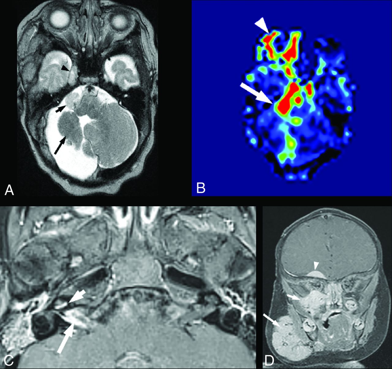
    Intracranial infantile hemangioma. A and B, A 1.5-month-old girl with segmental IH of the right face and PHACE syndrome. A, Axial T2-weighted image shows an extra-axial T2-hyperintense mass in the right cerebellopontine angle cistern (small arrow) and a similar lesion in the enlarged right MC (arrowhead). A long arrow points to an asymmetrically small right cerebellar hemisphere, which is an additional common manifestation of PHACE syndrome. Both lesions reveal diffuse enhancement on postcontrast imaging and markedly increased perfusion on arterial spin-labeling imaging (B). C, A 3-month-old girl with right orbital segmental IH and PHACE syndrome. Axial postcontrast T1-weighted image with fat suppression shows linear enhancement in the right IAC (large arrow) and linear enhancement of the cochlear basal turn (arrowhead). D, A 2-month-old girl with segmental IH of the right face and PHACE syndrome. Coronal postcontrast T1-weighted image with fat suppression shows a dural base extra-axial enhancing mass in the right frontal lobe undersurface (arrowhead). Additional IHs are demonstrated in the right face (large arrow) and the right skull base and inferior orbit (small arrow).

  • FIG 3.
    • Download figure
    • Open in new tab
    • Download powerpoint
    FIG 3.

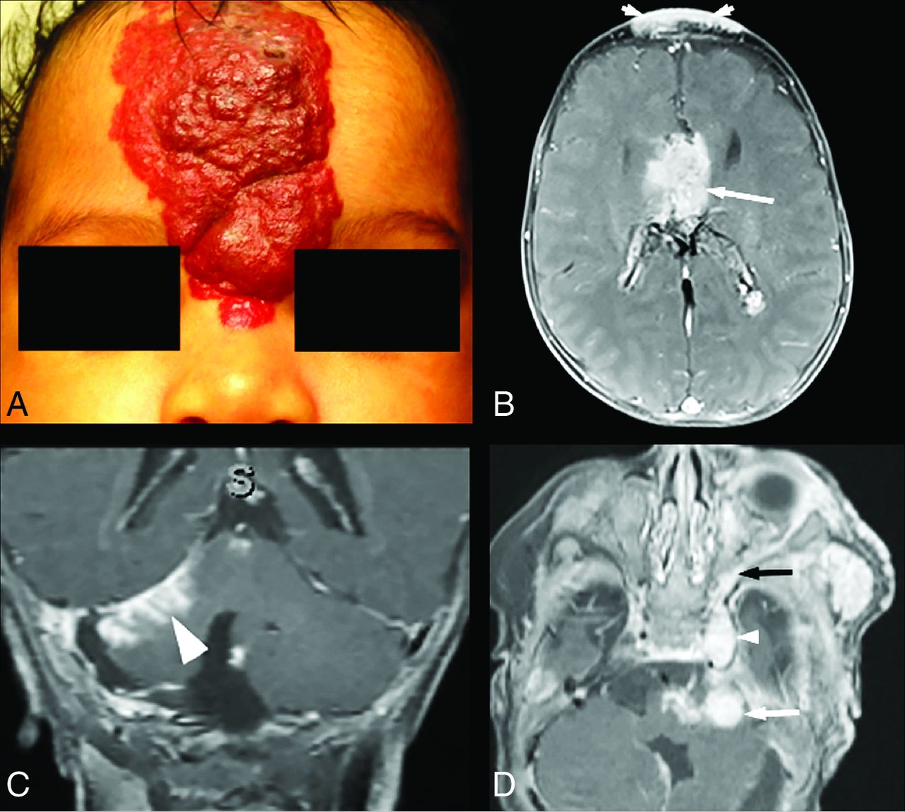
    Intracranial infantile hemangioma. A and B, A 2-month-old girl, born with a very large segmental IH of the forehead. A, Clinical photograph shows an IH in the S4 segment (according to the Haggstrom classification). B, Axial postcontrast T1-weighted image with fat suppression of the same patient shows a lobulated enhancing interhemispheric mass (arrow). A superficial forehead IH is indicated by arrowheads. C, A different 2-month-old girl with right facial IH and PHACE syndrome. Coronal postcontrast T1-weighted image with fat suppression shows focal leptomeningeal enhancement along the right tentorium (arrowhead). D, A 1.5-month-old boy with left facial IH. Axial postcontrast T1-weighted image with fat suppression shows enhancing hemangiomas in the left cerebellopontine angle cistern (white arrow) and in the enlarged left MC (small white arrowhead) and enhancing vascular tissue along the lateral orbital wall (black arrow), which seems to communicate between the intra- and extracranial components.

  • FIG 4.
    • Download figure
    • Open in new tab
    • Download powerpoint
    FIG 4.

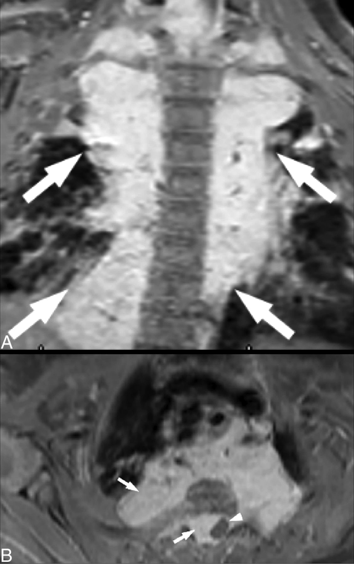
    Paraspinal IH. A 2-month-old girl with face and neck segmental IH. Coronal (A) and axial (B) postcontrast T1-weighted images with fat suppression show extensive bilateral enhancing paraspinal masses along the thoracic spine (arrows), with extension into the spinal canal and focal cord compression (arrowhead).

  • FIG 5.
    • Download figure
    • Open in new tab
    • Download powerpoint
    FIG 5.

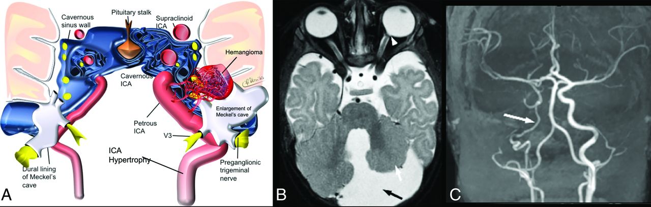
    A, Asymmetrical enlargement of Meckel's cave in patients with PHACE syndrome, an illustration showing a relationship between skull base hemangioma and adjacent nerves and vascular structures. Modified from a previously published image.33 B, Posterior fossa abnormality in patients with PHACE syndrome. A 6-week-old girl with a left facial IH. An axial T2-weighted image shows an enlarged retrocerebellar CSF space (black arrow), an asymmetrically small left cerebellar hemisphere (white arrow), and a small posterior vitreous coloboma in the left globe (arrowhead), which is another infrequent feature of PHACE syndrome. C, Arteriopathy of PHACE syndrome. A 6-month-old girl with a right-face IH and PHACE syndrome. Coronal TOF-MRA reconstructed image shows the diffusely small caliber of intracranial right ICA (arrow). Fig 5A courtesy of Malhotra, A., Tu, L., Kalra, V.B. et al. Neuroimaging of Meckel's cave in normal and disease conditions. Insights Imaging 9, 499–510 (2018). https://doi.org/10.1007/s13244-018-0604-7.

Tables

  • Figures
    • View popup
    Table 1:

    Characteristics of subjects with IH as part of a hemangioma syndrome compared with isolated IH

    Hemangioma Syndrome (n = 42)Isolated Superficial IH (n = 53)P Value
    Age (median) (IQR) (mo)2 (3–2)2 (3-2).53
    Female sex (%) (n)74% (31)86% (46).12
    Side/location of cutaneous IH (%) (n).78
        Right face only45% (19)40% (21)
        Left face only33% (14)41% (22)
        Bilateral face14% (6)15% (8)
        Other7% (3)4% (2)
    Facial segment (%) (n)
        S165% (27)30% (16).002
        S250% (21)66% (35).14
        S343% (18)28% (15).19
        S421% (9)4% (2).010
        Multiple facial segments57% (24)34% (18).037
        All facial segments14% (6)0% (0).006
    Intracranial IH (%) (n)45% (19)0% (0)<.001
    Paraspinal IH (%) (n)12% (5)0% (0).015
    MC enlargement (%) (n)67% (28)0% (0)<.001
    Posterior fossa abnormality (%) (n)48% (20)0% (0)<.001
    Cardiovascular abnormalities (%) ((n)95% (40)0% (0)<.001
    • Note:—IQR indicates Interquartile range.

    • View popup
    Table 2:

    Locations of neuroaxial IH and agreement between 3 readers

    Neuroaxial IH Location (n = 95)Reader 1Reader 2Reader 3κP ValueConsensus
    IAC1515151.00<.00115
    Cochlea3430.85<.0013
    Cerebellopontine angle5551.00<.0015
    MC2221.00<.0012
    Cavernous sinus2221.00<.0012
    Pterygopalatine fossa1111.00<.0011
    Vidian canal1111.00<.0011
    Leptomeningeal101080.89<.00110
    Dural base4441.00<.0014
    Intra-/paraspinal5551.00<.0015
PreviousNext
Back to top

In this issue

American Journal of Neuroradiology: 42 (8)
American Journal of Neuroradiology
Vol. 42, Issue 8
1 Aug 2021
  • Table of Contents
  • Index by author
  • Complete Issue (PDF)
Advertisement
Print
Download PDF
Email Article

Thank you for your interest in spreading the word on American Journal of Neuroradiology.

NOTE: We only request your email address so that the person you are recommending the page to knows that you wanted them to see it, and that it is not junk mail. We do not capture any email address.

Enter multiple addresses on separate lines or separate them with commas.
Neuroaxial Infantile Hemangiomas: Imaging Manifestations and Association with Hemangioma Syndromes
(Your Name) has sent you a message from American Journal of Neuroradiology
(Your Name) thought you would like to see the American Journal of Neuroradiology web site.
CAPTCHA
This question is for testing whether or not you are a human visitor and to prevent automated spam submissions.
Cite this article
T. Feygin, A.E. Goldman-Yassen, D.J. Licht, J.E. Schmitt, A. Mian, A. Vossough, L. Castelo-Soccio, J.R. Treat, A. Bhatia, A.N. Pollock
Neuroaxial Infantile Hemangiomas: Imaging Manifestations and Association with Hemangioma Syndromes
American Journal of Neuroradiology Aug 2021, 42 (8) 1520-1527; DOI: 10.3174/ajnr.A7204

Citation Manager Formats

  • BibTeX
  • Bookends
  • EasyBib
  • EndNote (tagged)
  • EndNote 8 (xml)
  • Medlars
  • Mendeley
  • Papers
  • RefWorks Tagged
  • Ref Manager
  • RIS
  • Zotero
0 Responses
Respond to this article
Share
Bookmark this article
Neuroaxial Infantile Hemangiomas: Imaging Manifestations and Association with Hemangioma Syndromes
T. Feygin, A.E. Goldman-Yassen, D.J. Licht, J.E. Schmitt, A. Mian, A. Vossough, L. Castelo-Soccio, J.R. Treat, A. Bhatia, A.N. Pollock
American Journal of Neuroradiology Aug 2021, 42 (8) 1520-1527; DOI: 10.3174/ajnr.A7204
del.icio.us logo Twitter logo Facebook logo Mendeley logo
  • Tweet Widget
  • Facebook Like
  • Google Plus One
Purchase

Jump to section

  • Article
    • Abstract
    • ABBREVIATIONS:
    • MATERIALS AND METHODS
    • RESULTS
    • DISCUSSION
    • CONCLUSIONS
    • Footnotes
    • References
  • Figures & Data
  • Info & Metrics
  • Responses
  • References
  • PDF

Related Articles

  • No related articles found.
  • PubMed
  • Google Scholar

Cited By...

  • No citing articles found.
  • Crossref (4)
  • Google Scholar

This article has been cited by the following articles in journals that are participating in Crossref Cited-by Linking.

  • Non‐vascular intracranial lesions in three children with PHACE association
    Mia A. Mologousis, Daniel M. Balkin, Edward R. Smith, Hart G. W. Lidov, Alice M. Li, Edward Yang, Marilyn G. Liang
    Pediatric Dermatology 2024 41 2
  • Feature gene selection and functional validation of SH3KBP1 in infantile hemangioma using machine learning
    Jiu Yin, Hui Gou, Jian Qi, Wenli Xing
    Biochemical and Biophysical Research Communications 2025 752
  • Hämangiome der Neuralachse bei Säuglingen oft im Rahmen von Syndromen
    Neuroradiologie Scan 2022 12 02
  • Update October 2021
    Francine Blei
    Lymphatic Research and Biology 2021 19 5

More in this TOC Section

  • Comparison of Image Quality and Radiation Dose in Pediatric Temporal Bone CT Using Photon-Counting Detector CT and Energy-Integrating Detector CT
  • SyMRI & MR Fingerprinting in Brainstem Myelination
  • Pons&Vermis Localization on Fetal MRI Using U-Net
Show more Pediatrics

Similar Articles

Advertisement

Indexed Content

  • Current Issue
  • Accepted Manuscripts
  • Article Preview
  • Past Issues
  • Editorials
  • Editors Choice
  • Fellow Journal Club
  • Letters to the Editor

Cases

  • Case Collection
  • Archive - Case of the Week
  • Archive - Case of the Month
  • Archive - Classic Case

Special Collections

  • Special Collections

Resources

  • News and Updates
  • Turn around Times
  • Submit a Manuscript
  • Author Policies
  • Manuscript Submission Guidelines
  • Evidence-Based Medicine Level Guide
  • Publishing Checklists
  • Graphical Abstract Preparation
  • Imaging Protocol Submission
  • Submit a Case
  • Become a Reviewer/Academy of Reviewers
  • Get Peer Review Credit from Publons

Multimedia

  • AJNR Podcast
  • AJNR SCANtastic
  • Video Articles

About Us

  • About AJNR
  • Editorial Board
  • Not an AJNR Subscriber? Join Now
  • Alerts
  • Feedback
  • Advertise with us
  • Librarian Resources
  • Permissions
  • Terms and Conditions

American Society of Neuroradiology

  • Not an ASNR Member? Join Now

© 2025 by the American Society of Neuroradiology All rights, including for text and data mining, AI training, and similar technologies, are reserved.
Print ISSN: 0195-6108 Online ISSN: 1936-959X

Powered by HighWire