Advertisement

Main menu

  • Home
  • Content
    • Current Issue
    • Accepted Manuscripts
    • Article Preview
    • Past Issue Archive
    • AJNR Case Collection
    • Case of the Week Archive
    • Classic Case Archive
    • Case of the Month Archive
  • Special Collections
    • Spinal CSF Leak Articles (Jan 2020-June 2024)
    • 2024 AJNR Journal Awards
    • Most Impactful AJNR Articles
  • Multimedia
    • AJNR Podcast
    • AJNR Scantastics
    • Video Articles
  • For Authors
    • Submit a Manuscript
    • Author Policies
    • Fast publishing of Accepted Manuscripts
    • Graphical Abstract Preparation
    • Manuscript Submission Guidelines
    • Imaging Protocol Submission
    • Submit a Case for the Case Collection
  • About Us
    • About AJNR
    • Editorial Board
  • More
    • Become a Reviewer/Academy of Reviewers
    • Subscribers
    • Permissions
    • Alerts
    • Feedback
    • Advertisers
    • ASNR Home
  • Other Publications
    • ajnr

User menu

  • Alerts
  • Log in

Search

  • Advanced search
American Journal of Neuroradiology

American Journal of Neuroradiology

ASHNR American Society of Functional Neuroradiology ASHNR American Society of Pediatric Neuroradiology ASSR
  • Alerts
  • Log in

Advanced Search

  • Home
  • Content
    • Current Issue
    • Accepted Manuscripts
    • Article Preview
    • Past Issue Archive
    • AJNR Case Collection
    • Case of the Week Archive
    • Classic Case Archive
    • Case of the Month Archive
  • Special Collections
    • Spinal CSF Leak Articles (Jan 2020-June 2024)
    • 2024 AJNR Journal Awards
    • Most Impactful AJNR Articles
  • Multimedia
    • AJNR Podcast
    • AJNR Scantastics
    • Video Articles
  • For Authors
    • Submit a Manuscript
    • Author Policies
    • Fast publishing of Accepted Manuscripts
    • Graphical Abstract Preparation
    • Manuscript Submission Guidelines
    • Imaging Protocol Submission
    • Submit a Case for the Case Collection
  • About Us
    • About AJNR
    • Editorial Board
  • More
    • Become a Reviewer/Academy of Reviewers
    • Subscribers
    • Permissions
    • Alerts
    • Feedback
    • Advertisers
    • ASNR Home
  • Follow AJNR on Twitter
  • Visit AJNR on Facebook
  • Follow AJNR on Instagram
  • Join AJNR on LinkedIn
  • RSS Feeds

Welcome to the new AJNR, Updated Hall of Fame, and more. Read the full announcements.


AJNR is seeking candidates for the position of Associate Section Editor, AJNR Case Collection. Read the full announcement.


Research ArticleAdult Brain

Brain Perfusion Alterations on 3D Pseudocontinuous Arterial Spin-Labeling MR Imaging in Patients with Autoimmune Encephalitis: A Case Series and Literature Review

R. Li, S. Jin, Y. Wang, J.-F. Li, H.-F. Xiao, Y.-L. Wang and L. Ma
American Journal of Neuroradiology May 2022, 43 (5) 701-706; DOI: https://doi.org/10.3174/ajnr.A7478
R. Li
aFrom the Department of Medical Imaging (R.L., S.J.), Tianjin Huanhu Hospital, Tianjin, China
bDepartment of Radiology (R.L., Y.W., J.-F.L., H.-F.X., Y.-L.W., L.M.), The First Medical Center of PLA General Hospital, Beijing, China
cDepartment of Medical Imaging (R.L., S.J.), Affiliated Huanhu Hospital of Nankai University, Tianjin, China
  • Find this author on Google Scholar
  • Find this author on PubMed
  • Search for this author on this site
  • ORCID record for R. Li
S. Jin
aFrom the Department of Medical Imaging (R.L., S.J.), Tianjin Huanhu Hospital, Tianjin, China
cDepartment of Medical Imaging (R.L., S.J.), Affiliated Huanhu Hospital of Nankai University, Tianjin, China
  • Find this author on Google Scholar
  • Find this author on PubMed
  • Search for this author on this site
  • ORCID record for S. Jin
Y. Wang
bDepartment of Radiology (R.L., Y.W., J.-F.L., H.-F.X., Y.-L.W., L.M.), The First Medical Center of PLA General Hospital, Beijing, China
  • Find this author on Google Scholar
  • Find this author on PubMed
  • Search for this author on this site
  • ORCID record for Y. Wang
J.-F. Li
bDepartment of Radiology (R.L., Y.W., J.-F.L., H.-F.X., Y.-L.W., L.M.), The First Medical Center of PLA General Hospital, Beijing, China
  • Find this author on Google Scholar
  • Find this author on PubMed
  • Search for this author on this site
  • ORCID record for J.-F. Li
H.-F. Xiao
bDepartment of Radiology (R.L., Y.W., J.-F.L., H.-F.X., Y.-L.W., L.M.), The First Medical Center of PLA General Hospital, Beijing, China
  • Find this author on Google Scholar
  • Find this author on PubMed
  • Search for this author on this site
  • ORCID record for H.-F. Xiao
Y.-L. Wang
bDepartment of Radiology (R.L., Y.W., J.-F.L., H.-F.X., Y.-L.W., L.M.), The First Medical Center of PLA General Hospital, Beijing, China
  • Find this author on Google Scholar
  • Find this author on PubMed
  • Search for this author on this site
  • ORCID record for Y.-L. Wang
L. Ma
bDepartment of Radiology (R.L., Y.W., J.-F.L., H.-F.X., Y.-L.W., L.M.), The First Medical Center of PLA General Hospital, Beijing, China
  • Find this author on Google Scholar
  • Find this author on PubMed
  • Search for this author on this site
  • ORCID record for L. Ma
  • Article
  • Figures & Data
  • Supplemental
  • Info & Metrics
  • Responses
  • References
  • PDF
Loading

Article Figures & Data

Figures

  • FIG 1.
    • Download figure
    • Open in new tab
    • Download powerpoint
    FIG 1.

    MR imaging of case 1. Findings on axial T2WI (A) and axial T1WI (B) are unremarkable. C, Axial T2 FLAIR reveals hyperintensity in the right hippocampus (white arrow). D, 3D pCASL demonstrates marked hyperperfusion in the corresponding region (black arrow).
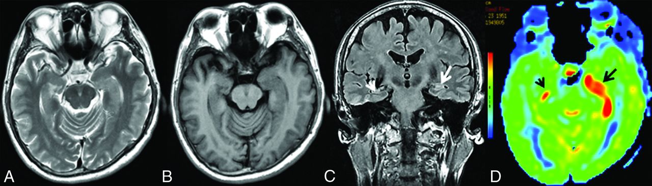
  • FIG 2.
    • Download figure
    • Open in new tab
    • Download powerpoint
    FIG 2.

    MR imaging of case 3. Findings on axial T2WI (A) and axial T1WI (B) are unremarkable. C, Coronal T2 FLAIR demonstrates hyperintensity in the right hippocampus (white arrow), whereas there are normal findings in the left hippocampus (white arrowhead). D, 3D pCASL depicts hyperperfusion in the corresponding regions on the FLAIR image, more obvious on the right side (black arrows).

  • FIG 3.
    • Download figure
    • Open in new tab
    • Download powerpoint
    FIG 3.

    MR imaging of case 5. Findings on axial T2WI (A) and axial T1WI (B) are unremarkable. C, Coronal T2 FLAIR shows subtle hyperintensity with mild swelling in the left hippocampus (white arrow), whereas it shows normal findings in the right hippocampus (white arrowhead). D, Increased CBF in the bilateral hippocampi could be detected on 3D pCASL, the left (black arrow) greater than the right (black arrowhead).

  • FIG 4.
    • Download figure
    • Open in new tab
    • Download powerpoint
    FIG 4.

    Serial MR imaging of case 7. On hospital day 3, the first conventional MR imaging examination findings were normal (not shown). On the second MR imaging check (26 days after admission), MR imaging shows swelling and hyperintensities on axial T2WI (A) and hypointensities on axial T1WI (B) in the bilateral hippocampi. C, Coronal T2 FLAIR shows hyperintensities in the bilateral hippocampi (white arrows). D, 3D pCASL reveals marked hyperperfusion in the bilateral medial temporal lobes (black arrows), the left greater than the right. On the third MR imaging check (96 days after admission), including axial T2WI (E), axial T1WI (F), and coronal T2 FLAIR (G), complete resolution of the inflammation in the bilateral medial temporal areas is seen, with enlargement of the bilateral temporal horns (white arrows), and 3D pCASL (H) shows decreased perfusion in the bilateral hippocampi (black arrows). The patient’s condition markedly improved with the administration of high-dose glucocorticoids, intravenous immunoglobulin, and plasma exchange (3 times).

  • FIG 5.
    • Download figure
    • Open in new tab
    • Download powerpoint
    FIG 5.

    Bar chart with the SD for the CBF (A) and rCBF (B) values of the lesions in the AE and control groups. The unit of CBF was mL/100 g/min.

PreviousNext
Back to top

In this issue

American Journal of Neuroradiology: 43 (5)
American Journal of Neuroradiology
Vol. 43, Issue 5
1 May 2022
  • Table of Contents
  • Index by author
  • Complete Issue (PDF)
Advertisement
Print
Download PDF
Email Article

Thank you for your interest in spreading the word on American Journal of Neuroradiology.

NOTE: We only request your email address so that the person you are recommending the page to knows that you wanted them to see it, and that it is not junk mail. We do not capture any email address.

Enter multiple addresses on separate lines or separate them with commas.
Brain Perfusion Alterations on 3D Pseudocontinuous Arterial Spin-Labeling MR Imaging in Patients with Autoimmune Encephalitis: A Case Series and Literature Review
(Your Name) has sent you a message from American Journal of Neuroradiology
(Your Name) thought you would like to see the American Journal of Neuroradiology web site.
CAPTCHA
This question is for testing whether or not you are a human visitor and to prevent automated spam submissions.
Cite this article
R. Li, S. Jin, Y. Wang, J.-F. Li, H.-F. Xiao, Y.-L. Wang, L. Ma
Brain Perfusion Alterations on 3D Pseudocontinuous Arterial Spin-Labeling MR Imaging in Patients with Autoimmune Encephalitis: A Case Series and Literature Review
American Journal of Neuroradiology May 2022, 43 (5) 701-706; DOI: 10.3174/ajnr.A7478

Citation Manager Formats

  • BibTeX
  • Bookends
  • EasyBib
  • EndNote (tagged)
  • EndNote 8 (xml)
  • Medlars
  • Mendeley
  • Papers
  • RefWorks Tagged
  • Ref Manager
  • RIS
  • Zotero
0 Responses
Respond to this article
Share
Bookmark this article
Brain Perfusion Alterations on 3D Pseudocontinuous Arterial Spin-Labeling MR Imaging in Patients with Autoimmune Encephalitis: A Case Series and Literature Review
R. Li, S. Jin, Y. Wang, J.-F. Li, H.-F. Xiao, Y.-L. Wang, L. Ma
American Journal of Neuroradiology May 2022, 43 (5) 701-706; DOI: 10.3174/ajnr.A7478
del.icio.us logo Twitter logo Facebook logo Mendeley logo
  • Tweet Widget
  • Facebook Like
  • Google Plus One
Purchase

Jump to section

  • Article
    • Abstract
    • ABBREVIATIONS:
    • CASE SERIES
    • RESULTS
    • DISCUSSION
    • CONCLUSIONS
    • Footnotes
    • References
  • Figures & Data
  • Supplemental
  • Info & Metrics
  • Responses
  • References
  • PDF

Related Articles

  • PubMed
  • Google Scholar

Cited By...

  • Arterial Spin Labeling Changes Parallel Asymmetric Perisylvian and Perirolandic Symptoms in 3 Pediatric Cases of Anti-NMDAR Encephalitis
  • REPLY:
  • Regarding "Brain Perfusion Alterations on 3D Pseudocontinuous Arterial Spin-Labeling MR Imaging in Patients with Autoimmune Encephalitis: A Case Series and Literature Review"
  • Crossref (13)
  • Google Scholar

This article has been cited by the following articles in journals that are participating in Crossref Cited-by Linking.

  • Arterial Spin Labeling Changes Parallel Asymmetric Perisylvian and Perirolandic Symptoms in 3 Pediatric Cases of Anti-NMDAR Encephalitis
    Alexander J. Sandweiss, Varun Kannan, Nilesh K. Desai, Stephen F. Kralik, Eyal Muscal, Kristen S. Fisher
    Neurology Neuroimmunology & Neuroinflammation 2023 10 4
  • Paraneoplastic neurological syndromes of the central nervous system: a single institution 7-year case series
    Fabrizio Giammello, Karol Galletta, Francesco Grillo, Teresa Brizzi, Marco Cavallaro, Enricomaria Mormina, Emma Scelzo, Cosimo Allegra, Claudia Stancanelli, Carmelo Rodolico, Olimpia Musumeci, Antonio Toscano, Francesca Granata
    Acta Neurologica Belgica 2023 123 4
  • Serial assessment of multimodality imaging in anti-leucine-rich glioma-inactivated 1 antibody encephalitis: A case report
    Takafumi Wada, Hitoshi Mori, Katsuro Shindo
    eNeurologicalSci 2022 29
  • Dynamic Contrast-enhanced MRI Quantification of Altered Vascular Permeability in Autoimmune Encephalitis
    So-Hyun Ji, Roh-Eul Yoo, Seung Hong Choi, Woo Jin Lee, Soon Tae Lee, Young Hun Jeon, Kyu Sung Choi, Ji Ye Lee, Inpyeong Hwang, Koung Mi Kang, Tae Jin Yun
    Radiology 2024 310 3
  • Experiences in implementing immunopsychiatry in real life
    Janet L. Cunningham, Gunnel Nordmark, David Fällmar, Simon Cervenka, Maike Gallwitz, Roland Säll, Peter T. Schmidt, Johan Rönnelid, Barbro Persson, Andreas Kindmark, Joachim Burman
    Journal of Affective Disorders Reports 2023 13
  • Acute pediatric encephalitis: etiology, course, and outcome of a 12-year single-center immunocompetent cohort
    Giacomo Brisca, Chiara Marini, Silvia Buratti, Marcello Mariani, Domenico Tortora, Giovanni Morana, Daniela Pirlo, Marta Romanengo, Giulia Cannizzaro, Ramona Cordani, Francesca Canzoneri, Maria Grazia Calevo, Lino Nobili, Diego Franciotta, Elio Castagnola, Andrea Moscatelli, Maria Margherita Mancardi
    Journal of Neurology 2023 270 10
  • FDG-PET and ASL MRI identify largely overlapping hypermetabolic and hyperperfusion changes in limbic autoimmune encephalitis
    Giacomo REBELLA, Denise CERNE, Luana BENEDETTI, Silvia MORBELLI, Martina RESAZ, Antonio UCCELLI, Lucio CASTELLAN, Flavio VILLANI, Enrico PEIRA, Federico MASSA, Luca ROCCATAGLIATA
    The Quarterly Journal of Nuclear Medicine and Molecular Imaging 2024 68 3
  • Autoimmune Encephalitis and Blood-Brain Barrier Permeability at Dynamic Contrast-enhanced MRI
    Massimo Filippi, Maria A. Rocca
    Radiology 2024 310 3
  • Evaluation of arterial spin labeling in the diagnosis and monitoring of myelin oligodendrocyte glycoprotein-associated cerebral cortical encephalitis
    Tomoya Shibahara, Kei Yamanaka, Mikiaki Matsuoka, Masaki Tachibana, Junya Kuroda, Hiroshi Nakane
    Multiple Sclerosis Journal 2024
  • Exploring brain perfusion in dogs with meningoencephalitis of unknown origin: A promising role for arterial spin labeling imaging
    Léa Arti, Kirsten Gnirs, Stella Papageorgiou, Yannick Ruel, Albert Agoulon, Nathalie Boddaert, Hugues Gaillot
    Journal of Veterinary Internal Medicine 2025 39 1

More in this TOC Section

Adult Brain

  • Diagnostic Neuroradiology of Monoclonal Antibodies
  • Clinical Outcomes After Chiari I Decompression
  • Cerebral ADC Changes in Fabry Disease
Show more Adult Brain

Functional

  • Kurtosis and Epileptogenic Tubers: A Pilot Study
  • Glutaric Aciduria Type 1: DK vs. Conventional MRI
  • Brain Iron in Niemann-Pick Type C: 7T Study
Show more Functional

Similar Articles

Advertisement

Indexed Content

  • Current Issue
  • Accepted Manuscripts
  • Article Preview
  • Past Issues
  • Editorials
  • Editors Choice
  • Fellow Journal Club
  • Letters to the Editor

Cases

  • Case Collection
  • Archive - Case of the Week
  • Archive - Case of the Month
  • Archive - Classic Case

Special Collections

  • Special Collections

Resources

  • News and Updates
  • Turn around Times
  • Submit a Manuscript
  • Author Policies
  • Manuscript Submission Guidelines
  • Evidence-Based Medicine Level Guide
  • Publishing Checklists
  • Graphical Abstract Preparation
  • Imaging Protocol Submission
  • Submit a Case
  • Become a Reviewer/Academy of Reviewers
  • Get Peer Review Credit from Publons

Multimedia

  • AJNR Podcast
  • AJNR SCANtastic
  • Video Articles

About Us

  • About AJNR
  • Editorial Board
  • Not an AJNR Subscriber? Join Now
  • Alerts
  • Feedback
  • Advertise with us
  • Librarian Resources
  • Permissions
  • Terms and Conditions

American Society of Neuroradiology

  • Not an ASNR Member? Join Now

© 2025 by the American Society of Neuroradiology All rights, including for text and data mining, AI training, and similar technologies, are reserved.
Print ISSN: 0195-6108 Online ISSN: 1936-959X

Powered by HighWire