Advertisement

Main menu

  • Home
  • Content
    • Current Issue
    • Accepted Manuscripts
    • Article Preview
    • Past Issue Archive
    • AJNR Case Collection
    • Case of the Week Archive
    • Classic Case Archive
    • Case of the Month Archive
  • Special Collections
    • Spinal CSF Leak Articles (Jan 2020-June 2024)
    • 2024 AJNR Journal Awards
    • Most Impactful AJNR Articles
  • Multimedia
    • AJNR Podcast
    • AJNR Scantastics
    • Video Articles
  • For Authors
    • Submit a Manuscript
    • Author Policies
    • Fast publishing of Accepted Manuscripts
    • Graphical Abstract Preparation
    • Manuscript Submission Guidelines
    • Imaging Protocol Submission
    • Submit a Case for the Case Collection
  • About Us
    • About AJNR
    • Editorial Board
  • More
    • Become a Reviewer/Academy of Reviewers
    • Subscribers
    • Permissions
    • Alerts
    • Feedback
    • Advertisers
    • ASNR Home
  • Other Publications
    • ajnr

User menu

  • Alerts
  • Log in

Search

  • Advanced search
American Journal of Neuroradiology

American Journal of Neuroradiology

ASHNR American Society of Functional Neuroradiology ASHNR American Society of Pediatric Neuroradiology ASSR
  • Alerts
  • Log in

Advanced Search

  • Home
  • Content
    • Current Issue
    • Accepted Manuscripts
    • Article Preview
    • Past Issue Archive
    • AJNR Case Collection
    • Case of the Week Archive
    • Classic Case Archive
    • Case of the Month Archive
  • Special Collections
    • Spinal CSF Leak Articles (Jan 2020-June 2024)
    • 2024 AJNR Journal Awards
    • Most Impactful AJNR Articles
  • Multimedia
    • AJNR Podcast
    • AJNR Scantastics
    • Video Articles
  • For Authors
    • Submit a Manuscript
    • Author Policies
    • Fast publishing of Accepted Manuscripts
    • Graphical Abstract Preparation
    • Manuscript Submission Guidelines
    • Imaging Protocol Submission
    • Submit a Case for the Case Collection
  • About Us
    • About AJNR
    • Editorial Board
  • More
    • Become a Reviewer/Academy of Reviewers
    • Subscribers
    • Permissions
    • Alerts
    • Feedback
    • Advertisers
    • ASNR Home
  • Follow AJNR on Twitter
  • Visit AJNR on Facebook
  • Follow AJNR on Instagram
  • Join AJNR on LinkedIn
  • RSS Feeds

Welcome to the new AJNR, Updated Hall of Fame, and more. Read the full announcements.


AJNR is seeking candidates for the position of Associate Section Editor, AJNR Case Collection. Read the full announcement.


Research ArticleHead & Neck
Open Access

Clinical Validation and Extension of an Automated, Deep Learning–Based Algorithm for Quantitative Sinus CT Analysis

C.J. Massey, L. Ramos, D.M. Beswick, V.R. Ramakrishnan and S.M. Humphries
American Journal of Neuroradiology September 2022, 43 (9) 1318-1324; DOI: https://doi.org/10.3174/ajnr.A7616
C.J. Massey
aFrom the Department of Otolaryngology-Head and Neck Surgery (C.J.M., L.R., V.R.R.), University of Colorado School of Medicine, Aurora, Colorado
  • Find this author on Google Scholar
  • Find this author on PubMed
  • Search for this author on this site
  • ORCID record for C.J. Massey
L. Ramos
aFrom the Department of Otolaryngology-Head and Neck Surgery (C.J.M., L.R., V.R.R.), University of Colorado School of Medicine, Aurora, Colorado
  • Find this author on Google Scholar
  • Find this author on PubMed
  • Search for this author on this site
  • ORCID record for L. Ramos
D.M. Beswick
bDepartment of Otolaryngology–Head and Neck Surgery (D.M.B.), University of California–Los Angeles School of Medicine, Los Angeles, California
  • Find this author on Google Scholar
  • Find this author on PubMed
  • Search for this author on this site
  • ORCID record for D.M. Beswick
V.R. Ramakrishnan
aFrom the Department of Otolaryngology-Head and Neck Surgery (C.J.M., L.R., V.R.R.), University of Colorado School of Medicine, Aurora, Colorado
  • Find this author on Google Scholar
  • Find this author on PubMed
  • Search for this author on this site
  • ORCID record for V.R. Ramakrishnan
S.M. Humphries
cQuantitative Imaging Laboratory (S.M.H.), Department of Radiology, National Jewish Health, Denver, Colorado
  • Find this author on Google Scholar
  • Find this author on PubMed
  • Search for this author on this site
  • ORCID record for S.M. Humphries
  • Article
  • Figures & Data
  • Info & Metrics
  • Responses
  • References
  • PDF
Loading

Article Figures & Data

Figures

  • Tables
  • FIG 1.
    • Download figure
    • Open in new tab
    • Download powerpoint
    FIG 1.

    3D reconstructed sinus CT scan with bony overlay (gray) and segmentation of the paranasal sinuses (colored) produced by the algorithm.
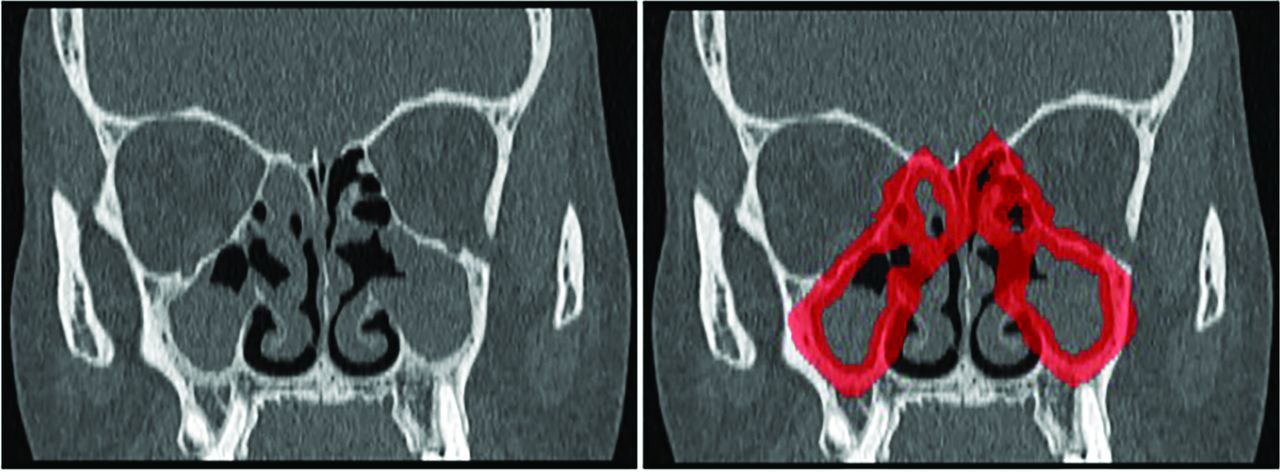
  • FIG 2.
    • Download figure
    • Open in new tab
    • Download powerpoint
    FIG 2.

    Illustration demonstrating application of osteitis segmentation rind. Left, Coronal CT with total GOSS score of 32. Right, Segmentation rind overlaid in red. Forty-eight percent of the rind is occupied by CT voxels corresponding to bone.

  • FIG 3.
    • Download figure
    • Open in new tab
    • Download powerpoint
    FIG 3.

    Flow diagram demonstrating the cohort-selection process.
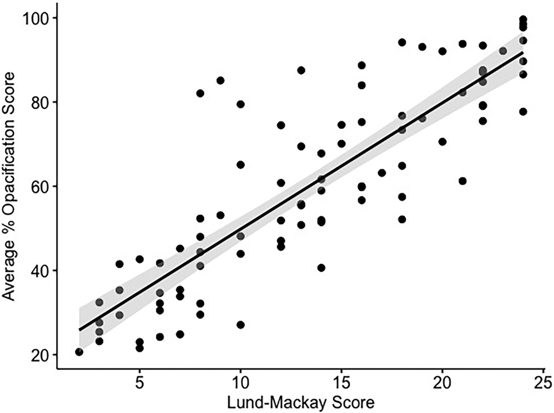
  • FIG 4.
    • Download figure
    • Open in new tab
    • Download powerpoint
    FIG 4.

    Scatterplot demonstrating algorithm-generated %SO versus LMS. The gray shaded area represents a 95% confidence interval for the regression line (ρ = 0.85, P < .001).
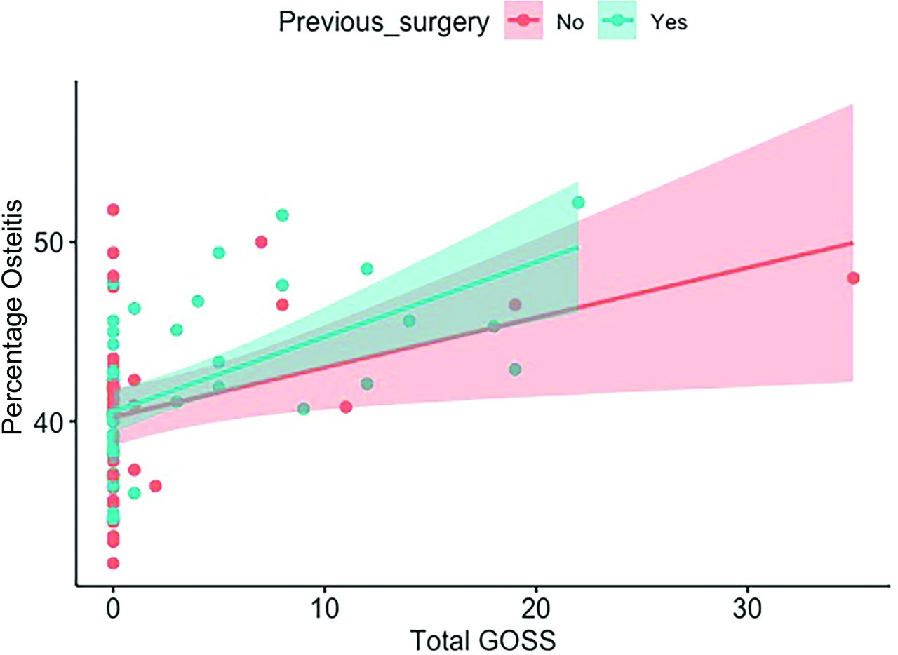
  • FIG 5.
    • Download figure
    • Open in new tab
    • Download powerpoint
    FIG 5.

    Algorithm-derived %OST versus total GOSS as stratified by surgical status. Overall ρ = 0.48 (P < .05). For surgically naïve patients, ρ = 0.29 (P = .08), and for postoperative subjects, ρ = 0.59 (P < .001).

Tables

  • Figures
    • View popup
    Table 1:

    Demographics of the study population

    Cohort Featuresn = 88 (%, or range)
    Female (%)52 (59)
    Avg. age (range) (yr)48.5 (22–78)
    White (%)70 (80)
    Polyps (%)59 (67)
    Asthma (%)53 (60)
    AERD (%)8 (9)
    Never smoker (%)55 (62)
    Prior surgery (%)48 (55)
    Avg. absolute serum eosinophils × 109/L0.31 (0–2.6)
    Avg. tissue eosinophils per HPF80 (0–413)
    Avg. SNOT-2249.6 (15–95)
    Avg. LKS6.8 (1–12)
    Avg. LMS13.5 (2–24)
    Avg. GOSS2.7 (0–35)
    Avg. sinus cavity volume (mL)60.8 (24.8–109.0)
    Avg. %SO60.1 (20.7–99.6)
    Avg. mHU10.5 (−139.2 to +57.4)
    Avg. %OST41.4 (32.1–52.2)
    • Note:—AERD indicates aspirin-exacerbated respiratory disease; HPF, high-power field; Avg., average.

    • View popup
    Table 2:

    CT scanner metrics and acquisition protocols used for the included scans

    Model (No.)Reconstruction Kernel (No.)Tube Potential (No.)Tube Current (No.)Pitch (No.)Axial Section Thickness (No.)Axial Section Spacing (No.)
    Siemens (n = 77)
     Sensation 64 (42)H31s (2)100 kVp (2)Modulated (22)0.7 (43)1.0 mm (77)0.6 mm (2)
     Definition (4)H60f (2)120 kVp (75)91 mA (41)0.8 (19)0.9 mm (63)
     Definition AS (3)H60s (1)100 mA (8)0.9 (14)1.0 mm (12)
     Definition AS+ (4)H70h (54)108 mA (6)1.0 (1)
     Definition Flash (18)J40s\\2 (1)
     Biograph 40 (6)J70h\\2 (17)
    GE Healthcare (n = 5)
     LightSpeed Pro 16 (1)Bone (5)120 kVp (5)110 mA (1)0.50–0.75 (2)0.625 mm (3)0.625 mm (3)
     LightSpeed VCT (1)150 mA (1)0.90–1.0 (3)1.25 mm (2)1.0 mm (1)
     Optima CT540 (2)160 mA (1)1.25 mm (1)
     Optima CT660 (1)>200 mA (2)
    Philips Healthcare (n = 4)
     Brilliance 64 (4)YC (3)120 kVp (4)85 mA (1)1.0 (4)1.0 mm (4)0.8 mm (1)
    YD (1)119 mA (2)0.9 mm (3)
    170 mA (1)
    Toshiba/Canon (n = 2)
     Aquilion ONE (1)FC30 (2)120 kVp (2)150 mA (1)1.0 (2)1.0 mm (2)0.8 mm (1)
     Aquilion Prime (1)200 mA (1)1.0 mm (1)
    • View popup
    Table 3:

    Correlation (ρ) values for CT-derived metrics and clinical parameters

    Clinical Parameter(%SO)LMSmHU of Opacified Regions
    LKS0.58a0.58a0.48a
    SNOT-220.05−0.020.08
    Absolute serum eosinophils0.26b0.34b0.14
    Tissue eosinophil count0.31b0.33b0.22b
    • ↵a P < .001.

    • ↵b P < .05.

    • View popup
    Table 4:

    Correlation (ρ) values for percentage sinus opacification stratified by sinus cavity and clinical parameters

    Clinical ParameterLeft MaxillaryRight MaxillaryAnterior EthmoidPosterior EthmoidFrontalSphenoid
    LKS0.43a0.34b0.64a0.56a0.53a0.35b
    SNOT-220.100.060.06−0.030.10−0.10
    Absolute serum eosinophils0.030.150.23b0.26b0.210.18
    Tissue eosinophil count0.130.210.37a0.39a0.23b0.11
    • ↵a P < .001.

    • ↵b P < .05.

PreviousNext
Back to top

In this issue

American Journal of Neuroradiology: 43 (9)
American Journal of Neuroradiology
Vol. 43, Issue 9
1 Sep 2022
  • Table of Contents
  • Index by author
  • Complete Issue (PDF)
Advertisement
Print
Download PDF
Email Article

Thank you for your interest in spreading the word on American Journal of Neuroradiology.

NOTE: We only request your email address so that the person you are recommending the page to knows that you wanted them to see it, and that it is not junk mail. We do not capture any email address.

Enter multiple addresses on separate lines or separate them with commas.
Clinical Validation and Extension of an Automated, Deep Learning–Based Algorithm for Quantitative Sinus CT Analysis
(Your Name) has sent you a message from American Journal of Neuroradiology
(Your Name) thought you would like to see the American Journal of Neuroradiology web site.
CAPTCHA
This question is for testing whether or not you are a human visitor and to prevent automated spam submissions.
Cite this article
C.J. Massey, L. Ramos, D.M. Beswick, V.R. Ramakrishnan, S.M. Humphries
Clinical Validation and Extension of an Automated, Deep Learning–Based Algorithm for Quantitative Sinus CT Analysis
American Journal of Neuroradiology Sep 2022, 43 (9) 1318-1324; DOI: 10.3174/ajnr.A7616

Citation Manager Formats

  • BibTeX
  • Bookends
  • EasyBib
  • EndNote (tagged)
  • EndNote 8 (xml)
  • Medlars
  • Mendeley
  • Papers
  • RefWorks Tagged
  • Ref Manager
  • RIS
  • Zotero
0 Responses
Respond to this article
Share
Bookmark this article
Automated Deep Learning for Sinus CT Analysis
C.J. Massey, L. Ramos, D.M. Beswick, V.R. Ramakrishnan, S.M. Humphries
American Journal of Neuroradiology Sep 2022, 43 (9) 1318-1324; DOI: 10.3174/ajnr.A7616
del.icio.us logo Twitter logo Facebook logo Mendeley logo
  • Tweet Widget
  • Facebook Like
  • Google Plus One
Purchase

Jump to section

  • Article
    • Abstract
    • ABBREVIATIONS:
    • MATERIALS AND METHODS
    • RESULTS
    • DISCUSSION
    • CONCLUSIONS
    • Footnotes
    • References
  • Figures & Data
  • Info & Metrics
  • Responses
  • References
  • PDF

Related Articles

  • No related articles found.
  • PubMed
  • Google Scholar

Cited By...

  • No citing articles found.
  • Crossref (13)
  • Google Scholar

This article has been cited by the following articles in journals that are participating in Crossref Cited-by Linking.

  • Deep learning radiomics-based preoperative prediction of recurrence in chronic rhinosinusitis
    Shaojuan He, Wei Chen, Xuehai Wang, Xinyu Xie, Fangying Liu, Xinyi Ma, Xuezhong Li, Anning Li, Xin Feng
    iScience 2023 26 4
  • Otolaryngologist perceptions of AI-based sinus CT interpretation
    Conner J. Massey, Annapoorani Asokan, Caroline Tietbohl, Megan Morris, Vijay R. Ramakrishnan
    American Journal of Otolaryngology 2023 44 5
  • Preliminary study on AI-assisted diagnosis of bone remodeling in chronic maxillary sinusitis
    Caiyun Zou, Hongbo Ji, Jie Cui, Bo Qian, Yu-Chen Chen, Qingxiang Zhang, Shuangba He, Yang Sui, Yang Bai, Yeming Zhong, Xu Zhang, Ting Ni, Zigang Che
    BMC Medical Imaging 2024 24 1
  • Surgical Navigation in the Anterior Skull Base Using 3-Dimensional Endoscopy and Surface Reconstruction
    Ryan A. Bartholomew, Haoyin Zhou, Maud Boreel, Krish Suresh, Saksham Gupta, Margaret B. Mitchell, Christopher Hong, Stella E. Lee, Timothy R. Smith, Jeffrey P. Guenette, C. Eduardo Corrales, Jayender Jagadeesan
    JAMA Otolaryngology–Head & Neck Surgery 2024 150 4
  • Deep Learning Techniques and Imaging in Otorhinolaryngology—A State-of-the-Art Review
    Christos Tsilivigkos, Michail Athanasopoulos, Riccardo di Micco, Aris Giotakis, Nicholas S. Mastronikolis, Francesk Mulita, Georgios-Ioannis Verras, Ioannis Maroulis, Evangelos Giotakis
    Journal of Clinical Medicine 2023 12 22
  • Imaging of chronic rhinosinusitis with nasal polyps in the era of biological therapies
    Remo Poto, Corrado Pelaia, Antonio di Salvatore, Hesham Saleh, Guy W. Scadding, Gilda Varricchi
    Current Opinion in Allergy & Clinical Immunology 2024 24 4
  • Deep Learning-Derived Quantitative Scores for Chronic Rhinosinusitis Assessment: Correlation With Quality of Life Outcomes
    Zhefan Shen, Ying Wei, Kexin Liu, Zhiqi Ma, Zhiliang Zhang, Xuechun Wang, Yong Li, Feng Shi, Zhongxiang Ding
    American Journal of Rhinology & Allergy 2025 39 3
  • Artificial intelligence optimizes the standardized diagnosis and treatment of chronic sinusitis
    Yang-Yang Liu, Shao-Peng Jiang, Ying-Bin Wang
    Frontiers in Physiology 2025 16
  • Blood Eosinophil Percentage and Improved Sinus CT Score as Diagnostic Tools for ECRS
    Fengzhen Li, Shenglei Wang, Xudong Cha, Tengfei Li, Yingqi Xie, Wenwen Wang, Wenwen Ren, Jianchun Liao, Huanhai Liu
    OTO Open 2024 8 1
  • Diagnostic accuracy of artificial intelligence in the detection of maxillary sinus pathology using computed tomography: A concise systematic review
    Asmaa T Uthman, Habiba Abouelenen, Shaheer Khan, Omar Bseiso, Natheer Al-Rawi
    Imaging Science in Dentistry 2025 55 1

More in this TOC Section

Head & Neck

  • NI-RADS for HEAD&NECK Cancer Recurrence
  • WHO Classification Update: Nasal&Skull Base Tumors
  • Peritumoral Signal in Vestibular Schwannomas
Show more Head & Neck

Functional

  • Glutaric Aciduria Type 1: DK vs. Conventional MRI
  • Kurtosis and Epileptogenic Tubers: A Pilot Study
  • Choroid Plexus Calcification&Microglial Activation
Show more Functional

Similar Articles

Advertisement

Indexed Content

  • Current Issue
  • Accepted Manuscripts
  • Article Preview
  • Past Issues
  • Editorials
  • Editors Choice
  • Fellow Journal Club
  • Letters to the Editor

Cases

  • Case Collection
  • Archive - Case of the Week
  • Archive - Case of the Month
  • Archive - Classic Case

Special Collections

  • Special Collections

Resources

  • News and Updates
  • Turn around Times
  • Submit a Manuscript
  • Author Policies
  • Manuscript Submission Guidelines
  • Evidence-Based Medicine Level Guide
  • Publishing Checklists
  • Graphical Abstract Preparation
  • Imaging Protocol Submission
  • Submit a Case
  • Become a Reviewer/Academy of Reviewers
  • Get Peer Review Credit from Publons

Multimedia

  • AJNR Podcast
  • AJNR SCANtastic
  • Video Articles

About Us

  • About AJNR
  • Editorial Board
  • Not an AJNR Subscriber? Join Now
  • Alerts
  • Feedback
  • Advertise with us
  • Librarian Resources
  • Permissions
  • Terms and Conditions

American Society of Neuroradiology

  • Not an ASNR Member? Join Now

© 2025 by the American Society of Neuroradiology All rights, including for text and data mining, AI training, and similar technologies, are reserved.
Print ISSN: 0195-6108 Online ISSN: 1936-959X

Powered by HighWire