Advertisement

Main menu

  • Home
  • Content
    • Current Issue
    • Accepted Manuscripts
    • Article Preview
    • Past Issue Archive
    • AJNR Case Collection
    • Case of the Week Archive
    • Classic Case Archive
    • Case of the Month Archive
  • Special Collections
    • Spinal CSF Leak Articles (Jan 2020-June 2024)
    • 2024 AJNR Journal Awards
    • Most Impactful AJNR Articles
  • Multimedia
    • AJNR Podcast
    • AJNR Scantastics
    • Video Articles
  • For Authors
    • Submit a Manuscript
    • Author Policies
    • Fast publishing of Accepted Manuscripts
    • Graphical Abstract Preparation
    • Manuscript Submission Guidelines
    • Imaging Protocol Submission
    • Submit a Case for the Case Collection
  • About Us
    • About AJNR
    • Editorial Board
  • More
    • Become a Reviewer/Academy of Reviewers
    • Subscribers
    • Permissions
    • Alerts
    • Feedback
    • Advertisers
    • ASNR Home
  • Other Publications
    • ajnr

User menu

  • Alerts
  • Log in

Search

  • Advanced search
American Journal of Neuroradiology

American Journal of Neuroradiology

ASHNR American Society of Functional Neuroradiology ASHNR American Society of Pediatric Neuroradiology ASSR
  • Alerts
  • Log in

Advanced Search

  • Home
  • Content
    • Current Issue
    • Accepted Manuscripts
    • Article Preview
    • Past Issue Archive
    • AJNR Case Collection
    • Case of the Week Archive
    • Classic Case Archive
    • Case of the Month Archive
  • Special Collections
    • Spinal CSF Leak Articles (Jan 2020-June 2024)
    • 2024 AJNR Journal Awards
    • Most Impactful AJNR Articles
  • Multimedia
    • AJNR Podcast
    • AJNR Scantastics
    • Video Articles
  • For Authors
    • Submit a Manuscript
    • Author Policies
    • Fast publishing of Accepted Manuscripts
    • Graphical Abstract Preparation
    • Manuscript Submission Guidelines
    • Imaging Protocol Submission
    • Submit a Case for the Case Collection
  • About Us
    • About AJNR
    • Editorial Board
  • More
    • Become a Reviewer/Academy of Reviewers
    • Subscribers
    • Permissions
    • Alerts
    • Feedback
    • Advertisers
    • ASNR Home
  • Follow AJNR on Twitter
  • Visit AJNR on Facebook
  • Follow AJNR on Instagram
  • Join AJNR on LinkedIn
  • RSS Feeds

Welcome to the new AJNR, Updated Hall of Fame, and more. Read the full announcements.


AJNR is seeking candidates for the position of Associate Section Editor, AJNR Case Collection. Read the full announcement.


Research ArticleSpine Imaging and Spine Image-Guided Interventions

Application of Spinal Subtraction and Bone Background Fusion CTA in the Accurate Diagnosis and Evaluation of Spinal Vascular Malformations

Xuehan Hu, Zhidong Yuan, Kaiyin Liang, Min Chen, Zhen Zhang, Hairong Zheng and Guanxun Cheng
American Journal of Neuroradiology March 2024, 45 (3) 351-357; DOI: https://doi.org/10.3174/ajnr.A8112
Xuehan Hu
aFrom the Department of Radiology (X.H., Z.Y., K.L., Z.Z., G.C.), Peking University Shenzhen Hospital, Shenzhen, China
bPaul C. Lauterbur Research Center for Biomedical Imaging (X.H., H.Z.), Shenzhen Institutes of Advanced Technology, Chinese Academy of Sciences
  • Find this author on Google Scholar
  • Find this author on PubMed
  • Search for this author on this site
  • ORCID record for Xuehan Hu
Zhidong Yuan
aFrom the Department of Radiology (X.H., Z.Y., K.L., Z.Z., G.C.), Peking University Shenzhen Hospital, Shenzhen, China
  • Find this author on Google Scholar
  • Find this author on PubMed
  • Search for this author on this site
  • ORCID record for Zhidong Yuan
Kaiyin Liang
aFrom the Department of Radiology (X.H., Z.Y., K.L., Z.Z., G.C.), Peking University Shenzhen Hospital, Shenzhen, China
  • Find this author on Google Scholar
  • Find this author on PubMed
  • Search for this author on this site
Min Chen
cDepartment of Radiology (M.C.), Southern University of Science and Technology Hospital, Shenzhen, China
  • Find this author on Google Scholar
  • Find this author on PubMed
  • Search for this author on this site
  • ORCID record for Min Chen
Zhen Zhang
aFrom the Department of Radiology (X.H., Z.Y., K.L., Z.Z., G.C.), Peking University Shenzhen Hospital, Shenzhen, China
  • Find this author on Google Scholar
  • Find this author on PubMed
  • Search for this author on this site
Hairong Zheng
bPaul C. Lauterbur Research Center for Biomedical Imaging (X.H., H.Z.), Shenzhen Institutes of Advanced Technology, Chinese Academy of Sciences
  • Find this author on Google Scholar
  • Find this author on PubMed
  • Search for this author on this site
Guanxun Cheng
aFrom the Department of Radiology (X.H., Z.Y., K.L., Z.Z., G.C.), Peking University Shenzhen Hospital, Shenzhen, China
  • Find this author on Google Scholar
  • Find this author on PubMed
  • Search for this author on this site
  • ORCID record for Guanxun Cheng
  • Article
  • Figures & Data
  • Supplemental
  • Info & Metrics
  • Responses
  • References
  • PDF
Loading

Article Figures & Data

Figures

  • Tables
  • FIG 1.
    • Download figure
    • Open in new tab
    • Download powerpoint
    FIG 1.

    Flow chart of the patient enrollment process and workflow of this study. Asterisk indicates accurate localization of critical lesions which defined as mostly correct when multiple feeding arteries are present, and the identification of drainage veins covering over 70% of the range.

  • FIG 2.
    • Download figure
    • Open in new tab
    • Download powerpoint
    FIG 2.

    Percentage of stacked bar chart of categorization of postprocessing SSBBF reconstruction accuracy. Types 3 and 4 are considered particularly beneficial in aiding clinical diagnosis and treatment.
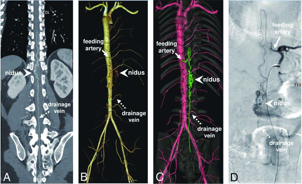
  • FIG 3.
    • Download figure
    • Open in new tab
    • Download powerpoint
    FIG 3.

    Spinal CTA images in a 28-year-old man with a spinal AVM. The crucial lesions, including nidus (arrowhead), feeding artery (solid arrow), and drainage vein (dotted arrow), are shown in MPR-CTA (A), RBS-CTA (B), SSBBF-CTA (C), and DSA images (D).
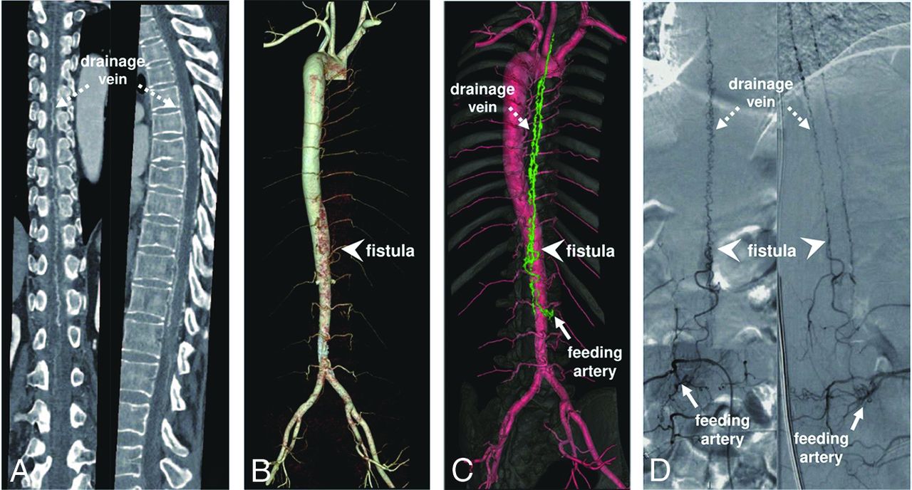
  • FIG 4.
    • Download figure
    • Open in new tab
    • Download powerpoint
    FIG 4.

    Spinal CTA images in a 55-year-old woman with a spinal AVF. The crucial lesions, including the fistula (arrowhead), feeding artery (solid arrow), and drainage vein (dotted arrow), are shown in an MPR-CTA image (A), a RBS-CTA image (B), SSBBF-CTA image (C), and a DSA image (D).

Tables

  • Figures
    • View popup
    Table 1:

    Patient characteristicsa

    Group AGroup BP Value
    Age (yr)b44.0 (SD, 14.3)44.6 (SD, 15.2).898
    Sexc.990
     Female911
     Male1316
    Sensory disturbance100% (22/22)88.9% (24/27).242
    Motor disturbance90.9% (20/22)77.8% (21/27).269
    Reflex abnormality68.2% (15/22)66.7% (18/27).910
    Urogenital disturbance72.7% (16/22)33.3% (9/27).005d
    Back pain9.1% (2/22)11.1% (3/27)1.000
    • ↵a Unless otherwise indicated, data are percentages, with numbers of patients in parentheses.

    • ↵b Age is presented as the mean.

    • ↵c Data are numbers of patients.

    • ↵d The clinical features of groups A and B showed statistically significant differences (P < .05).

    • View popup
    Table 2:

    Diagnostic efficacy of original images with MPR for primary screening of SVMs by different readersa

    ReaderSensitivity (%)Specificity (%)Accuracy (%)PPV (%)NPV (%)AUC [95% CI]Youden
    Reader 1b77.3 (17/22)92.6 (25/27)85.7 (42/49)89.5 (17/19)83.3 (25/30)0.849 (0.718–0.935)0.699
    Reader 290.9 (20/22)96.3 (26/27)93.9 (46/49)95.2 (20/21)92.9 (26/28)0.936 (0.828–0.986)0.872
    Reader 395.5 (21/22)100 (27/27)98.0 (48/49)100 (21/21)96.4 (27/28)0.977 (0.888–0.999)0.955
    • Note:—PPV indicates positive predictive value; NPV, negative predictive value; Reader 1, a radiologist with one year of experience; Reader 2, a junior physician who submitted the original report for the first time;Reader 3, a reviewing physician who also submitted the original report for the first time.

    • ↵a Data in brackets are 95% CIs, and data in parentheses are numbers of patients.

    • ↵b Readers 1 and 3 showed statistically significant differences (P < .05).

    • View popup
    Table 3:

    Scoring of lesion localization and visualization of SVMs using different postprocessing techniques of CTA imagesa

    MPR-CTARBS-CTAbSSBBF-CTAP Valuec
    Visualization of lesions (total points)6.00 (1.00)4.00 (2.75)6.00 (0.63).000
     Nidus/fistula2.00 (0.00)1.50 (1.00)2.00 (1.25).000
     Feeding artery2.00 (0.63)1.00 (1.50)2.00 (0.00).000
     Drainage vein2.00 (0.00)1.00 (1.00)2.00 (0.00).000
    Localization of lesions (total points)6.00 (0.00)3.25 (3.00)6.00 (1.00).000
     Nidus/fistula2.00 (0.00)1.00 (1.00)2.00 (0.00).000
     Feeding artery2.00 (0.00)1.00 (2.00)2.00 (0.00).000
     Drainage vein2.00 (0.00)1.00 (1.13)2.00 (0.00).00
    Overall morphology4.00 (0.00)2.75 (2.63)4.00 (0.50).000
    • ↵a The data are presented as median (interquartile range).

    • b There is a statistically significant difference between RBS-CTA and the other 2 groups (P < .05).

    • c There is a significant statistical difference among the 3 groups (P < .05).

    • View popup
    Table 4:

    Assessment of lesions of SVMs and diagnostic time using different postprocessing CTA techniques by different readersa

    Reader 1Reader 4P Value
    MPR-CTA
     Visualization of lesions4.00 (1.25)6.00 (1.00).001b
     Localization of lesions4.50 (2.00)6.00 (0.00).003b
     Overall morphology4.00 (1.00)4.00 (0.00).025b
     Diagnostic time9.00 (6.75)7.00 (3.00).017b
    RBS-CTA
     Visualization of lesions3.50 (3.00)3.50 (3.00).353
     Localization of lesions2.25 (3.00)3.00 (2.50).178
     Overall morphology2.50 (1.25)3.00 (2.00).070
     Diagnostic timeNANANA
    SSBBF-CTA
     Visualization of lesions6.00 (1.00)6.00 (1.00).705
     Localization of lesions6.00 (1.00)6.00 (1.00).206
     Overall morphology4.00 (0.25)4.00 (0.13).655
     Diagnostic time3.00 (2.00)c3.00 (1.25)c.943
    • Note:—NA indicates not applicable; Reader 1, a radiologist with one year of experience; Reader 4, a radiologist with 4 years of experience.

    • a The data are presented as median (interquartile range). The visualization and localization of lesions represent the total score of the nidus/fistula, feeding artery, and drainage vein.

    • ↵b There is a significant statistical difference between readers 1 and 4 (P < .05).

    • ↵c There is a statistically significant difference in diagnostic time between the use of MPR-CTA and SSBBF-CTA (P = .000).

PreviousNext
Back to top

In this issue

American Journal of Neuroradiology: 45 (3)
American Journal of Neuroradiology
Vol. 45, Issue 3
1 Mar 2024
  • Table of Contents
  • Index by author
  • Complete Issue (PDF)
Advertisement
Print
Download PDF
Email Article

Thank you for your interest in spreading the word on American Journal of Neuroradiology.

NOTE: We only request your email address so that the person you are recommending the page to knows that you wanted them to see it, and that it is not junk mail. We do not capture any email address.

Enter multiple addresses on separate lines or separate them with commas.
Application of Spinal Subtraction and Bone Background Fusion CTA in the Accurate Diagnosis and Evaluation of Spinal Vascular Malformations
(Your Name) has sent you a message from American Journal of Neuroradiology
(Your Name) thought you would like to see the American Journal of Neuroradiology web site.
CAPTCHA
This question is for testing whether or not you are a human visitor and to prevent automated spam submissions.
Cite this article
Xuehan Hu, Zhidong Yuan, Kaiyin Liang, Min Chen, Zhen Zhang, Hairong Zheng, Guanxun Cheng
Application of Spinal Subtraction and Bone Background Fusion CTA in the Accurate Diagnosis and Evaluation of Spinal Vascular Malformations
American Journal of Neuroradiology Mar 2024, 45 (3) 351-357; DOI: 10.3174/ajnr.A8112

Citation Manager Formats

  • BibTeX
  • Bookends
  • EasyBib
  • EndNote (tagged)
  • EndNote 8 (xml)
  • Medlars
  • Mendeley
  • Papers
  • RefWorks Tagged
  • Ref Manager
  • RIS
  • Zotero
0 Responses
Respond to this article
Share
Bookmark this article
Spinal Subtraction CTA for Vascular Malformations
Xuehan Hu, Zhidong Yuan, Kaiyin Liang, Min Chen, Zhen Zhang, Hairong Zheng, Guanxun Cheng
American Journal of Neuroradiology Mar 2024, 45 (3) 351-357; DOI: 10.3174/ajnr.A8112
del.icio.us logo Twitter logo Facebook logo Mendeley logo
  • Tweet Widget
  • Facebook Like
  • Google Plus One
Purchase

Jump to section

  • Article
    • Abstract
    • ABBREVIATIONS:
    • MATERIALS AND METHODS
    • RESULTS
    • DISCUSSION
    • CONCLUSIONS
    • Footnotes
    • References
  • Figures & Data
  • Supplemental
  • Info & Metrics
  • Responses
  • References
  • PDF

Related Articles

  • PubMed
  • Google Scholar

Cited By...

  • No citing articles found.
  • Crossref (2)
  • Google Scholar

This article has been cited by the following articles in journals that are participating in Crossref Cited-by Linking.

  • The holistic and local perspectives in teaching spinal arteriovenous malformations
    Xianli Lv
    The Neuroradiology Journal 2025
  • Vascular Pathologic Conditions in and around the Spinal Cord
    Jennifer McCarty, Charlotte Chung, Rohan Samant, Clark Sitton, Eliana Bonfante, Peng Roc Chen, Eytan Raz, Maksim Shapiro, Roy Riascos, Jose Gavito-Higuera
    RadioGraphics 2024 44 9

More in this TOC Section

  • Imaging of Myxopapillary Ependymomas
  • Image-Guided Interventions for Low Back Pain
  • Technical Tips for CT-Guided Fibrin Glue Patching of CSF-Venous Fistulas
Show more Spine Imaging and Spine Image-Guided Interventions

Similar Articles

Advertisement

Indexed Content

  • Current Issue
  • Accepted Manuscripts
  • Article Preview
  • Past Issues
  • Editorials
  • Editors Choice
  • Fellow Journal Club
  • Letters to the Editor

Cases

  • Case Collection
  • Archive - Case of the Week
  • Archive - Case of the Month
  • Archive - Classic Case

Special Collections

  • Special Collections

Resources

  • News and Updates
  • Turn around Times
  • Submit a Manuscript
  • Author Policies
  • Manuscript Submission Guidelines
  • Evidence-Based Medicine Level Guide
  • Publishing Checklists
  • Graphical Abstract Preparation
  • Imaging Protocol Submission
  • Submit a Case
  • Become a Reviewer/Academy of Reviewers
  • Get Peer Review Credit from Publons

Multimedia

  • AJNR Podcast
  • AJNR SCANtastic
  • Video Articles

About Us

  • About AJNR
  • Editorial Board
  • Not an AJNR Subscriber? Join Now
  • Alerts
  • Feedback
  • Advertise with us
  • Librarian Resources
  • Permissions
  • Terms and Conditions

American Society of Neuroradiology

  • Not an ASNR Member? Join Now

© 2025 by the American Society of Neuroradiology All rights, including for text and data mining, AI training, and similar technologies, are reserved.
Print ISSN: 0195-6108 Online ISSN: 1936-959X

Powered by HighWire