
Article Figures & Data
Figures
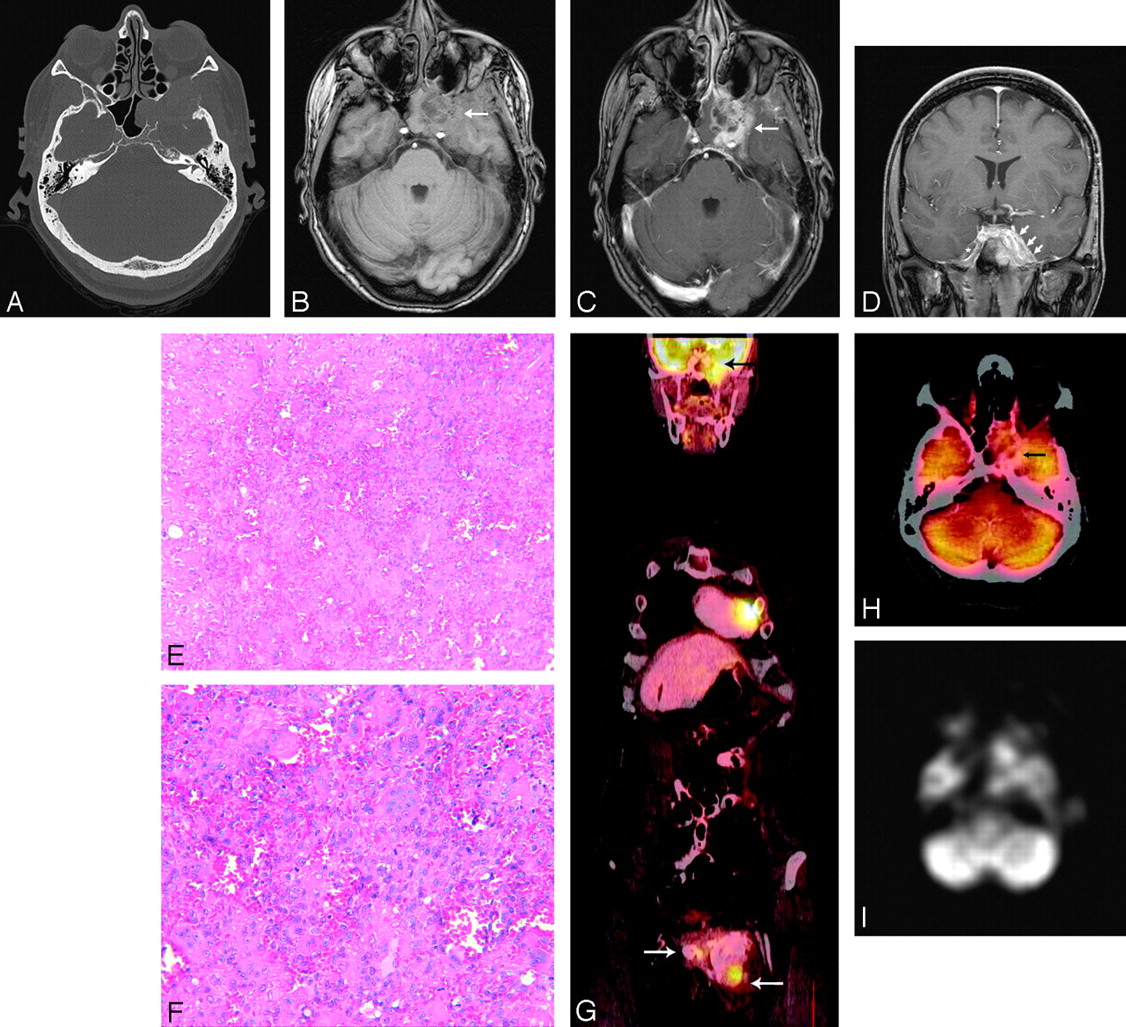
- Fig 1.
A 44-year-old woman presented with a history of pelvic GCT recurrence, with a mass of the left ischium and left inferior pubic ramus, identified and treated at that site 15 years previously. The patient presented with new left facial numbness, indicating likely trigeminal nerve sensory root deficits. Contrast-enhanced CT of the skull base was performed (A, bone windows) to evaluate a suspected lesion. A lytic and contrast-enhancing mass was seen involving the lesser and greater wings of the sphenoid bone, with possible invasion of the cavernous sinus and Meckel cave. Brain and skull base MR imaging demonstrated a heterogeneously enhancing mass obliterating the sphenoid sinus to the left of midline, with invasion of the left cavernous sinus and Meckel cave on axial pre- and postcontrast T1-weighted images (B and C, arrows), explaining the patient’s symptoms (note the lack of well-visualized nerve roots within an obliterated left Meckel’s cave on coronal T1-weighted postcontrast images [D, arrows], compared with the roots on the more normal opposite side [asterisk]). Microscopic examination (E, hematoxylin-eosin, original magnification ×10) from the sphenoid bone biopsy sample and partially resected tumor shows a highly vascular tumor comprising abundant osteoclast-like giant cells intermixed with mononuclear epitheloid cells. Reactive osteoid was present focally (left), likely at the advancing border of the tumor. Higher power view (F, original magnification ×20) demonstrated that the nuclear features of the epitheloid cells resembled those of the multinucleated cells, with mitotic figures being common throughout the sample. Findings were consistent with GCT (and not metastatic disease) and were similar to those of the pelvic lesion. Coregistered fused PET/CT coronal (G) and axial (H) reformations performed 3 months later (to evaluate the remainder of the body) demonstrated high metabolic uptake within the sphenoid mass (black arrow; SUV, 7.4) and the recurrent left pubic lesion at the primary site (with associated surrounding bone cement; white arrows; SUV, 8.4). Note that it may be difficult to distinguish a discrete sphenoid lesion adjacent to the normal relatively high metabolic activity of the cerebral cortex in the adjacent middle cranial fossa, particularly on the nonfused PET data (I).

In this issue


Jump to section
Related Articles
Cited By...
- No citing articles found.