Advertisement

Main menu

  • Home
  • Content
    • Current Issue
    • Accepted Manuscripts
    • Article Preview
    • Past Issue Archive
    • AJNR Case Collection
    • Case of the Week Archive
    • Classic Case Archive
    • Case of the Month Archive
  • Special Collections
    • Spinal CSF Leak Articles (Jan 2020-June 2024)
    • 2024 AJNR Journal Awards
    • Most Impactful AJNR Articles
  • Multimedia
    • AJNR Podcast
    • AJNR Scantastics
    • Video Articles
  • For Authors
    • Submit a Manuscript
    • Author Policies
    • Fast publishing of Accepted Manuscripts
    • Graphical Abstract Preparation
    • Manuscript Submission Guidelines
    • Imaging Protocol Submission
    • Submit a Case for the Case Collection
  • About Us
    • About AJNR
    • Editorial Board
  • More
    • Become a Reviewer/Academy of Reviewers
    • Subscribers
    • Permissions
    • Alerts
    • Feedback
    • Advertisers
    • ASNR Home
  • Other Publications
    • ajnr

User menu

  • Alerts
  • Log in

Search

  • Advanced search
American Journal of Neuroradiology

American Journal of Neuroradiology

ASHNR American Society of Functional Neuroradiology ASHNR American Society of Pediatric Neuroradiology ASSR
  • Alerts
  • Log in

Advanced Search

  • Home
  • Content
    • Current Issue
    • Accepted Manuscripts
    • Article Preview
    • Past Issue Archive
    • AJNR Case Collection
    • Case of the Week Archive
    • Classic Case Archive
    • Case of the Month Archive
  • Special Collections
    • Spinal CSF Leak Articles (Jan 2020-June 2024)
    • 2024 AJNR Journal Awards
    • Most Impactful AJNR Articles
  • Multimedia
    • AJNR Podcast
    • AJNR Scantastics
    • Video Articles
  • For Authors
    • Submit a Manuscript
    • Author Policies
    • Fast publishing of Accepted Manuscripts
    • Graphical Abstract Preparation
    • Manuscript Submission Guidelines
    • Imaging Protocol Submission
    • Submit a Case for the Case Collection
  • About Us
    • About AJNR
    • Editorial Board
  • More
    • Become a Reviewer/Academy of Reviewers
    • Subscribers
    • Permissions
    • Alerts
    • Feedback
    • Advertisers
    • ASNR Home
  • Follow AJNR on Twitter
  • Visit AJNR on Facebook
  • Follow AJNR on Instagram
  • Join AJNR on LinkedIn
  • RSS Feeds

Welcome to the new AJNR, Updated Hall of Fame, and more. Read the full announcements.


AJNR is seeking candidates for the position of Associate Section Editor, AJNR Case Collection. Read the full announcement.


Research ArticleHead and Neck

Carcinoid Tumor Metastases to the Extraocular Muscles: MR Imaging and CT Findings and Review of the Literature

A. Gupta, J.L. Chazen and C.D. Phillips
American Journal of Neuroradiology August 2011, 32 (7) 1208-1211; DOI: https://doi.org/10.3174/ajnr.A2470
A. Gupta
  • Find this author on Google Scholar
  • Find this author on PubMed
  • Search for this author on this site
J.L. Chazen
  • Find this author on Google Scholar
  • Find this author on PubMed
  • Search for this author on this site
C.D. Phillips
  • Find this author on Google Scholar
  • Find this author on PubMed
  • Search for this author on this site
  • Article
  • Figures & Data
  • Info & Metrics
  • Responses
  • References
  • PDF
Loading

Article Figures & Data

Figures

  • Tables
  • Fig 1.
    • Download figure
    • Open in new tab
    • Download powerpoint
    Fig 1.

    Coronal postcontrast T1-weighted image of the orbits in patient 1 demonstrates a heterogeneously enhancing ovoid lesion involving the right medial rectus (arrow). A similarly enhancing but larger lobulated lesion involves the left lateral rectus (arrowhead).
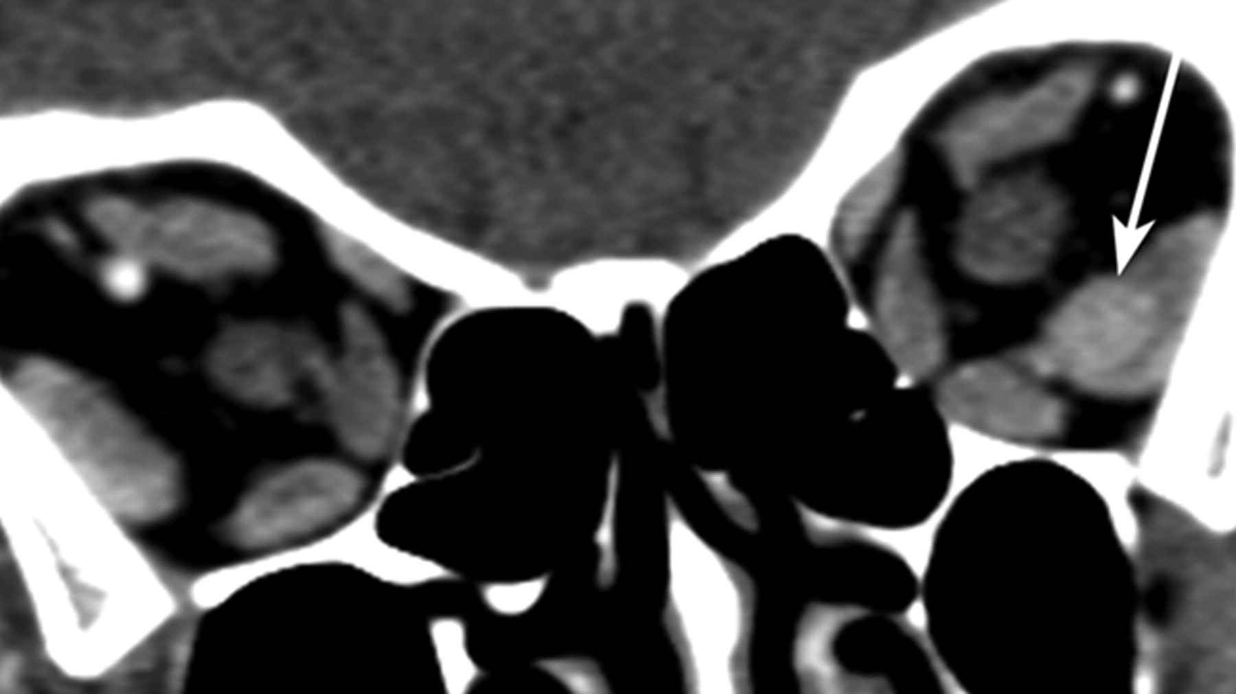
  • Fig 2.
    • Download figure
    • Open in new tab
    • Download powerpoint
    Fig 2.

    Coronal reformat contrast-enhanced CT image of the orbits in patient 2 demonstrates a homogeneously enhancing well-circumscribed rounded mass within the left lateral rectus (arrow).

  • Fig 3.
    • Download figure
    • Open in new tab
    • Download powerpoint
    Fig 3.

    A, Coronal fat-saturated T2-weighted image of the orbits in patient 5 demonstrates a heterogeneously hyperintense well-circumscribed masses involving the right lateral (arrow) and left inferior recti (arrowhead). B, Axial fat-saturated T1-weighted postgadolinium image in this same patient demonstrates heterogeneous hypoenhancement of the fusiform masses of the right lateral (arrow) and left inferior recti (arrowhead).

  • Fig 4.
    • Download figure
    • Open in new tab
    • Download powerpoint
    Fig 4.

    Axial contrast-enhanced CT image of the orbits in patient 6 shows ovoid well-defined lesions of the right medial (black arrow) and lateral (white arrow) recti.

Tables

  • Figures
  • Demographic, clinical, and imaging findings for the patient cohort

    Patient No.Age (yr)SexClinical PresentationCT or MRI AvailableEOMs InvolvedLesion MorphologyLesion MarginsInvolvement of Insertion of EOM on GlobeDensity/Intensity on Postcontrast Relative to Normal EOM
    164FProptosis and CN VI palsyMRIRt. MRFusiformWell-definedNoMildly hyperintense
    Lt. LR
    242FGaze difficulty and tearingCTLt. LRFusiformWell-definedNoModerately hyperdense
    371FN/AMRIRt. SORoundWell-definedNoMildly hypointense
    471FN/AMRIRt. LR, MR, SO, Lt. LR and IRRound and fusiformWell-definedNoMildly hypointense
    562FProptosis and painMRIRt. LRRoundWell-definedNoModerately hypointense
    Lt. IR
    653FProptosis and gaze difficultyCTRt. LR, SR, MRRound and fusiformWell-definedNoMildly hyperdense
    Lt. LR
    770MBlurry visionMRIRt. IRFusiformWell-definedNoIsointense
    Lt. IO
PreviousNext
Back to top

In this issue

American Journal of Neuroradiology: 32 (7)
American Journal of Neuroradiology
Vol. 32, Issue 7
1 Aug 2011
  • Table of Contents
  • Index by author
Advertisement
Print
Download PDF
Email Article

Thank you for your interest in spreading the word on American Journal of Neuroradiology.

NOTE: We only request your email address so that the person you are recommending the page to knows that you wanted them to see it, and that it is not junk mail. We do not capture any email address.

Enter multiple addresses on separate lines or separate them with commas.
Carcinoid Tumor Metastases to the Extraocular Muscles: MR Imaging and CT Findings and Review of the Literature
(Your Name) has sent you a message from American Journal of Neuroradiology
(Your Name) thought you would like to see the American Journal of Neuroradiology web site.
CAPTCHA
This question is for testing whether or not you are a human visitor and to prevent automated spam submissions.
Cite this article
A. Gupta, J.L. Chazen, C.D. Phillips
Carcinoid Tumor Metastases to the Extraocular Muscles: MR Imaging and CT Findings and Review of the Literature
American Journal of Neuroradiology Aug 2011, 32 (7) 1208-1211; DOI: 10.3174/ajnr.A2470

Citation Manager Formats

  • BibTeX
  • Bookends
  • EasyBib
  • EndNote (tagged)
  • EndNote 8 (xml)
  • Medlars
  • Mendeley
  • Papers
  • RefWorks Tagged
  • Ref Manager
  • RIS
  • Zotero
0 Responses
Respond to this article
Share
Bookmark this article
Carcinoid Tumor Metastases to the Extraocular Muscles: MR Imaging and CT Findings and Review of the Literature
A. Gupta, J.L. Chazen, C.D. Phillips
American Journal of Neuroradiology Aug 2011, 32 (7) 1208-1211; DOI: 10.3174/ajnr.A2470
del.icio.us logo Twitter logo Facebook logo Mendeley logo
  • Tweet Widget
  • Facebook Like
  • Google Plus One
Purchase

Jump to section

  • Article
    • Abstract
    • Materials and Methods
    • Results
    • Discussion
    • Acknowledgments
    • Footnotes
    • References
  • Figures & Data
  • Info & Metrics
  • Responses
  • References
  • PDF

Related Articles

  • No related articles found.
  • PubMed
  • Google Scholar

Cited By...

  • No citing articles found.
  • Crossref (33)
  • Google Scholar

This article has been cited by the following articles in journals that are participating in Crossref Cited-by Linking.

  • Multimodality Imaging Findings in Carcinoid Tumors: A Head-to-Toe Spectrum
    Ameya Jagdish Baxi, Kedar Chintapalli, Amol Katkar, Carlos S. Restrepo, Sonia L. Betancourt, Abhijit Sunnapwar
    RadioGraphics 2017 37 2
  • Orbital metastases from neuroendocrine neoplasms: clinical implications and outcomes
    Lukasz Kamieniarz, Eleni Armeni, Luke Furtado O’Mahony, Charlotte Leigh, Lukon Miah, Akshay Narayan, Ankit Bhatt, Nicholas Cox, Dalvinder Mandair, Shaunak Navalkissoor, Martyn Caplin, Christos Toumpanakis
    Endocrine 2020 67 2
  • Extraocular muscle enlargement
    Khizar Rana, Valerie Juniat, Sandy Patel, Dinesh Selva
    Graefe's Archive for Clinical and Experimental Ophthalmology 2022 260 11
  • The eye of the beholder: orbital metastases from midgut neuroendocrine tumors, a two institution experience
    Satya Das, Gino Pineda, Laura Goff, Rachel Sobel, Jordan Berlin, George Fisher
    Cancer Imaging 2018 18 1
  • Imaging Anatomy and Pathology of Extraocular Muscles in Adults
    Christian B. van der Pol, Santanu Chakraborty, Jennifer Gao, Thanh Nguyen, Carlos Torres, Rafael Glikstein
    Canadian Association of Radiologists Journal 2014 65 4
  • Intraorbital Metastasis From Solitary Fibrous Tumor
    Mrinali M. Patel, Frederick A. Jakobiec, Fouad R. Zakka, Rose Du, Donald J. Annino, Sheila Borboli-Gerogiannis, Anthony B. Daniels
    Ophthalmic Plastic & Reconstructive Surgery 2013 29 3
  • Orbital Imaging
    Daniel E. Meltzer
    Radiologic Clinics of North America 2015 53 1
  • Orbital ultrasonography in the diagnosis of neoplastic extraocular muscle enlargement
    Giovanni H. Greaves, Kym Livingston, Grant T. Liu, Kenneth S. Shindler, Nicholas J. Volpe, Maxwell Pistilli, Sonul Mehta, Madhura A. Tamhankar
    Orbit 2017 36 5
  • Carcinoid Tumors of the Orbit and Ocular Adnexa
    Hamzah Mustak, Wenjing Liu, Fabiola Murta, Omar Ozgur, Steven Couch, James Garrity, Roman Shinder, Michael Kazim, Alison Callahan, Brent Hayek, HeeJoon Kim, Fei Yu, Bita Esmaeli, Geoffrey Rose, Daniel Rootman
    Ophthalmic Plastic & Reconstructive Surgery 2021 37 3
  • Pseudocystic appearance of an orbital carcinoid metastasis
    Luke A. Halliday, David Curragh, Paul Ikgan Sia, Dinesh Selva
    Orbit 2020 39 1

More in this TOC Section

  • Correlation of Apparent Diffusion Coefficient at 3T with Prognostic Parameters of Retinoblastoma
  • Parathyroid Lesions: Characterization with Dual-Phase Arterial and Venous Enhanced CT of the Neck
  • Comparison of Contrast Effect on the Cochlear Perilymph after Intratympanic and Intravenous Gadolinium Injection
Show more Head and Neck

Similar Articles

Advertisement

Indexed Content

  • Current Issue
  • Accepted Manuscripts
  • Article Preview
  • Past Issues
  • Editorials
  • Editors Choice
  • Fellow Journal Club
  • Letters to the Editor

Cases

  • Case Collection
  • Archive - Case of the Week
  • Archive - Case of the Month
  • Archive - Classic Case

Special Collections

  • Special Collections

Resources

  • News and Updates
  • Turn around Times
  • Submit a Manuscript
  • Author Policies
  • Manuscript Submission Guidelines
  • Evidence-Based Medicine Level Guide
  • Publishing Checklists
  • Graphical Abstract Preparation
  • Imaging Protocol Submission
  • Submit a Case
  • Become a Reviewer/Academy of Reviewers
  • Get Peer Review Credit from Publons

Multimedia

  • AJNR Podcast
  • AJNR SCANtastic
  • Video Articles

About Us

  • About AJNR
  • Editorial Board
  • Not an AJNR Subscriber? Join Now
  • Alerts
  • Feedback
  • Advertise with us
  • Librarian Resources
  • Permissions
  • Terms and Conditions

American Society of Neuroradiology

  • Not an ASNR Member? Join Now

© 2025 by the American Society of Neuroradiology All rights, including for text and data mining, AI training, and similar technologies, are reserved.
Print ISSN: 0195-6108 Online ISSN: 1936-959X

Powered by HighWire