
Article Figures & Data
Figures
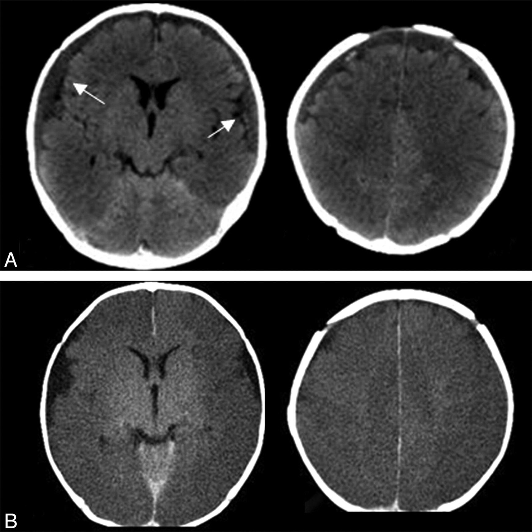
- Fig 1.
Type 1 discrepancy (major, life-threatening). A 15-week-old male infant found unresponsive. A, CT at the time of presentation (top) was initially interpreted by the trainee as a small falcine subdural hematoma and normal brain parenchyma. The staff final report documented bilateral subdural collections with regions of hemorrhages (arrows) and diffuse loss of gray-white matter differentiation consistent with edema/ischemia. B, Follow-up head CT (bottom) 7 hours later demonstrates evolution of diffuse cortical edema consistent with diffuse ischemic injury. At the time of clinician notification of the discrepancy, the patient was already being treated for presumed diffuse brain injury clinically and was on ventilator respiratory support and intracranial pressure management. Further investigation of the clinical scenario coupled with the imaging abnormalities were concerning for non-accidental trauma with a subsequent confession of inflicted injury by a caretaker.
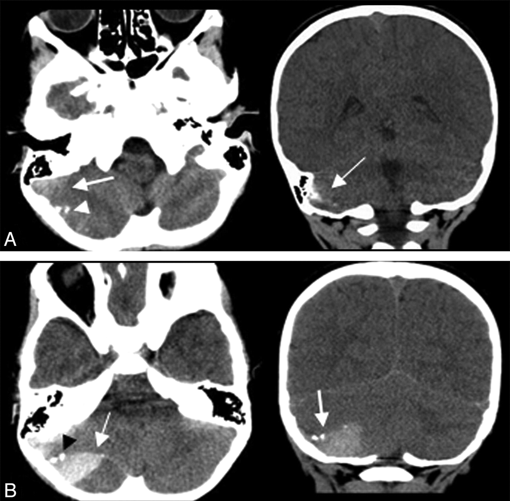
- Fig 2.
Type 1 discrepancy (major, life-threatening). A 3-year-old boy with a head injury and vomiting. A, Initial head CT axial images (left) and coronal reformat (right) were interpreted by the trainee as possible ICH versus streak artifacts (arrows). The attending radiologist thought the finding was artifacts, marking this as a discrepancy. The patient was monitored clinically for 24 hours in the hospital with no concerning symptoms and was discharged without a repeat head CT. The patient presented 3 days later with worsening headache and vomiting. B, Repeat head CT demonstrates a large mixed-attenuation posterior fossa epidural hematoma with mass effect in the location of the previously questioned artifacts (arrows). Small foci of bone attenuation are identified, displaced from the inner table (black arrowhead), retrospectively identified on the previous scan (A, white arrowhead). The patient was immediately taken to surgery for evacuation and made a complete recovery with no permanent deficits.
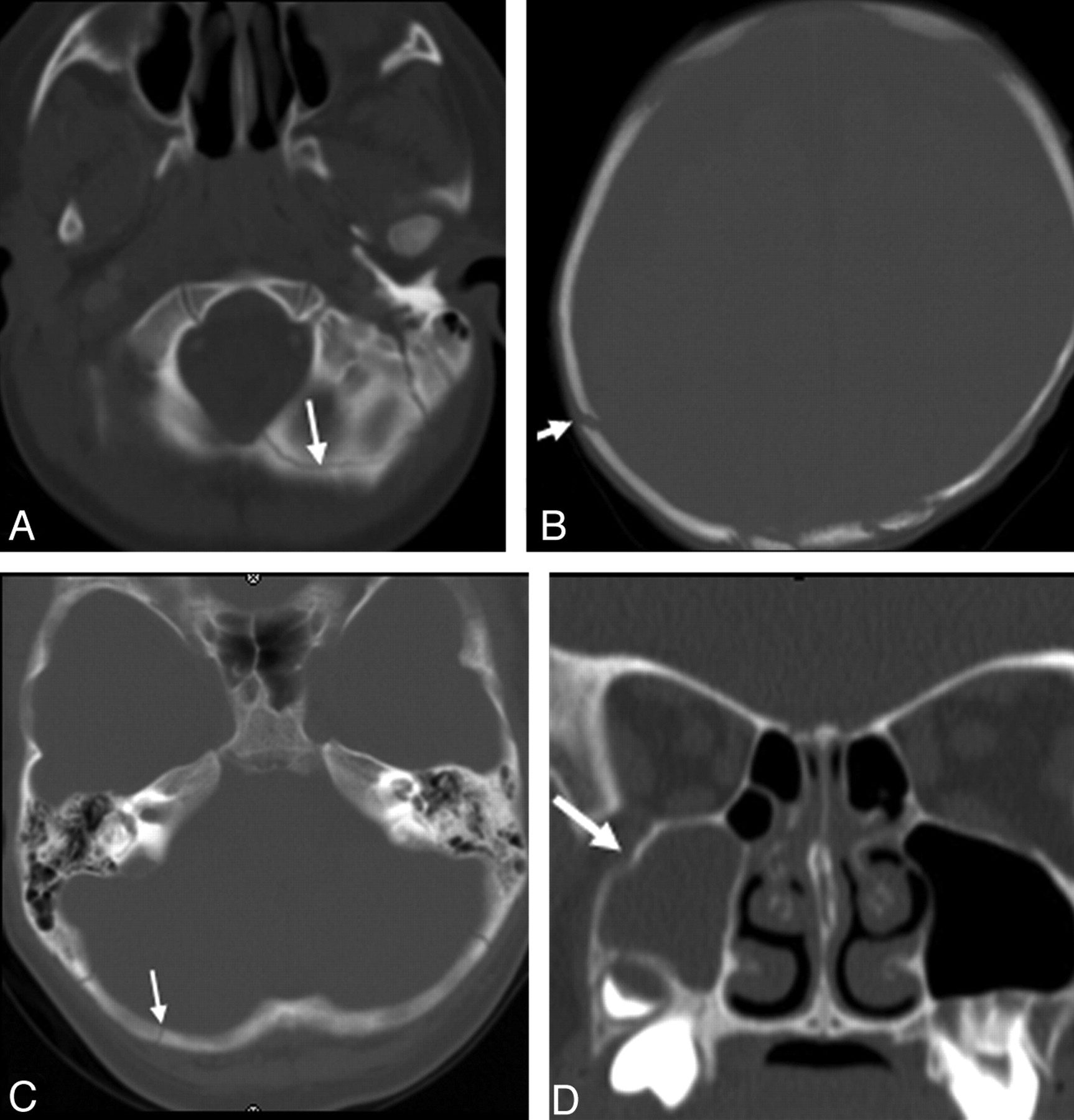
- Fig 3.
Examples of misinterpreted fractures (arrows). A, Overcall of the normal posterior intraoccipital synchondrosis as a fracture. B, Right parietal calvarial fracture. C, Nondisplaced right occipital fracture. D, Minimally displaced right maxillary fracture.
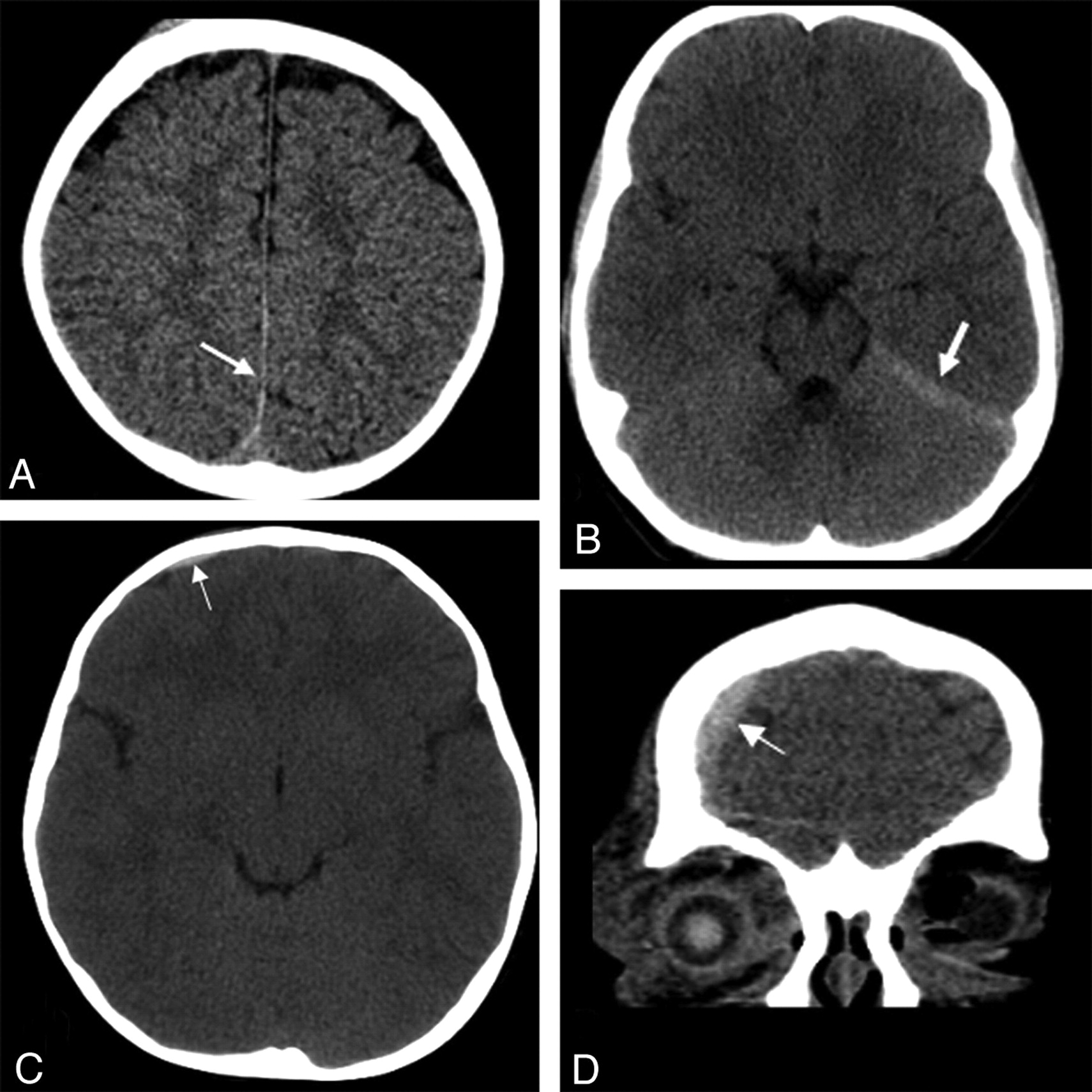
- Fig 4.
Examples of misinterpreted ICH (arrows). A, Small subdural hemorrhage along the posterior interhemispheric fissure. B, Small subdural hemorrhage along the left tentorial leaflet seen as asymmetric hyperattenuation compared with the contralateral side. C and D, Small right frontal extra-axial hemorrhage (C), more obvious on subsequently performed coronal reformats (D).
- Fig 5.
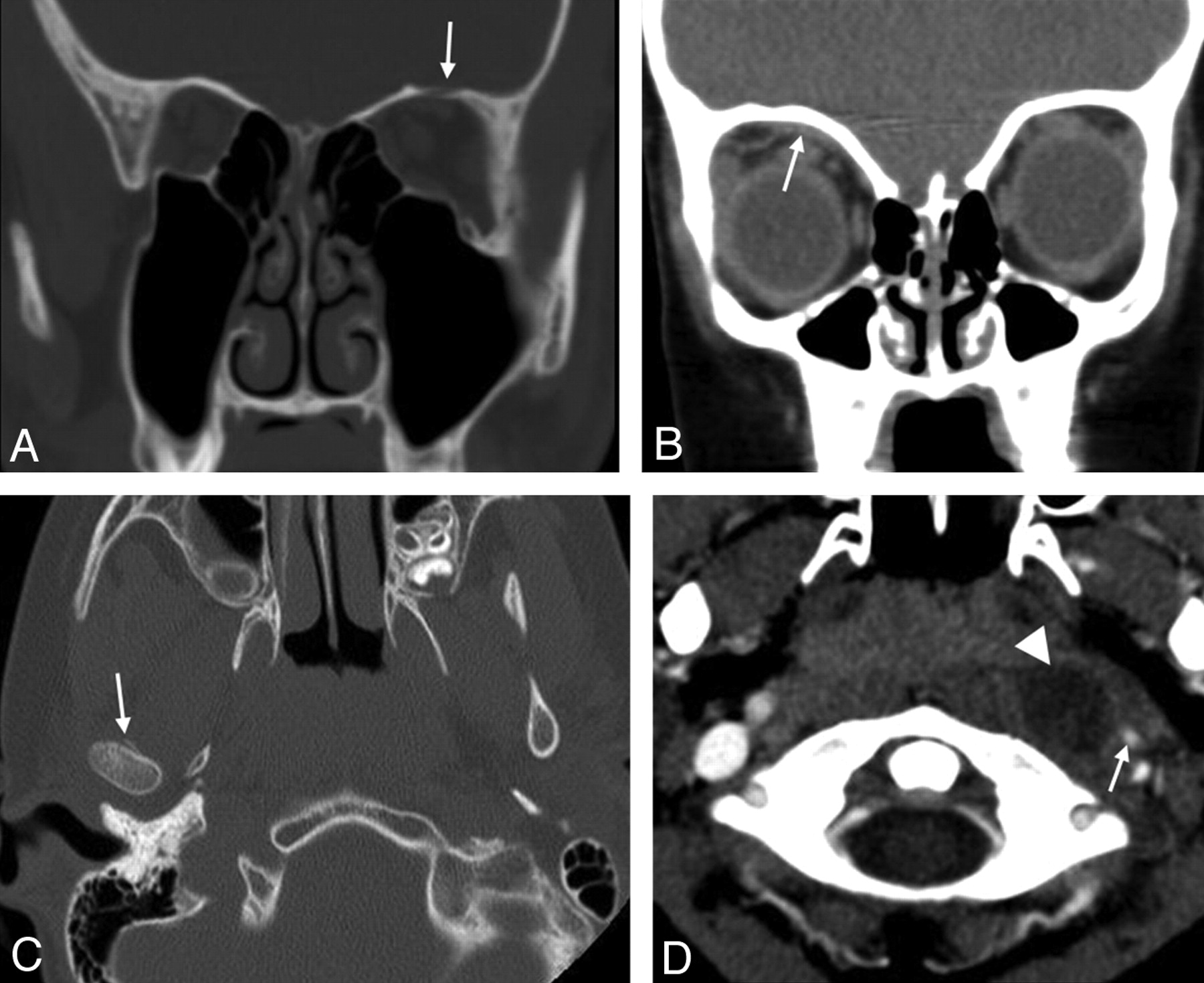
Examples of misinterpreted examinations of the face, neck, and orbits. A, Nondisplaced orbital roof fracture (arrow). B, Small extraconal hematoma (arrow) in a patient with an orbital roof fracture. C, Subtle fracture of the right mandibular condyle. D, Missed caliber change of the left internal carotid artery, secondary to retropharyngeal inflammatory disease and abscess (correctly identified by the trainee, arrowhead).
- Fig 6.
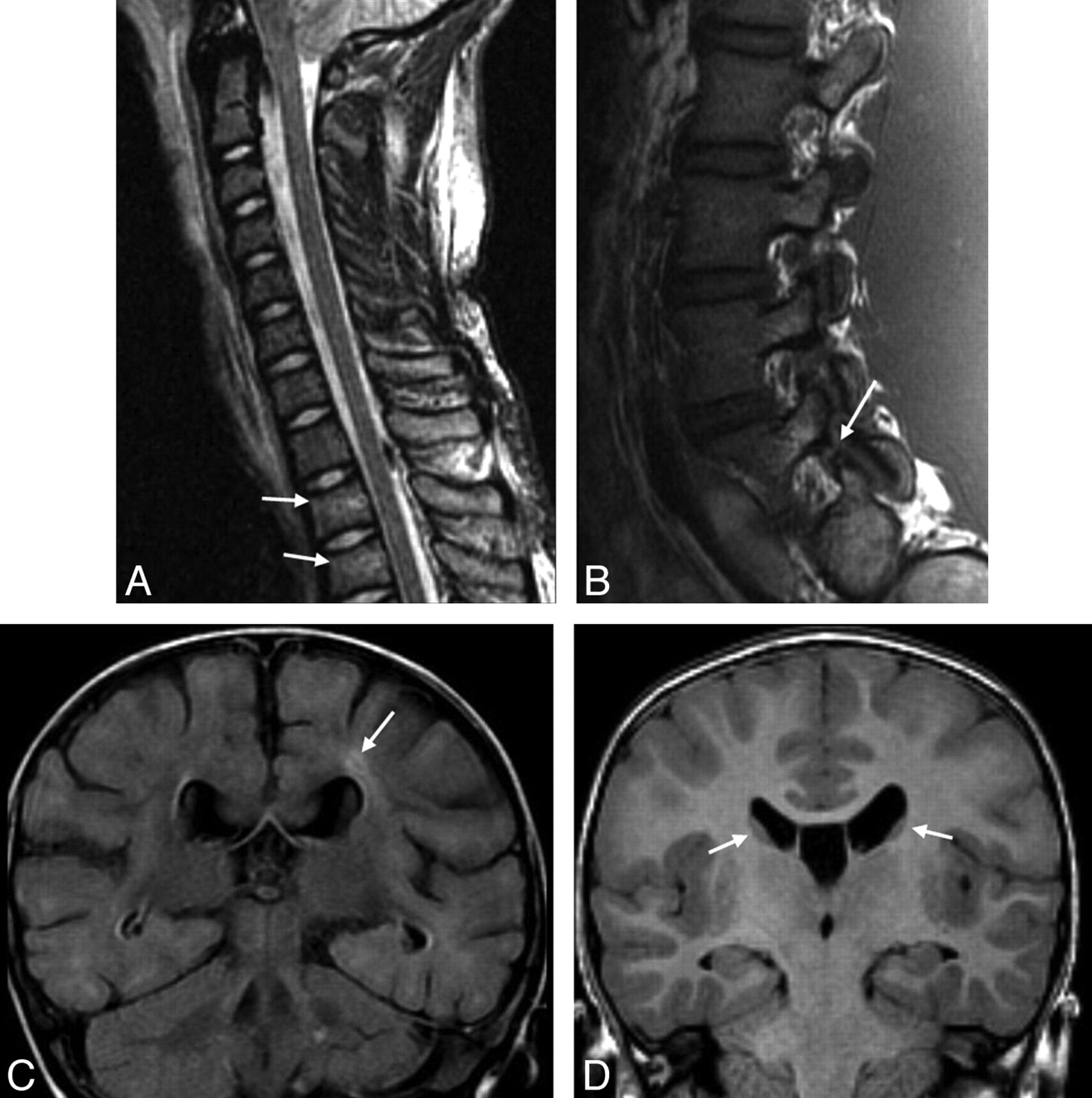
Examples of misinterpreted MR imaging examinations. A, Missed subtle upper thoracic vertebral body edema and slight height loss consistent with mild compression fractures (arrows). B, Missed L5 pars defect (arrow). C, Missed focal periventricular signal intensity in a 12-month-old child (fluid-attenuated inversion recovery sequence, arrow). D, Normal caudate nuclei misinterpreted as periventricular nodular heterotopia (arrows).


Tables
Type Number of Discrepant Trainee Reports (Rate)a 1: Major, life-threatening 6 (0.17%) 1A: Finding not originally identified 5 1B: Finding identified, incorrectly characterized 1 2: Minor, related to clinical presentation 75 (2.1%) 2A: Finding not originally identified 56 2B: Finding identified, incorrectly characterized 19 3: Minor, unrelated to clinical presentation 8 (0.23%) 3A: Finding not originally identified 8 3B: Finding identified, incorrectly characterized 0 4: Possible abnormality 17 (0.49%) 4A: Confirmed on follow-up imaging 2 4B: Not confirmed on follow-up imaging 8 4C: No follow-up imaging performed 7 5: Abnormality called when none present (overcall) 37 (1.1%) 5A: Resulting in inappropriate therapy 2 5B: Not resulting in inappropriate therapy 35 -
↵a % of total exams interpreted (N = 3496).
-
Type Number of Discrepant Reports (% of Discrepancies) 1: No effect on clinical management/outcome 97 (68) 2: No direct treatment change but imaging or clinical follow-up performed related to the discrepancy 43 (30) 3: Direct treatment change, no sequelae 3 (2.1) 4: Direct treatment change (morbidity) 0 (0) 5: Death potentially related 0 (0) Discrepancy Subclass No. (N = 143) Fracture 28 ICH 23 Focal brain parenchymal attenuation/signal abnormality 16 Other 15 Ventricle size 11 Diffuse edema/attenuation/signal 9 Mass lesion 7 Vascular 7 Abscess/fluid/edema 6 Extra-axial collection 5 Osseous, nonfracture 5 Cerebellar tonsil position 3 Intracranial/soft-tissue air 3 Vertebral alignment 3 Intraspinal hemorrhage 2 Exam Type Discrepant Exams (% of Discrepant Exams) Total Exams Read (% Discrepant)a CT Total 131 (91.6) 3102 (4.22 [3.49–4.94]) CT head 103 (72.0) 2748 (3.75 [3.02–4.48]) CT face/orbit/neck 20 (14.0) 213 (9.39 [5.24–13.54]) CT spine 8 (5.6) 141 (5.67 [1.5–9.84]) MRI total 12 (8.4) 394 (3.04 [1.22–4.86]) MRI brain 7 (4.9) 211 (3.32 [0.66–5.98]) MRI spine 4 (2.8) 113 (3.54 [0.0–7.39]) MRA 1 (0.7) 70 (1.43 [0.0–4.92]) Total exams 143 3496 -
↵a Discrepancy rate (% [95% CI]). The discrepancy rates for CT scans of the face, orbit, and neck were significantly larger compared with CT head, combined CT head and spine, and MRI. The discrepancy rate was not significantly different comparing MRI and CT, or MRI subcategories.
-
Year of Training Discrepancies/Exams Read Discrepancy Rate (95% CI)a 1 0/23 0% 2 2/80 2.5% (0.0–6.54) 3 15/128 11.7% (5.76–17.68) 4 7/50 14.0% (5.38–24.62) Fellow 119/3215 3.7% (3.03–4.37) -
↵a The combined discrepancy rate for residents was 8.54% (95% CI, 5.09%–11.99%). The combined discrepancy rate for first- and second-year residents was 1.94% (95% CI, 0.0%- 5.08%). The combined discrepancy rate for third- and fourth-year residents was 12.36%, (95% CI, 7.24%–17.48%). There were statistically significant discrepancy rates comparing residents and fellows, third- and fourth-year residents and fellows, and third- and fourth-year residents and first- and second-year residents.
-


