
Article Figures & Data
Figures
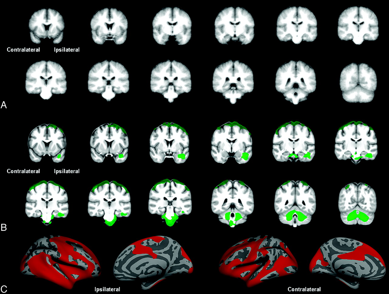
- Fig 1.
Controls versus TLE-mts. A, VBM-SPM GM differences. B, DBM Jacobian differences. C, FS-CT differences between groups. All results corrected for multiple comparisons by using permutation analysis (P < .05).
- Fig 2.
Controls versus TLE-no. A, VBM-SPM GM differences. B, DBM Jacobian differences. C, FS-CT differences between groups. All results corrected for multiple comparisons by using permutation analysis (P < .05).
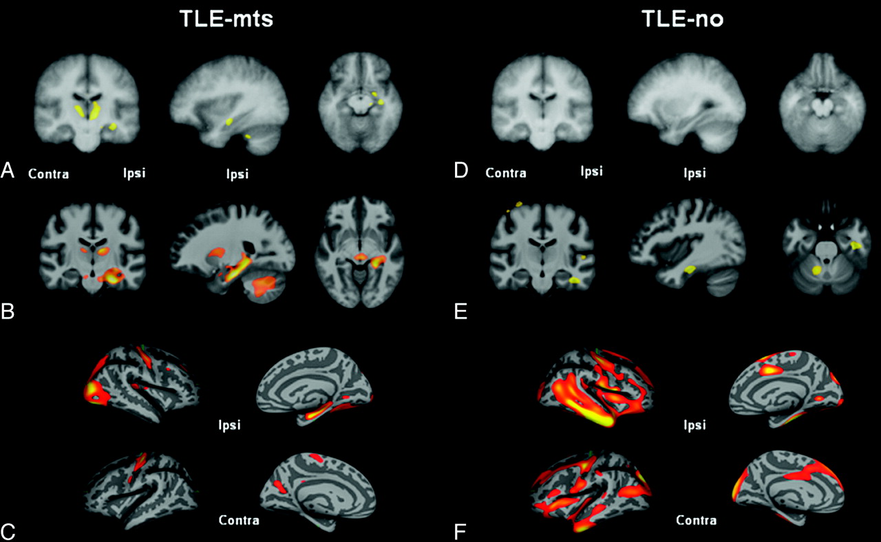
- Fig 3.
Controls versus TLE-mts and controls versus TLE-no after FDR < .05 correction for multiple comparisons. A, VBM-SPM GM differences. B, DBM Jacobian differences. C, FS-CT differences between controls and TLE-mts. D, VBM-SPM GM differences. E, DBM Jacobian differences. F, FS-CT differences between controls and TLE-no.


Tables
Method Region Cluster Size Cluster t-Statistic Freesurfer Ipsilateral temporo-occipital 20 414.11 mm2 (70 691 vertices) 6.139 DBM Bilateral mesial temporal lobe, bilateral thalamus, basal ganglia, subcortical white matter, cerebellum, and brain stem 259 230 mm2 (259 230 vertices) 7.529 VBM Ipsilateral mesial temporal lobe, bilateral thalamus, and ipsilateral basal ganglia 282 288 mm3 (282 288 voxels) 5.693 Ipsilateral temporal and bilateral occipital cortex Cerebellum and brain stem -
Note:—Clusters representing significant differences between controls and TLE-mts for each method after correction for multiple comparisons by using permutation testing.
-
Method Region Cluster Size Cluster t-Statistic Freesurfer Ipsilateral inferior and lateral temporal lobe; insula, posterior, and superior frontal lobe; and lateral and medial occipital region 62 071 mm2 (70 691 vertices) 7.679 Contralateral inferior and lateral temporal lobe; insula, posterior, and superior frontal lobe; and lateral and medial occipital region 58 961 mm2 (66 554 vertices) 4.883 DBM Cerebellum, brain stem, and ipsilateral temporal lobe 74 453 mm3 (74 453 voxels) 5.281 Bilateral superior frontal cortex, pre- and postcentral cortex, and superior pariental cortex 54 397 mm3 (54 397 voxels) 4.572 -
Note:—Clusters representing significant differences between controls and TLE-no for each method after correction for multiple comparisons by using permutation testing. No significant clusters were found using the VBM method.
-


