Advertisement

Main menu

  • Home
  • Content
    • Current Issue
    • Accepted Manuscripts
    • Article Preview
    • Past Issue Archive
    • AJNR Case Collection
    • Case of the Week Archive
    • Classic Case Archive
    • Case of the Month Archive
  • Special Collections
    • Spinal CSF Leak Articles (Jan 2020-June 2024)
    • 2024 AJNR Journal Awards
    • Most Impactful AJNR Articles
  • Multimedia
    • AJNR Podcast
    • AJNR Scantastics
    • Video Articles
  • For Authors
    • Submit a Manuscript
    • Author Policies
    • Fast publishing of Accepted Manuscripts
    • Graphical Abstract Preparation
    • Manuscript Submission Guidelines
    • Imaging Protocol Submission
    • Submit a Case for the Case Collection
  • About Us
    • About AJNR
    • Editorial Board
  • More
    • Become a Reviewer/Academy of Reviewers
    • Subscribers
    • Permissions
    • Alerts
    • Feedback
    • Advertisers
    • ASNR Home
  • Other Publications
    • ajnr

User menu

  • Alerts
  • Log in

Search

  • Advanced search
American Journal of Neuroradiology

American Journal of Neuroradiology

ASHNR American Society of Functional Neuroradiology ASHNR American Society of Pediatric Neuroradiology ASSR
  • Alerts
  • Log in

Advanced Search

  • Home
  • Content
    • Current Issue
    • Accepted Manuscripts
    • Article Preview
    • Past Issue Archive
    • AJNR Case Collection
    • Case of the Week Archive
    • Classic Case Archive
    • Case of the Month Archive
  • Special Collections
    • Spinal CSF Leak Articles (Jan 2020-June 2024)
    • 2024 AJNR Journal Awards
    • Most Impactful AJNR Articles
  • Multimedia
    • AJNR Podcast
    • AJNR Scantastics
    • Video Articles
  • For Authors
    • Submit a Manuscript
    • Author Policies
    • Fast publishing of Accepted Manuscripts
    • Graphical Abstract Preparation
    • Manuscript Submission Guidelines
    • Imaging Protocol Submission
    • Submit a Case for the Case Collection
  • About Us
    • About AJNR
    • Editorial Board
  • More
    • Become a Reviewer/Academy of Reviewers
    • Subscribers
    • Permissions
    • Alerts
    • Feedback
    • Advertisers
    • ASNR Home
  • Follow AJNR on Twitter
  • Visit AJNR on Facebook
  • Follow AJNR on Instagram
  • Join AJNR on LinkedIn
  • RSS Feeds

Welcome to the new AJNR, Updated Hall of Fame, and more. Read the full announcements.


AJNR is seeking candidates for the position of Associate Section Editor, AJNR Case Collection. Read the full announcement.


Research ArticlePediatrics

Optimizing the Detection of Subtle Insular Lesions on MRI When Insular Epilepsy Is Suspected

J. Blustajn, S. Krystal, D. Taussig, S. Ferrand-Sorbets, G. Dorfmüller and M. Fohlen
American Journal of Neuroradiology September 2019, 40 (9) 1581-1585; DOI: https://doi.org/10.3174/ajnr.A6143
J. Blustajn
aFrom the Diagnostic Neuroradiology Department (J.B., S.K.)
  • Find this author on Google Scholar
  • Find this author on PubMed
  • Search for this author on this site
  • ORCID record for J. Blustajn
S. Krystal
aFrom the Diagnostic Neuroradiology Department (J.B., S.K.)
  • Find this author on Google Scholar
  • Find this author on PubMed
  • Search for this author on this site
  • ORCID record for S. Krystal
D. Taussig
bPediatric Neurosurgery Department (D.T., S.F.-S., G.D., M.F.), Rothschild Foundation Hospital, Paris, France.
  • Find this author on Google Scholar
  • Find this author on PubMed
  • Search for this author on this site
  • ORCID record for D. Taussig
S. Ferrand-Sorbets
bPediatric Neurosurgery Department (D.T., S.F.-S., G.D., M.F.), Rothschild Foundation Hospital, Paris, France.
  • Find this author on Google Scholar
  • Find this author on PubMed
  • Search for this author on this site
  • ORCID record for S. Ferrand-Sorbets
G. Dorfmüller
bPediatric Neurosurgery Department (D.T., S.F.-S., G.D., M.F.), Rothschild Foundation Hospital, Paris, France.
  • Find this author on Google Scholar
  • Find this author on PubMed
  • Search for this author on this site
  • ORCID record for G. Dorfmüller
M. Fohlen
bPediatric Neurosurgery Department (D.T., S.F.-S., G.D., M.F.), Rothschild Foundation Hospital, Paris, France.
  • Find this author on Google Scholar
  • Find this author on PubMed
  • Search for this author on this site
  • ORCID record for M. Fohlen
  • Article
  • Figures & Data
  • Info & Metrics
  • Responses
  • References
  • PDF
Loading

Article Figures & Data

Figures

  • Tables
  • Fig 1.
    • Download figure
    • Open in new tab
    • Download powerpoint
    Fig 1.

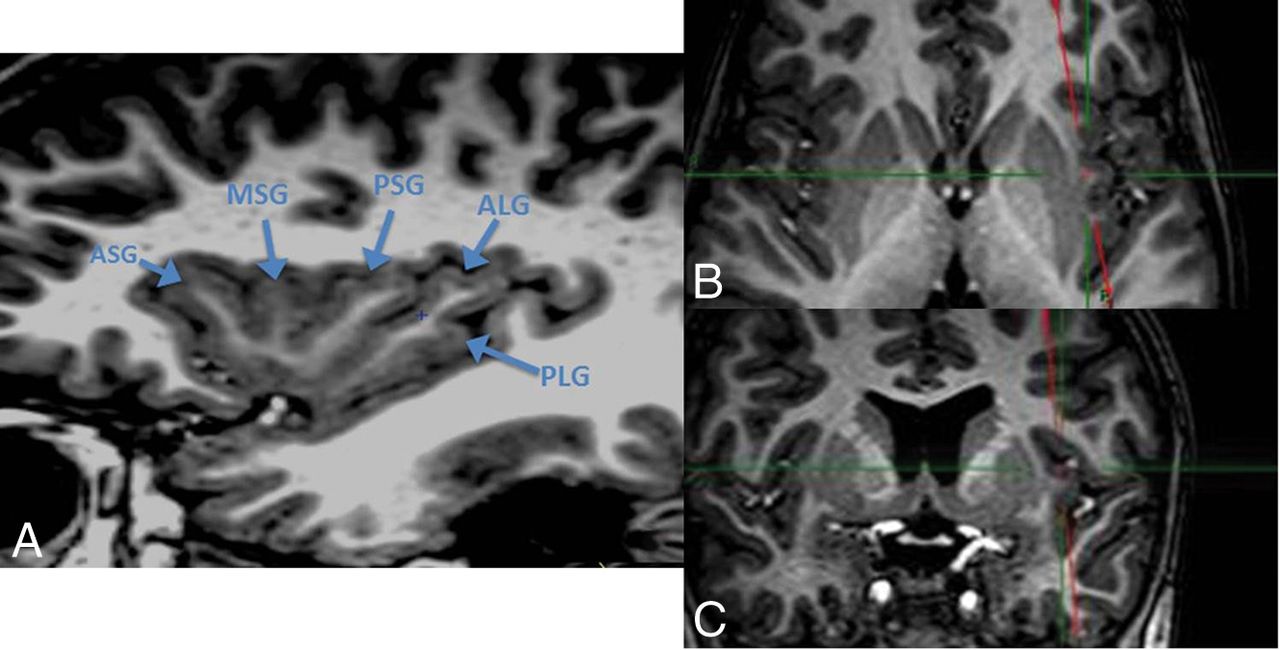
    3D-T1-weighted sequence with MPR processing showing the common gyral pattern of the insula: reference view to analyze the insula. A, T1WI oblique sagittal view, parallel to the base of the insula, anterior lobule. ASG indicates anterior short gyrus; MSG, middle short gyrus; PSG, posterior short gyrus. Posterior lobule: ALG indicates anterior long gyrus; PLG, posterior long gyrus, which appears classically shorter than the ALG. B, T1WI axial view shows the oblique cut plane parallel to the base of the insula. C, T1WI coronal view shows the oblique cut plane parallel to the base of the insula.

  • Fig 2.
    • Download figure
    • Open in new tab
    • Download powerpoint
    Fig 2.

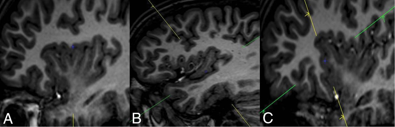
    T1WI sagittal view representation of the peri-insular sulci. A, Superior peri-insular sulcus. B, Inferior peri-insular sulcus. C, Anterior peri-insular sulcus.

  • Fig 3.
    • Download figure
    • Open in new tab
    • Download powerpoint
    Fig 3.

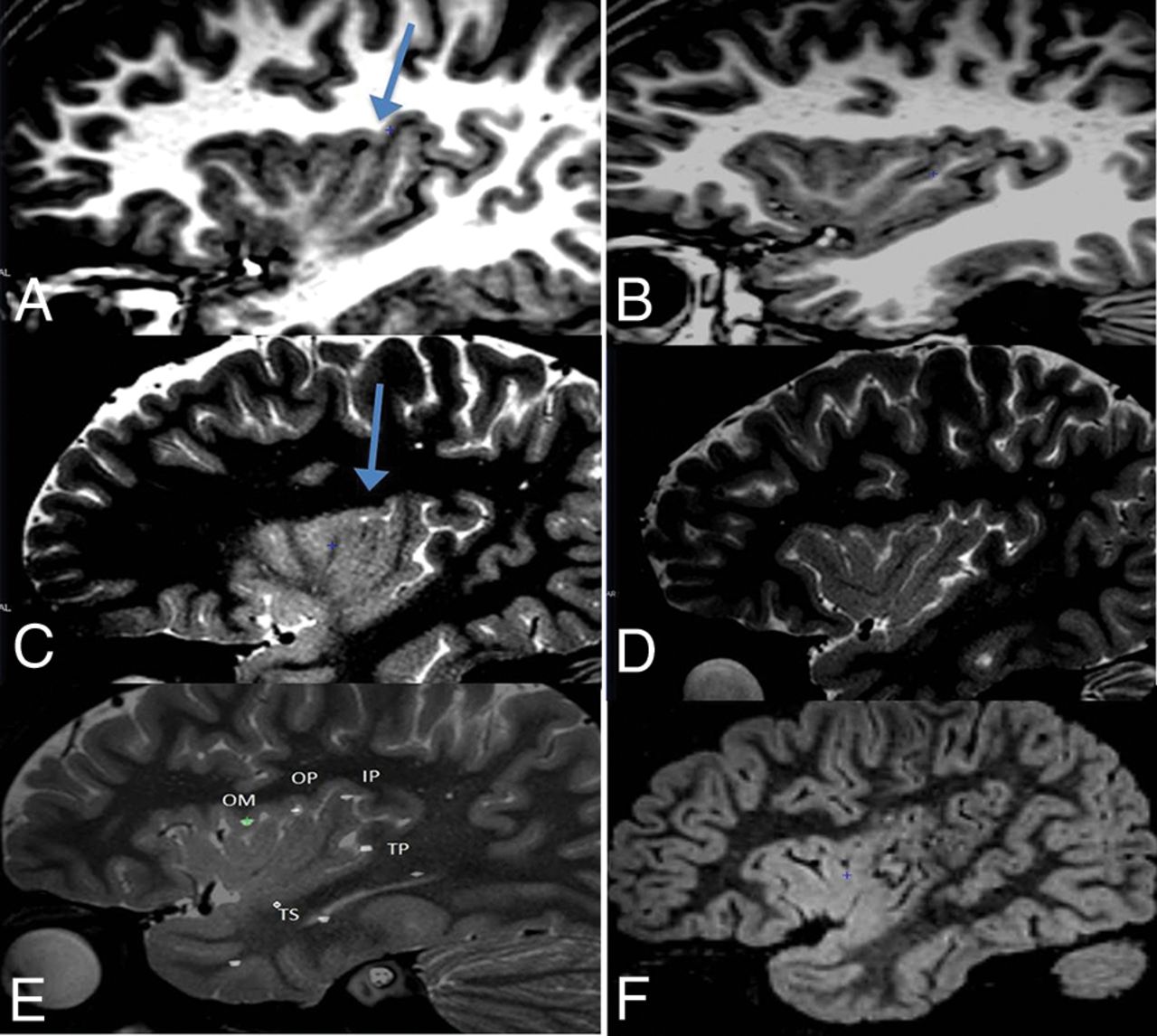
    Patient 1. Isolated insular lesion. Oblique sagittal view parallel to the base of the insula. A, T1WI oblique sagittal view shows an unusual gyral pattern of the posterior lobule of the right insula, which shows 3 gyri (arrow). B, T1WI oblique sagittal view shows a normal gyral pattern of the contralateral insula. C, T2WI oblique sagittal view shows blurring of the most anterior long gyrus of the right insula (arrow). D, T2WI oblique sagittal view shows no blurring of the contralateral insula. E, T2WI oblique sagittal view with SEEG electrodes (ictal onset zone around electrode OP). OM, OP, IP, TP, and TS indicate the names of depth electrodes. F, FLAIR sagittal view of the right insula after an operation.

  • Fig 4.
    • Download figure
    • Open in new tab
    • Download powerpoint
    Fig 4.

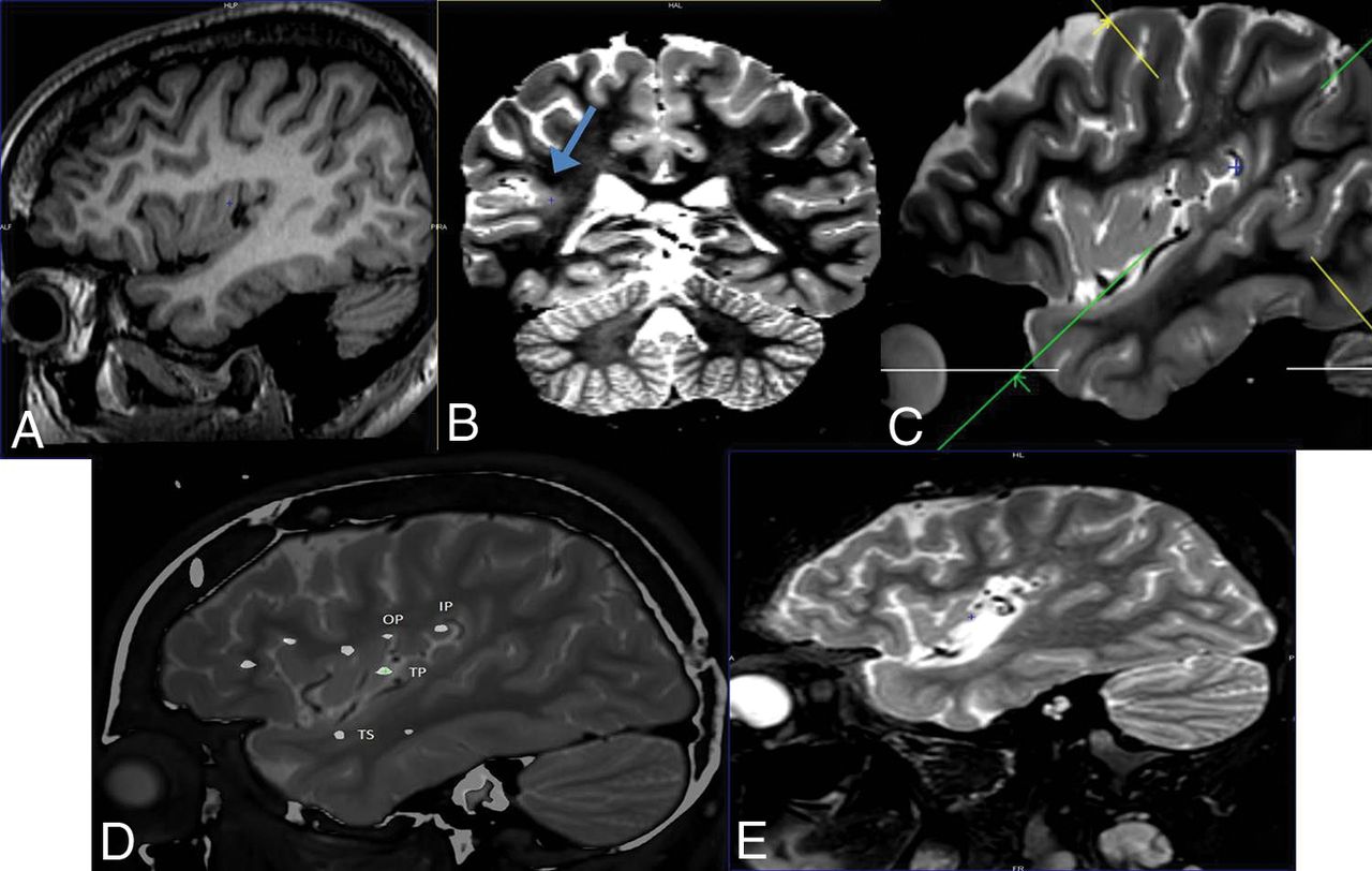
    Patient 3. Insular lesion extending into the peri-insular sulcus. A, T1WI oblique sagittal view, parallel to the base of the insula, shows hypoplasia of the posterior insular lobule. B, T2WI coronal view perpendicular to the inferior peri-insular sulcus shows blurring of the posterior portion of the right inferior peri-insular sulcus (arrow). C, T2WI sagittal view shows the cut plane perpendicular to the posterior portion of the inferior peri-insular sulcus, used to obtain the coronal view in B. D, T2WI sagittal view with SEEG electrodes (ictal onset zone around OP, IP, TP, and TS, representing the depth electrodes). E, T2WI sagittal view after surgical resection of the posterior lobule of the insula and the superior temporal gyrus.

Tables

  • Figures
    • View popup
    Table 1:

    Clinical data

    PatientSexAge at Seizure Onset (yr)Neurologic Status before SurgeryAge at Last Surgery (yr)Topography of Resective SurgeryPostsurgical DeficitPathologyFU Duration (yr)Engela
    1M2.5LH, Special education13.0Posterior Ins and posterior Op0FCD IIa4.2I
    2M3.2RH, MoMR6.2Anterior Ins and Op, + frontal DiscLeft facial paresisFCD Ib4.2I
    3F3.5RH, Normal cognitive function14.7Posterior Ins and STG0Negative2.4I
    4F0.8RH, MoMR3.0Anterior Ins+ frontal Disc0FCD IIa2.3III
    5M0.4Left hemiparesis SMR, ASD6.1Anterior Ins+ frontal Disc0FCD IIa0.9I
    6F2.0LH, MiMR7.6Posterior Ins and parietal Op, temporal Op0FCD IIb3.0III
    7M1.0LH, MoMR, ASD9.7Anterior Ins and Inferior Frontal0FCD IIb1.8II
    • Note:—LH indicates left-handed; RH, right-handed; MiMR, mild mental retardation; MoMR, moderate mental retardation; SMR, severe mental retardation; ASD, autism spectrum disorder; Op, operculum; Ins, insula; STG, superior temporal gyrus; Disc, disconnection; FU, follow-up; yr, year.

    • ↵a Engel Surgical Outcome Scale.

    • View popup
    Table 2:

    Review of presurgical MRIs in patients with intractable insular epilepsy with no initial detection of insular lesions

    Patient No.SideInsulaPeri-Insular SulcusSurrounding Structures
    Gyral PatternBlurringSulcal FormBlurringGyral PatternBlurring
    1RSupernumerary ALG+––––
    2RPoorly defined AL+Irregular anterior portion of SPSAnterior SPS–Frontal opercula, orbitofrontal
    3RHypoplasia PL−–Posterior IPSHypoplasia STGSTG, HG
    4RIrregular AL+Irregular anterior portion of SPSAnterior SPS––
    5RIrregular AL+–APS–Frontal opercula, orbitofrontal
    6LThick PL+–Posterior IPS–HG, temporal stem
    7LPoorly defined AL+Poorly defined APSAPS–Pars orbitalis
    • Note:—ALG indicates the anterior long gyrus; R, right; L, left; SPS, superior peri-insular sulcus; PL, posterior lobule; IPS, inferior peri-insular sulcus; STG, superior temporal gyrus; HG, Heschl gyrus; AL, anterior lobule; APS, anterior peri-insular sulcus; –, normal; +, present; −, absent.

PreviousNext
Back to top

In this issue

American Journal of Neuroradiology: 40 (9)
American Journal of Neuroradiology
Vol. 40, Issue 9
1 Sep 2019
  • Table of Contents
  • Index by author
  • Complete Issue (PDF)
Advertisement
Print
Download PDF
Email Article

Thank you for your interest in spreading the word on American Journal of Neuroradiology.

NOTE: We only request your email address so that the person you are recommending the page to knows that you wanted them to see it, and that it is not junk mail. We do not capture any email address.

Enter multiple addresses on separate lines or separate them with commas.
Optimizing the Detection of Subtle Insular Lesions on MRI When Insular Epilepsy Is Suspected
(Your Name) has sent you a message from American Journal of Neuroradiology
(Your Name) thought you would like to see the American Journal of Neuroradiology web site.
CAPTCHA
This question is for testing whether or not you are a human visitor and to prevent automated spam submissions.
Cite this article
J. Blustajn, S. Krystal, D. Taussig, S. Ferrand-Sorbets, G. Dorfmüller, M. Fohlen
Optimizing the Detection of Subtle Insular Lesions on MRI When Insular Epilepsy Is Suspected
American Journal of Neuroradiology Sep 2019, 40 (9) 1581-1585; DOI: 10.3174/ajnr.A6143

Citation Manager Formats

  • BibTeX
  • Bookends
  • EasyBib
  • EndNote (tagged)
  • EndNote 8 (xml)
  • Medlars
  • Mendeley
  • Papers
  • RefWorks Tagged
  • Ref Manager
  • RIS
  • Zotero
0 Responses
Respond to this article
Share
Bookmark this article
Optimizing the Detection of Subtle Insular Lesions on MRI When Insular Epilepsy Is Suspected
J. Blustajn, S. Krystal, D. Taussig, S. Ferrand-Sorbets, G. Dorfmüller, M. Fohlen
American Journal of Neuroradiology Sep 2019, 40 (9) 1581-1585; DOI: 10.3174/ajnr.A6143
del.icio.us logo Twitter logo Facebook logo Mendeley logo
  • Tweet Widget
  • Facebook Like
  • Google Plus One
Purchase

Jump to section

  • Article
    • Abstract
    • ABBREVIATIONS:
    • Methodology
    • Results
    • Discussion
    • Conclusions
    • REFERENCES
  • Figures & Data
  • Info & Metrics
  • Responses
  • References
  • PDF

Related Articles

  • No related articles found.
  • PubMed
  • Google Scholar

Cited By...

  • No citing articles found.
  • Crossref (3)
  • Google Scholar

This article has been cited by the following articles in journals that are participating in Crossref Cited-by Linking.

  • What is the cognitive footprint of insular glioma?
    Noah M. Nichols, Bahie Ezzat, Allison C. Waters, Fedor Panov, Raymund L. Yong, Isabelle M. Germano
    Frontiers in Human Neuroscience 2024 18
  • A Case of Hypermotor Seizures in Posterior Insular Cortex Epilepsy
    Dinesh M Chaudhari, Priyal ., Pushpendra Nath Renjen, Kamal Ahmad
    Cureus 2023
  • Case Report: Ictal hypersalivation: a stereoelectroencephalography exploration
    Sumika Ouchida, Armin Nikpour, David Neville Wilson, Greg Fairbrother
    Frontiers in Surgery 2025 12

More in this TOC Section

  • Comparison of Image Quality and Radiation Dose in Pediatric Temporal Bone CT Using Photon-Counting Detector CT and Energy-Integrating Detector CT
  • SyMRI & MR Fingerprinting in Brainstem Myelination
  • Venous Sinus Stenosis in Mucopolysaccharidosis I
Show more Pediatrics

Similar Articles

Advertisement

Indexed Content

  • Current Issue
  • Accepted Manuscripts
  • Article Preview
  • Past Issues
  • Editorials
  • Editors Choice
  • Fellow Journal Club
  • Letters to the Editor

Cases

  • Case Collection
  • Archive - Case of the Week
  • Archive - Case of the Month
  • Archive - Classic Case

Special Collections

  • Special Collections

Resources

  • News and Updates
  • Turn around Times
  • Submit a Manuscript
  • Author Policies
  • Manuscript Submission Guidelines
  • Evidence-Based Medicine Level Guide
  • Publishing Checklists
  • Graphical Abstract Preparation
  • Imaging Protocol Submission
  • Submit a Case
  • Become a Reviewer/Academy of Reviewers
  • Get Peer Review Credit from Publons

Multimedia

  • AJNR Podcast
  • AJNR SCANtastic
  • Video Articles

About Us

  • About AJNR
  • Editorial Board
  • Not an AJNR Subscriber? Join Now
  • Alerts
  • Feedback
  • Advertise with us
  • Librarian Resources
  • Permissions
  • Terms and Conditions

American Society of Neuroradiology

  • Not an ASNR Member? Join Now

© 2025 by the American Society of Neuroradiology All rights, including for text and data mining, AI training, and similar technologies, are reserved.
Print ISSN: 0195-6108 Online ISSN: 1936-959X

Powered by HighWire